Bacterial aggregation in infected root canal
DOI:
https://doi.org/10.17305/bjbms.2005.3228Keywords:
root canal infection, bacterial morphotype, microscopic examinationAbstract
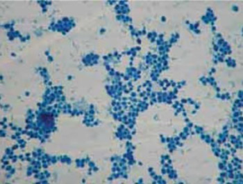
The aim of this study was to investigate different microbial morphotypes in the root canal infection associated with chronic diffuse periapical lesion. In forty cases of asymptomatic teeth with radiographically diagnosed diffuse periapical lesion we took specimens of infected tissue from the root canals at the beginning of endodontic treatment. Fixation and four different staining methods of the specimens were obtained to provide microscope examination. All examined root canal specimens were heavily infected by bacteria. The most commonly identified were cocci 92 %, small mostly G+ diplococci and large G+cocci in clusters and grapelike groups, bacilli found in 67%, coccobacilli 37%, fungi 17%, and spirochetes in 5%.
Citations
Downloads
Download data is not yet available.
Downloads
Published
20-11-2005
Issue
Section
Short Communication
Categories
How to Cite
1.
Bacterial aggregation in infected root canal. Biomol Biomed [Internet]. 2005 Nov. 20 [cited 2025 Nov. 16];5(4):35-9. Available from: https://www.bjbms.org/ojs/index.php/bjbms/article/view/3228
Received 2018-02-20
Accepted 2018-02-20
Published 2005-11-20
Accepted 2018-02-20
Published 2005-11-20