Lymphatic and blood vessels in parathyroid tumors: Immunohistochemical study with LYVE-1 and von Willebrand factor
DOI:
https://doi.org/10.17305/bb.2024.10547Keywords:
Adenoma, blood vessels, carcinoma, immunohistochemistry, lymphatic vessels, lymphatic vessel endothelial receptor, parathyroidAbstract
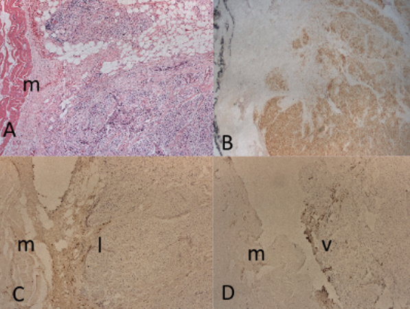
Parathyroid tumors exhibit distinct histopathological features that differentiate benign from malignant lesions. This study aimed to study characteristic distribution of lymphatic and blood vessels in normal parathyroid gland and parathyroid tumors to distinguish cancer from benign tumors. Immunohistochemical staining for lymphatic and blood vessels and parathyroid hormone was performed with parathyroid proliferating lesions including adenoma, multiglandular multiple adenomas, atypical tumor, and carcinoma. Lymphatic vessels were immunostained with lymphatic vessel endothelial receptor 1 (LYVE-1) and blood vessels were immunostained with von Willebrand factor (vWf). Normal parathyroid gland contained several round blood vessels with less linear lymphatic vessels. The less parathormone-immunostained adenomas weighing less than 2 g revealed larger blood vessels with perivascular small linear lymphatic vessels, and the larger adenomas contained proportionally larger blood vessels. Smaller multiglandular multiple adenomas were like smaller adenomas with less parathormone staining and contained larger, round blood vessels with perivascular small linear lymphatic vessels. Larger multiglandular multiple adenomas weighing more than 4 g and one atypical tumor contained nodular pattern consisted of alternating strongly parathormone-positive lobes and negative lobes with numerous, dilated blood vessels and perivascular linear lymphatic vessels. Both primary and metastatic carcinomas were strongly and diffusely positive for parathyroid hormone with numerous lymphatic and blood vessels at the invading margin. Thus, normal parathyroid has rich blood vessels which provide accessibility of minced tissues seeding for auto-transplantation. Immunostaining patterns of parathyroid hormone, lymphatic and blood vessels help to distinguish carcinoma from benign parathyroid proliferating lesions. The negative immunohistochemical staining for parafibromin will detect carriers of hyperparathyroidism-jaw tumor (HPT-JT) syndrome and would help in diagnosing parathyroid cancer in both HPT-JT carriers and sporadic patients.
Citations
Downloads
Downloads
Published
License
Copyright (c) 2024 Tatsuo Tomita

This work is licensed under a Creative Commons Attribution 4.0 International License.


