Fetal heart quantification technique improves the prenatal prediction of coarctation of the aorta: A retrospective analysis
DOI:
https://doi.org/10.17305/bb.2024.10988Keywords:
Fetal heart quantification, cardiac function, prenatal prediction, coarctation of the aorta (CoA), fetal heart analysisAbstract
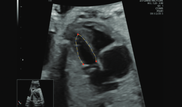
Coarctation of the aorta (CoA) ranks among the most prevalent congenital heart defects and poses a life-threatening risk if left undiagnosed. Herein, we utilized fetal heart quantification (HQ) technology to improve the prenatal prediction of CoA. A retrospective analysis was conducted on 64 fetal cases with suspected aortic arch constriction, identified through prenatal ultrasound findings between November 2020 and March 2022 at the Department of Ultrasound, Sir Run Run Shaw Hospital, Zhejiang University. According to the follow-up results, these cases were divided into two groups: 35 cases confirmed as CoA by postpartum surgery or induction, and 29 cases initially suspected of CoA prenatally but subsequently ruled out postnatally. Additionally, 88 cases of normal fetuses were randomly selected as the control group. Both conventional M-mode ultrasound techniques and Fetal HQ software were utilized for fetal analysis across all groups. Parameters related to the heart were measured, including fetal 4-CV length, width, Global Spherical Index (GSI), Mitral Annular Plane Systolic Excursion (MAPSE), areas and ratios of the left and right ventricles, as well as lengths and ratios of the left and right ventricles. Functional measurements of the left and right ventricles included ejection fraction (EF), fractional area change (FAC), global longitudinal strain (GLS), fractional shortening (FS), end-diastolic diameter (ED), and sphericity index (SI). Left ventricular (LV)-GLS, LV-FAC, LV-EF, and LV-EF Z-score could potentially differentiate between true CoA and false CoA or normal groups and serve as potential indicators for the clinical diagnosis of CoA. The receiver operating characteristic (ROC) curves indicated that LV-GLS and LV-EF Z-score have the greatest predictive power for CoA diagnosis. The segments 6-12 of FS in the confirmed CoA group were significantly lower than those in the false CoA and normal groups. Fetal HQ technology, by assessing changes in the size and shape of the heart, can provide relatively reliable parameter support for the prenatal diagnosis of fetal aortic coarctation.
Citations
Downloads
Downloads
Published
Issue
Section
Categories
License
Copyright (c) 2024 Xiaoxi Lu , Bowen Zhao, Mei Pan, Lijian Huang , Xiaomin Zhang, Xiaohui Peng, Ran Chen, Xiangdong Zhang

This work is licensed under a Creative Commons Attribution 4.0 International License.


