Microsatellite instability and B-type Raf proto-oncogene mutation in colorectal cancer: Clinicopathological characteristics and effects on survival

Authors

  • Sebnem Batur Cerrahpasa Medical Faculty, Istanbul University
  • Dogu Vuralli Bakkaloglu Cerrahpasa Medical Faculty, Istanbul University
  • Nuray Kepil Cerrahpasa Medical Faculty, Istanbul University
  • Sibel Erdamar Cerrahpasa Medical Faculty, Istanbul University

DOI:

https://doi.org/10.17305/bjbms.2016.1238

Keywords:

Microsatellite instability, DNA mismatch repair genes, B-type Raf proto-oncogene mutation, survival, polymerase chain reaction, immunohistochemistry

Abstract

Prognostic significance of microsatellite instability (MSI) status and B-type Raf proto-oncogene (BRAF) mutation in colorectal cancer is controversial. The aim of this study was to examine the clinical and pathological characteristics associated with microsatellite stability and the effect of MSI and BRAF mutation on the survival of patients with colorectal cancer. The study included 145 colorectal cancer cases. All the patients were examined for DNA mismatch repair (MMR) proteins with an immunohistochemical method. Molecular assessment of MSI was available in a subset of 41 patients. In addition, BRAF mutation analysis was performed in 30 cases. Immunohistochemically, MMR deficiency was present in 28 (19.3%) patients. Female gender (p = 0.001), lesion size ≥5 cm (p = 0.013), Crohn-like response (p = 0.035), and right-sided localization (p < 0.001) were significantly more frequent among MMR-deficient patients. The overall survival was 44.1 ± 5.1 months (95% confidence interval [CI], 33.7-54.4). Multivariate analyses identified only high tumor grade as an independent predictor of poor overall survival: odd ratio, 6.7 (95% CI 2.1-21.7), p = 0.002. In the subset of patients with available BRAF assessment (n = 30), a negative BRAF status was associated with better survival when compared to a positive BRAF status (36.7 ± 2.1 vs. 34.1 ± 7.2 months, p = 0.048). The sensitivity and specificity of the immunohistochemical method in predicting positive MSI status, with the molecular method as a reference, were 85.7% (95% CI: 56.2%-97.5%) and 88.9% (95% CI: 69.7%-97.1%), respectively. BRAF appears to be a significant predictor of a worse outcome in patients with colorectal cancer. Further studies with a large spectrum of clinical and biological variables are warranted.

Downloads

Download data is not yet available.

Author Biographies

Sebnem Batur, Cerrahpasa Medical Faculty, Istanbul University

Department of Pathology

Dogu Vuralli Bakkaloglu, Cerrahpasa Medical Faculty, Istanbul University

Department of Pathology

Nuray Kepil, Cerrahpasa Medical Faculty, Istanbul University

Department of Pathology

Sibel Erdamar, Cerrahpasa Medical Faculty, Istanbul University

Department of Pathology

References

Ferlay J, Shin HR, Bray F, Forman D, Mathers C, Parkin DM. Estimates of worldwide burden of cancer in 2008: GLOBOCAN 2008. Int J Cancer. 2010;127(12):2893-917.

http://dx.doi.org/10.1002/ijc.25516.

Center MM, Jemal A, Ward E. International trends in colorectal cancer incidence rates. Cancer Epidemiol Biomarkers Prev. 2009;18(6):1688-94.

http://dx.doi.org/10.1158/1055-9965.EPI-09-0090.

Siegel R, Ma J, Zou Z, Jemal A. Cancer statistics, 2014. CA Cancer J Clin. 2014;64(1):9-29.

http://dx.doi.org/10.3322/caac.21208.

Herzig DO, Tsikitis VL. Molecular markers for colon diagnosis, prognosis and targeted therapy. J Surg Oncol. 2015;111(1):96-102. http://dx.doi.org/10.1002/jso.23806.

Boland CR, Thibodeau SN, Hamilton SR, Sidransky D, Eshleman JR, Burt RW, et al. A National Cancer Institute Workshop on Microsatellite Instability for cancer detection and familial predisposition: development of international criteria for the determination of microsatellite instability in colorectal cancer. Cancer Res. 1998;58(22):5248-57.

Lynch HT, Smyrk T, Lynch JF. Molecular genetics and clinical-pathology features of hereditary nonpolyposis colorectal carcinoma (Lynch syndrome): historical journey from pedigree anecdote to molecular genetic confirmation. Oncology. 1998;55(2):103-8.

http://dx.doi.org/10.1159/000011843.

Thibodeau SN, Bren G, Schaid D. Microsatellite instability in cancer of the proximal colon. Science. 1993;260(5109):816-19. http://dx.doi.org/10.1126/science.8484122.

Aaltonen LA, Peltomaki P, Leach FS, Sistonen P, Pylkkanen L, Mecklin JP, et al. Clues to the pathogenesis of familial colorectal cancer. Science. 1993;260(5109):812-16.

http://dx.doi.org/10.1126/science.8484121.

Merok MA, Ahlquist T, Royrvik EC, Tufteland KF, Hektoen M, Sjo OH, et al. Microsatellite instability has a positive prognostic impact on stage II colorectal cancer after complete resection: results from a large, consecutive Norwegian series. Ann Oncol. 2013;24(5):1274-82. http://dx.doi.org/10.1093/annonc/mds614.

Lothe RA, Peltomaki P, Meling GI, Aaltonen LA, Nystrom-Lahti M, Pylkkanen L, et al. Genomic instability in colorectal cancer: relationship to clinicopathological variables and family history. Cancer Res. 1993;53(24):5849-52.

Popat S, Hubner R, Houlston RS. Systematic review of microsatellite instability and colorectal cancer prognosis. J Clin Oncol. 2005;23(3):609-18.

http://dx.doi.org/10.1200/JCO.2005.01.086.

Guastadisegni C, Colafranceschi M, Ottini L, Dogliotti E. Microsatellite instability as a marker of prognosis and response to therapy: a meta-analysis of colorectal cancer survival data. Eur J Cancer. 2010;46(15):2788-98.

http://dx.doi.org/10.1016/j.ejca.2010.05.009.

Benatti P, Gafa R, Barana D, Marino M, Scarselli A, Pedroni M, et al. Microsatellite instability and colorectal cancer prognosis. Clin Cancer Res. 2005;11(23):8332-40.

http://dx.doi.org/10.1158/1078-0432.CCR-05-1030.

Malesci A, Laghi L, Bianchi P, Delconte G, Randolph A, Torri V, et al. Reduced likelihood of metastases in patients with microsatellite-unstable colorectal cancer. Clin Cancer Res. 2007;13(13):3831-39.

http://dx.doi.org/10.1158/1078-0432.CCR-07-0366.

Samowitz WS, Curtin K, Ma KN, Schaffer D, Coleman LW, Leppert M, et al. Microsatellite instability in sporadic colon cancer is associated with an improved prognosis at the population level. Cancer Epidemiol Biomarkers Prev. 2001;10(9):917-23.

Gatalica Z, Vranic S, Xiu J, Swensen J, Reddy S. High microsatellite instability (MSI-H) colorectal carcinoma: a brief review of predictive biomarkers in the era of personalized medicine. Fam Cancer. 2016 Feb 13 (Epub ahead of print).

http://dx.doi.org/10.1007/s10689-016-9884-6.

Fransen K, Klintenas M, Osterstrom A, Dimberg J, Monstein HJ, Soderkvist P. Mutation analysis of the BRAF, ARAF and RAF-1 genes in human colorectal adenocarcinomas. Carcinogenesis. 2004;25(4):527-33. http://dx.doi.org/10.1093/carcin/bgh049.

Bokemeyer C, Van Cutsem E, Rougier P, Ciardiello F, Heeger S, Schlichting M, et al. Addition of cetuximab to chemotherapy as first-line treatment for KRAS wild-type metastatic colorectal cancer: pooled analysis of the CRYSTAL and OPUS randomised clinical trials. Eur J Cancer. 2012;48(10):1466-75. http://dx.doi.org/10.1016/j.ejca.2012.02.057.

French AJ, Sargent DJ, Burgart LJ, Foster NR, Kabat BF, Goldberg R, et al. Prognostic significance of defective mismatch repair and BRAF V600E in patients with colon cancer. Clin Cancer Res. 2008;14(11):3408-15.

http://dx.doi.org/10.1158/1078-0432.CCR-07-1489.

Ogino S, Shima K, Meyerhardt JA, McCleary NJ, Ng K, Hollis D, et al. Predictive and prognostic roles of BRAF mutation in stage III colon cancer: results from intergroup trial CALGB 89803. Clin Cancer Res. 2012;18(3):890-900.

http://dx.doi.org/10.1158/1078-0432.CCR-11-2246.

Yokota T, Ura T, Shibata N, Takahari D, Shitara K, Nomura M, et al. BRAF mutation is a powerful prognostic factor in advanced and recurrent colorectal cancer. Br J Cancer. 2011;104(5):856-62. http://dx.doi.org/10.1038/bjc.2011.19.

Vogelaar F, Van Erning F, Reimers M, Van Der Linden J, Pruijt J, Van Den Brule A, et al. The prognostic value of Microsatellite Instability, KRAS, BRAF and PIK3CA mutations in stage II colon cancer patients. Mol Med. 2015:1-26. DOI: 10.2119/molmed.2015.00220.

Lin CC, Lai YL, Lin TC, Chen WS, Jiang JK, Yang SH, et al. Clinicopathologic features and prognostic analysis of MSI-high colon cancer. Int J Colorectal Dis. 2012;27(3):277-86.

http://dx.doi.org/10.1007/s00384-011-1341-2.

Kaur G, Masoud A, Raihan N, Radzi M, Khamizar W, Kam LS. Mismatch repair genes expression defects & association with clinicopathological characteristics in colorectal carcinoma. Indian J Med Res. 2011;134:186-92.

Alexander J, Watanabe T, Wu TT, Rashid A, Li S, Hamilton SR. Histopathological identification of colon cancer with microsatellite instability. Am J Pathol. 2001;158(2):527-35. http://dx.doi.org/10.1016/S0002-9440(10)63994-6.

Lin CC, Lin JK, Lin TC, Chen WS, Yang SH, Wang HS, et al. The prognostic role of microsatellite instability, codon-specific KRAS, and BRAF mutations in colon cancer. J Surg Oncol. 2014;110(4):451-7. http://dx.doi.org/10.1002/jso.23675.

Iachetta F, Domati F, Reggiani-Bonetti L, Barresi V, Magnani G, Marcheselli L, et al. Prognostic relevance of microsatellite instability in pT3N0M0 colon cancer: a population-based study. Intern Emerg Med. 2016;11(1):41-6. DOI: 10.1007/s11739-015-1285-6.

Deschoolmeester V, Van Damme N, Baay M, Claes K, Van Marck E, Baert FJ, et al. Microsatellite instability in sporadic colon carcinomas has no independent prognostic value in a Belgian study population. Eur J Cancer. 2008;44(15):2288-95.

http://dx.doi.org/10.1016/j.ejca.2008.06.043.

Shin US, Cho SS, Moon SM, Park SH, Jee SH, Jung EJ, et al. Is microsatellite instability really a good prognostic factor of colorectal cancer? Ann Coloproctol. 2014;30(1):28-34.

http://dx.doi.org/10.3393/ac.2014.30.1.28.

Sargent DJ, Marsoni S, Monges G, Thibodeau SN, Labianca R, Hamilton SR, et al. Defective mismatch repair as a predictive marker for lack of efficacy of fluorouracil-based adjuvant therapy in colon cancer. J Clin Oncol. 2010;28(20):3219-26.

http://dx.doi.org/10.1200/JCO.2009.27.1825.

Saridaki Z, Souglakos J, Georgoulias V. Prognostic and predictive significance of MSI in stages II/III colon cancer. World J Gastroenterol. 2014;20(22):6809-14.

http://dx.doi.org/10.3748/wjg.v20.i22.6809.

Sinicrope FA, Foster NR, Thibodeau SN, Marsoni S, Monges G, Labianca R, et al. DNA mismatch repair status and colon cancer recurrence and survival in clinical trials of 5-fluorouracil-based adjuvant therapy. J Natl Cancer Inst. 2011;103(11):863-75.

http://dx.doi.org/10.1093/jnci/djr153.

Samowitz WS, Curtin K, Wolff RK, Tripp SR, Caan BJ, Slattery ML. Microsatellite instability and survival in rectal cancer. Cancer Causes Control. 2009;20(9):1763-8.

http://dx.doi.org/10.1007/s10552-009-9410-3.

Farina-Sarasqueta A, van Lijnschoten G, Moerland E, Creemers GJ, Lemmens VE, Rutten HJ, et al. The BRAF V600E mutation is an independent prognostic factor for survival in stage II and stage III colon cancer patients. Ann Oncol. 2010;21(12):2396-402.

http://dx.doi.org/10.1093/annonc/mdq258.

Roth AD, Tejpar S, Delorenzi M, Yan P, Fiocca R, Klingbiel D, et al. Prognostic role of KRAS and BRAF in stage II and III resected colon cancer: results of the translational study on the PETACC-3, EORTC 40993, SAKK 60-00 trial. J Clin Oncol. 2010;28(3):466-74.

http://dx.doi.org/10.1200/JCO.2009.23.3452.

Taieb J, Zaanan A, Le Malicot K, Julie C, Blons H, Mineur L, et al. Prognostic effect of BRAF and KRAS mutations in patients with Stage III colon cancer treated with Leucovorin, Fluorouracil, and Oxaliplatin with or without Cetuximab: a post hoc analysis of the PETACC-8 Trial. JAMA Oncol. 2016;14:1-11.
http://dx.doi.org/10.1001/jamaoncol.2015.5225.

Microsatellite instability and B-type Raf proto-oncogene mutation in colorectal cancer: Clinicopathological characteristics and effects on survival

Downloads

Additional Files

Published

2016-11-10

How to Cite

1.
Batur S, Vuralli Bakkaloglu D, Kepil N, Erdamar S. Microsatellite instability and B-type Raf proto-oncogene mutation in colorectal cancer: Clinicopathological characteristics and effects on survival. Biomol Biomed [Internet]. 2016Nov.10 [cited 2023Mar.26];16(4):254-60. Available from: https://www.bjbms.org/ojs/index.php/bjbms/article/view/1238

Issue

Section

Pathology

INTRODUCTION

Colorectal cancer (CRC) is one of the most common malignancies in the developed world, and it is on the rise in developing countries; thus, representing a global public health challenge [1-3]. Most CRCs develop sporadically without evidence of family history or genetic predisposition [4].

The microsatellite instability (MSI) pathway is among the several proposed mechanisms for the development of colon cancer. Microsatellites are repeats of short DNA motifs, 1-5 base pairs in length, distributed throughout the genome, particularly in noncoding regions. MSI is a change in the composition of these microsatellites within tumor tissue [5]. Such instabilities result from deficient function of DNA mismatch repair (MMR) genes, thus indicating a tendency toward increased genetic mutations [6-8]. Actually, DNA MSI represents a molecular manifestation of DNA MMR deficiency. Deficiency of MMR can be detected by immunohistochemical methods targeting MMR proteins in the tumor or using molecular methods utilizing polymerase chain reaction (PCR). Several studies examined the association between MSI and prognosis of CRC, most demonstrating better prognosis in MSI patients [9-16].

The B-type Raf proto-oncogene (BRAF) encodes an enzyme that takes part in intracellular signaling and cell growth, and BRAF mutation has been frequently observed in CRC, supporting its role in tumorigenesis [4,17]. BRAF mutation is also associated with worse outcomes in colorectal cancer, although conflicting results have been reported [18-21].

This study aimed to examine the clinicopathological characteristics associated with microsatellite stability and the effect of MSI and BRAF mutations on the survival of patients with colorectal cancer. In addition, we examined the reliability of immunohistochemical evaluation of microsatellite stability status in comparison with PCR-based assessment.

MATERIALS AND METHODS

Patients

This retrospective study included 145 colorectal cancer cases diagnosed between 2012 and 2013, with available MSI findings. The medical records were reviewed for demographical and clinical characteristics. In addition, hematoxylin and eosin stained preparations were re-examined. Cases with subserosa (T3) and peritoneal invasion (T4) were selected. All patients were examined for DNA MMR proteins with an immunohistochemical method, and molecular assessment of MSI was available in a subset of 41 patients. In addition, BRAF mutation analysis was performed in 30 cases. During follow-up, all patients or relatives were assessed by phone to get information on survival status.

Immunohistochemical assessment of DNA mismatch repair proteins

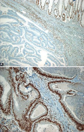
All immunohistochemical staining procedures were performed using an automated device (BenchMark XT IHK/ISH Staining Module, Ventana Medical Systems Inc., Medical Systems, Tucson, AZ, US). About 4 µm thick sections of 10% paraffin-fixed blocks were obtained from a site best representing the tumor and left overnight at 40°C on positively charged slides. Deparaffinization was attained using solutions compatible with the staining system, and the preparations were rehydrated using decreasing concentrations of alcohol solutions and distilled water. To retrieve antigens, they were left in a 10 mmol/l buffered citrate solution at 36°C for 30 minutes. Then commercial primary monoclonal antibodies were applied to the sections: MLH1 (Clone GM011, Genemed Biotechnologies, South San Francisco, USA), MSH2 (Clone G219-1129, Cell Marque Corporation. Rocklin, California, USA), MSH6 (Clone GM024, Genemed Biotechnologies, South San Francisco, USA), and PMS2 (Clone A16-4, Biocare Medical, Pike Lane, Concord, USA 1:50). After that, the preparations were counterstained with 0.01% hematoxylin and rinsed. Negative protein expression was defined as the absence of nuclear staining in tumor cells with concurrent internal positive controls, which were non-neoplastic colonic mucosa, stromal cells, infiltrating lymphocytes, or the centers of lymphoid follicles. If at least one marker showed negative expression, then the tumor was considered DNA MMR protein deficient. Figure 1 shows example images for the immunohistochemical evaluation of the MMR protein status.

FIGURE 1: Immunohistochemical evaluation of mismatch repair protein status in colorectal carcinoma. (A) Loss of MLH-1 expression is evident in carcinoma tissue (lower part of the figure) while it is preserved in normal tissue (upper part of the figure) (100× magnification). (B) Normal expression of MSH2 in carcinoma tissue. (200× magnification)

Analysis of microsatellite instability with PCR

MSI was examined using molecular methods in 41 patients. For each patient, a pair of tissue samples (one from the tumor and one from a tumor-free site) was selected from formalin fixed paraffin embedded (FFPE) tissue. Using microtome, 5-6 sections, 5-10 µm thick, were obtained from each tissue and placed in Eppendorf tubes separately. The Qiagen FFPE Tissue Kit was used for DNA extraction. Following deparaffinization and lysis, released DNA was passed through nitrocellulose filter colons, purified with rinsing solutions, and prepared for use or storage with elusion solution. The purity and concentration of DNA were confirmed with spectrophotometry. Next, samples from normal and tumor tissues were amplified using a Techne 3200 PCR device and appropriate software. For the amplification reaction, forward primers were labeled with a WellRED Dye while reverse primers were unlabeled; thus, both strands of the target DNA segments were amplified and became visible on the device. The following MSI markers were used: BAT26, BAT25, NR24, NR21, and NR27. DNA fragment analysis was carried out on a Beckman-Coulter DNA sequence analysis device using the capillary electrophoresis method. The samples were loaded on a 96-well plate, and the analysis was initiated with the sequence analysis device. If allelic profiles of the microsatellite markers of normal DNA and tumor DNA are similar, then there is no MSI. But if the allelic profiles do not match each other, this indicates MSI.

Results were interpreted as follows: High-frequency MSI (MSI-H) was considered when two or more of the 5 markers showed instability; low-frequency MSI (MSI-L) was considered when instability was observed at a single locus; microsatellite stable (MSS) was determined when none of the markers showed instability.

BRAF analysis with real-time PCR

Appropriately prepared DNA from the tumor region of the samples (as described above) from 30 patients was used for BRAF analysis. Conditions for the analysis were optimized for a Light Cycler480 Real Time PCR device (Roche Diagnostics GmbH). Identification of mutations was performed using the Entrogen BRAF Codon600 Mutation Analysis Kit (V600E) (Entrogen Inc.). Master mixture for the reaction was prepared as instructed by the kit manufacturer with following constituents: ×2 PCR reaction solution 15 µl, primer solution 6 µl, DNA sample 20 ng, completed to 30 µl with water. A positive control (PC) was used for the construction of reference curve, and contamination was checked with a negative control (NTC). After loading the device, PCR program was initiated: denaturation, at 95°C for 10 minutes; quantification, during denaturation at 95°C for 15 seconds through 40 cycles; annealing, at 60°C for 30 seconds. The amplification and quantification of target DNA were carried out simultaneously throughout the cycles. Complete conformity of target DNA and fluorescent-labeled primer pairs, specifically designed to detect the mutation site, results in a fluorescent emission which is recorded and analyzed using a specific software and based on cycle threshold (Ct) values. If a mutation is present, the fluorescence emission occurs before the 37th cycle. In the case of a wild-type sequence (no mutation), only basal fluorescent emission is detected.

Statistical analysis

SPSS version 21 was used for the analyses of data. Continuous variables were compared using the Student t-test for independent samples, and categorical variables were compared using the Pearson’s Chi-square test or Fisher’s exact test, where appropriate. Overall survival estimates were calculated using the Kaplan-Meier test and univariate comparisons were performed using the log-rank test. Overall survival was defined as the time elapsed between the date of diagnosis and death from any cause, and patients alive at the last follow-up were censored. For multivariate analysis, potential univariate predictors of overall survival were entered into a Cox proportional hazards model to identify independent predictors. In addition, diagnostic performance of the immunohistochemical method in predicting MSI status was calculated in reference to the molecular method for the patients with both assessments available. Two-sided p < 0.05 was considered statistically significant.

RESULTS

Patients

The mean age was 64.0 ± 12.1 years. Immunohistochemically, there was MMR protein deficiency in 28 (19.3%) patients. The distribution of the clinical parameters by the MMR status is given in Table 1. The MMR status did not differ with regard to the age, histological type, tumor grade, or lymph node involvement (p > 0.05 for all, respectively). However, the female gender (p = 0.001), lesion size ≥5 cm (p = 0.013), and Crohn-like response (p = 0.035) were significantly more frequent among the MMR protein deficient patients; in addition, the tumor was located more commonly at the right colon in these patients (p < 0.001). The distribution of clinical parameters by the MSI status, summarized in Table 2, was similar to the distribution of the MMR status.

TABLE 1: Distribution of clinical parameters by MMR status assessed by the immunohistochemical method
TABLE 2: Distribution of clinical parameters by MSI status

Survival

During the mean 23.8 ± 10.1 months of the follow-up, 33 deaths occurred (22.8%). The overall survival was 44.1 ± 5.1 months (95% confidence interval [CI], 33.7-54.4). Figure 2A shows the Kaplan-Meier curve of the overall survival for the entire study population. Table 3 shows the univariate analysis results for potential predictors including the MMR status for the overall survival. A high tumor grade was associated with shorter overall survival (26.0 ± 3.6 vs. 51.8 ± 6.1 months, p = 0.002). The multivariate analyses identified only high tumor grade as an independent predictor of poor overall survival: Odd ratio, 6.7 (95% CI 2.1-21.7), p = 0.002. Figure 2B shows Kaplan-Meier curves by the tumor grade status. In the subset of the patients with available BRAF assessment (n = 30), the percentage of BRAF mutation was 33.3% (10/30) and a negative BRAF status was associated with better survival when compared to positive BRAF status (36.7 ± 2.1 vs. 34.1 ± 7.2 months, p = 0.048) (Figure 2C). About 4 of 10 BRAF positive cases and 7 of 20 BRAF negative cases were MSI, all of which were MSI-H. When combinations with microsatellite stability status were further analyzed, BRAF positive plus MSI tumors had poorer overall survival (14.4 ± 7.6 months) when compared to BRAF negative plus MSI tumors (36.8 ± 1.2, p = 0.001) and BRAF negative plus MSS tumors (35.8 ± 3.7, p = 0.011).

FIGURE 2: (A) Kaplan-Meier curve of the overall survival for the entire study population, (B) Kaplan-Meier curves by the tumor grade status. The blue curve indicates low grade and green curve indicates high-grade tumor, (C) Kaplan-Meier curves by B-type Raf proto-oncogene (BRAF) status. The blue curve indicates BRAF negative status and green curve indicates BRAF positive status
TABLE 3: Univariate analysis for overall survival with the log-rank test

Molecular versus immunohistochemical analysis of MSI status

The molecular analysis of the MSI status was also available in a subset of patients (n = 41). Twenty-seven cases were MSS, 14 cases were MSI-H and there were no MSI-L cases. The sensitivity and specificity of the immunohistochemical method in predicting positive MSI status, with the molecular method as a reference, were 85.7% (95% CI: 56.2%-97.5%) and 88.9% (95% CI: 69.7%-97.1%), respectively. False positivity and false negativity rates were 7.3% and 4.9%, respectively.

DISCUSSION

This study examined the microsatellite stability status of colorectal cancer patients along with its relation to clinicopathological variables and effects on survival. Although no effect of MSI on survival was detected, BRAF mutation was significantly associated with worse survival. In addition, the immunohistochemical and molecular methods were in good agreement in identifying the MSI status of the patients.

The overall prevalence of the MSI status evaluated by the MMR immunohistochemistry was 19.3% in this study, showing significant association with the female gender, large lesion size at diagnosis, Crohn-like response, and right-sided localization. These findings are largely in agreement with previous studies although different methods have been utilized for the assessment of MSI. Merok et al. found an overall MSI prevalence of 14% among colorectal cancer patients who underwent resection, with highest prevalence in females (19%) and in proximal colon cancer (29%) [9]. Another study by Vogelaar et al. found an overall prevalence of 23%, where MSI positivity was significantly associated with female sex, right-sided location, and poor differentiation [22]. Another study examining specifically the clinicopathological features of MSI-H colon cancers showed significant association with poor differentiation, proximal location, high mucin content, and female predominance [23]. However, in the study by Kaur et al. where the overall prevalence of abnormal DNA MMR protein expression was 18.7%, this condition was associated with proximal lesion, right-sided location, mucinous type, and poor differentiation, but not with the age, gender, or ethnic status of the patient [24]. In our study, we could not find a statistically meaningful correlation between the MSI status and histological grade; this is most probably due to low/high-grade case ratio which is high in this study. Regarding peritumoral Crohn’s-like lymphoid reaction, it emerged as an intensive marker for MSI-H tumors in the study by Alexander et al. [25].

To date, several studies with diverse methodologies have examined the role of MSI status in predicting survival and treatment outcomes in colorectal cancer, mostly supporting favorable outcomes in association with MSI. The survival advantage of MSI tumors has been demonstrated in a number of studies [15,23,26], and two large meta-analyses confirmed these findings. One of these meta-analyses examined data from a large pool of colorectal cancer patients (n = 12782) and showed survival advantage of MSI both in terms of overall survival and disease-free survival (DFS) [12]. In another meta-analysis with pooled data from 7642 cases, MSI had advantage in terms of overall survival and survival advantage persisted for the subgroups of patients with locally advanced disease and patients treated with adjuvant fluorouracil (FU) [11].

Several studies have relatively distinct findings with regard to different subgroups and different outcomes. For example, a study from Italy examined the prognostic significance of MSI particularly in patients with pT3N0M0 tumors and found an advantage for MSI-H tumors in terms of relapse-free survival [27]. Benatti et al. found survival advantage of MSI-H particularly for stage II and III cancers [13]. In another study, MSI was strongly associated with a decreased likelihood of lymph node and distant organ metastases at diagnosis, regardless of the pathologic features of the tumors [14]. A Norwegian study found survival advantage in favor of MSI tumors, but this finding was only confined to stage II tumors [9].

On the other hand, several studies did not find advantage for MSI tumors. In a Belgian study, MSI status did not emerge as an independent predictor of survival, instead tumor stage and differentiation significantly affected survival outcome [28], similar to the findings of our study. In a study by Shin et al., overall survival and DFS did not differ with regard to MSI status. However, in the subgroup of patients with stage II disease, DFS was worse for patients with MSI-H tumors [29]. In a recent study, with stage II colon cancer patients, a trend toward worse overall survival was seen in patients with an MSI tumor when compared to patients with an MSS tumor; however, the difference did not reach statistical significance [22].

Another issue to be addressed regarding the outcomes for MSI-H tumors is their response to therapy relative to microsatellite stable tumors. Some evidence suggests that MSI patients may not benefit or may get reduced benefit from FU-based adjuvant therapy, despite overall better survival of these patients [30-32]. Thus, some of the discrepancies for survival advantage of MSI status across different studies may well arise from the differences in the treatment protocols (i.e., with or without adjuvant treatment).

In contrast to the findings for colorectal cancer, Samowitz et al. found more than two times risk of dying of rectal cancer for patients with MSI-H tumors, and the authors attributed this finding to the relatively high frequency of Lynch-associated cancers among rectal MSI-H tumors [33].

Similar to several abovementioned studies, the MSI status did not emerge as an independent predictor of survival in this study. Previous studies suggest that the effect of microsatellite stability status on survival seem to be substantially affected by several other parameters; thus, an accurate stratification of MSI patients for other parameters such as tumor stage, treatment type, tumor site, and other molecular markers seems to be necessary to better interpret MSI status and to utilize it for decision-making.

Several studies tested the predictive value of BRAF mutation in diverse settings of colorectal cancer. Farina-Sarasqueta et al. examined the prognostic significance of BRAF mutations in stage II and stage III disease, where BRAF mutation was an independent prognostic factor indicating worse outcomes in terms of overall survival and cancer-specific survival [34]. Another study found an association with BRAF mutation and worse survival in stage III colon cancer [20]. Yokota et al. identified BRAF mutation as a powerful prognostic factor in advanced and recurrent colorectal cancer treated with anti-epidermal growth factor receptor therapies [21]. However, studies stratifying cases according to both BRAF mutation and MSI status obtained diverse results. In the study by French et al., BRAF V600E mutation status did not affect disease-free or overall survival. However, a subgroup analysis of patients without BRAF mutation but with deficient MMR genes had significantly improved overall survival compared to other subgroups with regard to combined BRAF and MMR status [19]. In another study, BRAF mutation was not predictive for recurrence-free survival, but associated with worse overall survival, particularly for MSI-low and MSI-stable tumors [35]. Taieb et al. examined the prognostic importance of BRAF mutation in stage III colon cancer patients that received adjuvant chemotherapy and found that in patients with microsatellite-stable tumors BRAF V600E mutation was independently associated with worse clinical outcomes. In contrast, in patients with MSI tumors, it was associated with significantly longer DFS [36]. The present study found an association between better overall survival and the MSI plus BRAF negative status and MSS plus BRAF negative status. However, it is important to emphasize that the number of patients in each subgroup was relatively small.

This study found relatively high diagnostic value of the immunohistochemical method when the PCR method is used as a reference, with almost 90% sensitivity and specificity and less than 5% false negativity rate. Although the use of PCR method is recommended in most settings, in situations where PCR is not available, the immunohistochemical method may be an alternative.

The main limitation of this study is the retrospective design and a low number of patients, which did not allow testing the effects of too many parameters on the study outcomes.

CONCLUSION

In our study, BRAF mutation seems to be a significant predictor of worse outcome in colorectal cancer patients with MSI. However, further studies with better design, taking a large spectrum of clinical and biological variables into consideration, are warranted to draw firm conclusions.

DECLARATION OF INTERESTS

The authors declare no conflict of interests.

REFERENCES

  1. , , , , , (). Estimates of worldwide burden of cancer in 2008: GLOBOCAN 2008. Int J Cancer. http://dx.doi.org/10.1002/ijc.25516
  2. , , (). International trends in colorectal cancer incidence rates. Cancer Epidemiol Biomarkers Prev. http://dx.doi.org/10.1158/1055-9965.EPI-09-0090
  3. , , , (). Cancer statistics, 2014. CA Cancer J Clin. http://dx.doi.org/10.3322/caac.21208
  4. , (). Molecular markers for colon diagnosis, prognosis and targeted therapy. J Surg Oncol. http://dx.doi.org/10.1002/jso.23806
  5. , , , , , (). A National Cancer Institute Workshop on Microsatellite Instability for cancer detection and familial predisposition: development of international criteria for the determination of microsatellite instability in colorectal cancer. Cancer Res.
  6. , , (). Molecular genetics and clinical-pathology features of hereditary nonpolyposis colorectal carcinoma (Lynch syndrome): historical journey from pedigree anecdote to molecular genetic confirmation. Oncology. http://dx.doi.org/10.1159/000011843
  7. , , (). Microsatellite instability in cancer of the proximal colon. Science. http://dx.doi.org/10.1126/science.8484122
  8. , , , , , (). Clues to the pathogenesis of familial colorectal cancer. Science. http://dx.doi.org/10.1126/science.8484121
  9. , , , , , (). Microsatellite instability has a positive prognostic impact on stage II colorectal cancer after complete resection: results from a large, consecutive Norwegian series. Ann Oncol. http://dx.doi.org/10.1093/annonc/mds614
  10. , , , , , (). Genomic instability in colorectal cancer: relationship to clinicopathological variables and family history. Cancer Res.
  11. , , (). Systematic review of microsatellite instability and colorectal cancer prognosis. J Clin Oncol. http://dx.doi.org/10.1200/JCO.2005.01.086
  12. , , , (). Microsatellite instability as a marker of prognosis and response to therapy: a meta-analysis of colorectal cancer survival data. Eur J Cancer. http://dx.doi.org/10.1016/j.ejca.2010.05.009
  13. , , , , , (). Microsatellite instability and colorectal cancer prognosis. Clin Cancer Res. http://dx.doi.org/10.1158/1078-0432.CCR-05-1030
  14. , , , , , (). Reduced likelihood of metastases in patients with microsatellite-unstable colorectal cancer. Clin Cancer Res. http://dx.doi.org/10.1158/1078-0432.CCR-07-0366
  15. , , , , , (). Microsatellite instability in sporadic colon cancer is associated with an improved prognosis at the population level. Cancer Epidemiol Biomarkers Prev.
  16. , , , , (). High microsatellite instability (MSI-H) colorectal carcinoma: a brief review of predictive biomarkers in the era of personalized medicine. Fam Cancer. (Epub ahead of print)http://dx.doi.org/10.1007/s10689-016-9884-6
  17. , , , , , (). Mutation analysis of the BRAF, ARAF and RAF-1 genes in human colorectal adenocarcinomas. Carcinogenesis. http://dx.doi.org/10.1093/carcin/bgh049
  18. , , , , , (). Addition of cetuximab to chemotherapy as first-line treatment for KRAS wild-type metastatic colorectal cancer: pooled analysis of the CRYSTAL and OPUS randomised clinical trials. Eur J Cancer. http://dx.doi.org/10.1016/j.ejca.2012.02.057
  19. , , , , , (). Prognostic significance of defective mismatch repair and BRAF V600E in patients with colon cancer. Clin Cancer Res. http://dx.doi.org/10.1158/1078-0432.CCR-07-1489
  20. , , , , , (). Predictive and prognostic roles of BRAF mutation in stage III colon cancer: results from intergroup trial CALGB 89803. Clin Cancer Res. http://dx.doi.org/10.1158/1078-0432.CCR-11-2246
  21. , , , , , (). BRAF mutation is a powerful prognostic factor in advanced and recurrent colorectal cancer. Br J Cancer. http://dx.doi.org/10.1038/bjc.2011.19
  22. , , , , , (). The prognostic value of Microsatellite Instability, KRAS, BRAF and PIK3CA mutations in stage II colon cancer patients. Mol Med. DOI: 10.2119/molmed.2015.00220
  23. , , , , , (). Clinicopathologic features and prognostic analysis of MSI-high colon cancer. Int J Colorectal Dis. http://dx.doi.org/10.1007/s00384-011-1341-2
  24. , , , , , (). Mismatch repair genes expression defects & association with clinicopathological characteristics in colorectal carcinoma. Indian J Med Res.
  25. , , , , , (). Histopathological identification of colon cancer with microsatellite instability. Am J Pathol. http://dx.doi.org/10.1016/S0002-9440(10)63994-6
  26. , , , , , (). The prognostic role of microsatellite instability, codon-specific KRAS, and BRAF mutations in colon cancer. J Surg Oncol. http://dx.doi.org/10.1002/jso.23675
  27. , , , , , (). Prognostic relevance of microsatellite instability in pT3N0M0 colon cancer: a population-based study. Intern Emerg Med. DOI: 10.1007/s11739-015-1285-6
  28. , , , , , (). Microsatellite instability in sporadic colon carcinomas has no independent prognostic value in a Belgian study population. Eur J Cancer. http://dx.doi.org/10.1016/j.ejca.2008.06.043
  29. , , , , , (). Is microsatellite instability really a good prognostic factor of colorectal cancer?. Ann Coloproctol. http://dx.doi.org/10.3393/ac.2014.30.1.28
  30. , , , , , (). Defective mismatch repair as a predictive marker for lack of efficacy of fluorouracil-based adjuvant therapy in colon cancer. J Clin Oncol. http://dx.doi.org/10.1200/JCO.2009.27.1825
  31. , , (). Prognostic and predictive significance of MSI in stages II/III colon cancer. World J Gastroenterol. http://dx.doi.org/10.3748/wjg.v20.i22.6809
  32. , , , , , (). DNA mismatch repair status and colon cancer recurrence and survival in clinical trials of 5-fluorouracil-based adjuvant therapy. J Natl Cancer Inst. http://dx.doi.org/10.1093/jnci/djr153
  33. , , , , , (). Microsatellite instability and survival in rectal cancer. Cancer Causes Control. http://dx.doi.org/10.1007/s10552-009-9410-3
  34. , , , , , (). The BRAF V600E mutation is an independent prognostic factor for survival in stage II and stage III colon cancer patients. Ann Oncol. http://dx.doi.org/10.1093/annonc/mdq258
  35. , , , , , (). Prognostic role of KRAS and BRAF in stage II and III resected colon cancer: results of the translational study on the PETACC-3, EORTC 40993, SAKK 60-00 trial. J Clin Oncol. http://dx.doi.org/10.1200/JCO.2009.23.3452
  36. , , , , , (). Prognostic effect of BRAF and KRAS mutations in patients with Stage III colon cancer treated with Leucovorin, Fluorouracil, and Oxaliplatin with or without Cetuximab: a post hoc analysis of the PETACC-8 Trial. JAMA Oncol. http://dx.doi.org/10.1001/jamaoncol.2015.5225