Anatomical and functional outcomes of scleral buckling versus primary vitrectomy in pseudophakic retinal detachment

Authors

  • Veysel Cankurtaran Department of Retina, SB Ankara Ulucanlar Eye Education and Research Hospital, Ankara, Turkey
  • Mehmet Citirik Department of Retina, SB Ankara Ulucanlar Eye Education and Research Hospital, Ankara, Turkey
  • Mert Simsek Department of Retina, SB Ankara Ulucanlar Eye Education and Research Hospital, Ankara, Turkey
  • Kemal Tekin Department of Retina, SB Ankara Ulucanlar Eye Education and Research Hospital, Ankara, Turkey
  • Mehmet Yasin Teke Department of Retina, SB Ankara Ulucanlar Eye Education and Research Hospital, Ankara, Turkey

DOI:

https://doi.org/10.17305/bjbms.2017.1560

Keywords:

Gas, pars plana vitrectomy, pseudophakic retinal detachment, scleral buckling, silicone oil

Abstract

Retinal detachment is the separation of the sensory retina from the retinal pigment epithelium by subretinal fluid. There are several types of retinal re-attachment surgery, including scleral buckling (SB), pneumatic retinopexy, and vitrectomy (with or without SB). The objective of this study was to compare anatomical and visual outcomes between patients with pseudophakic rhegmatogenous retinal detachment (RRD) who underwent pars plana vitrectomy (PPV) with silicone oil (SO) or perfluoropropane (C3F8) gas tamponade and pseudophakic RRD patients who underwent SB surgery. We evaluated retrospectively 101 pseudophakic RRD patients from a single center. The patients were classified into three groups according to the surgical procedure performed: PPV + Silicone - patients who underwent PPV with SO tamponade; PPV + Gas - patients who underwent PPV with perfluoropropane gas tamponade; and SB group - patients who underwent SB surgery. The groups were compared with regard to primary and final anatomical and visual outcomes. The number of patients in PPV + Silicone, PPV + Gas, and SB group was 39 (38.6%), 32 (31.7%), and 30 (29.7%), respectively. The mean follow-up period in PPV + Silicone, PPV + Gas, and SB group was 33.95 ± 23.58, 32.62 ± 10.95, and 33.76 ± 16.62 months, respectively. No significant difference was observed between the groups neither with regard to primary and final anatomical and visual success rates nor in relation to the recurrence rate of retinal detachment. According to our anatomical and visual outcome results, either of the three methods (i.e., PPV + Silicone, PPV + Gas, or SB) can be used in the treatment of pseudophakic retinal detachment.

Downloads

Download data is not yet available.

Author Biographies

Veysel Cankurtaran, Department of Retina, SB Ankara Ulucanlar Eye Education and Research Hospital, Ankara, Turkey

Department of Retina

Mehmet Citirik, Department of Retina, SB Ankara Ulucanlar Eye Education and Research Hospital, Ankara, Turkey

Department of Retina

Mert Simsek, Department of Retina, SB Ankara Ulucanlar Eye Education and Research Hospital, Ankara, Turkey

Department of Retina

Kemal Tekin, Department of Retina, SB Ankara Ulucanlar Eye Education and Research Hospital, Ankara, Turkey

Department of Retina

Mehmet Yasin Teke, Department of Retina, SB Ankara Ulucanlar Eye Education and Research Hospital, Ankara, Turkey

Department of Retina

References

Olsen G, Olson RJ. Update on a long-term, prospective study of capsulotomy and retinal detachment rates after cataract surgery. J Cataract Refract Surg 2000;26(7):1017-21. https://doi.org/10.1016/S0886-3350(00)00304-7.

Javitt JC, Vitale S, Canner JK. National outcomes of cataract extraction. I. Retinal detachment after inpatient surgery. Ophthalmology 1991;98(6):895-902. https://doi.org/10.1016/S0161-6420(91)32204-8.

Ah-Fat FG, Sharma MC, Majid MA, McGalliard JN, Wong D. Trends in vitreoretinal surgery at a tertiary referral center: 1987 to 1996. Br J Ophthalmol 1999;83(4):396-8. https://doi.org/10.1136/bjo.83.4.396.

Lois N, Wong D. Pseudophakic retinal detachment. Surv Ophthalmol 2003;48(5):467-87. https://doi.org/10.1016/S0039-6257(03)00083-3.

Yoshida A, Ogasawara H, Jalkh AE, Sanders RJ, McMeel JW, Schepens CL. Retinal detachment after cataract surgery. Surgical results. Ophthalmology 1992;99(3):460-5. http://dx.doi.org/10.1016/S0161-6420(92)31952-9.

Cousins S, Boniuk I, Okun E, Johnston GP, Arribas NP, Escoffery RF, et al. Pseudophakic retinal detachments in the presence of various IOL types. Ophthalmology 1986;93(9):1198-208.

https://doi.org/10.1016/S0161-6420(86)33596-6.

Yoshida A, Ogasawara H, Jalkh AE, Sanders RJ, McMeel JW, Schepens CL. Retinal detachment after cataract surgery. Predisposing factors. Ophthalmology 1992;99(3):453-9. https://doi.org/10.1016/S0161-6420(92)31953-0.

Bartz-Schmidt KU, Kirchhof B, Heimann K. Primary vitrectomy for pseudophakic retinal detachment. Br J Ophthalmol 1996;80(4):34634-9. https://doi.org/10.1136/bjo.80.4.346.

Desai UR, Strassman IB. Combined pars plana vitrectomy and scleral buckling for pseudophakic and aphakic retinal detachments in which a break is not seen preoperatively. Ophthalmic Surg Lasers 1997;28(9):718-22.

Speicher MA, Fu AD, Martin JP, von Fricken MA. Primary vitrectomy alone for repair of retinal detachments following cataract surgery. Retina 2000;20(5):459-64. https://doi.org/10.1097/00006982-200005000-00005.

Schwartz SG, Flynn HW. Primary retinal detachment: scleral buckle or pars plana vitrectomy? Curr Opin Ophthalmol 2006;17(3):245-50. https://doi.org/10.1097/01.icu.0000193097.28798.fc.

Saw SM, Gazzard G, Wagle AM, Lim J, Au Eong KG. An evidence-based analysis of surgical interventions for uncomplicated rhegmatogenous retinal detachment. Acta Ophthalmol Scand 2006;84(5):606-12.

https://doi.org/10.1111/j.1600-0420.2006.00715.x.

Benson WE, Chan P, Sharma S, Snyder WB, Bloome MA, Birch DG. Current popularity of pneumatic retinopexy. Retina 1999;19(3):238-41.

https://doi.org/10.1097/00006982-199903000-00010.

Stone TW, Mittra RA. ASRS 2013 Preferences and Trends Membership Survey. American Society of Retina Specialists; Chicago, IL, USA: 2013.

Cohen E, Zerach A, Mimouni M, Barak A. Reassessment of pneumatic retinopexy for primary treatment of rhegmatogenous retinal detachment. Clin Ophthalmol 2015;9(4):2033-7. DOI: 10.2147/OPTH.S91486.

Ling J, Noori J, Safi F, Eller AW. Pneumatic retinopexy for rhegmatogenous retinal detachment in pseudophakia. Semin Ophthalmol 2016;6:1-4. [Epub ahead of print]. https://doi.org/10.1080/08820538.2016.1190849.

Lake S, Dereklis D, Georgiadis N, Alexandridis A, Economidis P. Management of pseudophakic retinal detachment with various intraocular lens types. Ann Ophthalmol 1993;25(10):381-4.

Ho PC, Tolentino FI. Pseudophakic retinal detachment. Surgical success rate with various types of IOLs. Ophthalmology 1984;91(7):847-52.

https://doi.org/10.1016/S0161-6420(84)34227-0.

Brazitikos PD, Androudi S, Christen WG, Stangos NT. Primary pars plana vitrectomy versus scleral buckle surgery for the treatment of pseudophakic retinal detachment: a randomized clinical trial. Retina 2005;25(8):957-64.

https://doi.org/10.1097/00006982-200512000-00001.

Weichel ED, Martidis A, Fineman MS, McNamara JA, Park CH, Vander JF, et al. Pars plana vitrectomy versus combined pars plana vitrectomy-scleral buckle for primary repair of pseudophakic retinal detachment. Ophthalmology 2006;113(11):2033-40. https://doi.org/10.1016/j.ophtha.2006.05.038.

Speicher MA, Fu AD, Martin JP, von Fricken MA. Primary vitrectomy alone for repair of retinal detachments following cataract surgery. Retina 2000;20(5):459-64. https://doi.org/10.1097/00006982-200005000-00005.

Campo RV, Sipperley JO, Sneed SR, Park DW, Dugel PU, Jacobsen J, et al. Pars plana vitrectomy without scleral buckle for pseudophakic retinal detachments. Ophthalmology 1999;106(9):1811-5. https://doi.org/10.1016/S0161-6420(99)90353-6.

Martinez-Castillo V, Zapata MA, Boixadera A, Fonollosa A, García-Arumí J. Pars plana vitrectomy, laser retinopexy, and aqueous tamponade for pseudophakic rhegmatogenous retinal detachment. Ophthalmology 2007;114(2):297-302. https://doi.org/10.1016/j.ophtha.2006.07.037.

Bovey EH, Gonvers M, Sahli O. Surgical treatment of retinal detachment in pseudophakia: comparison between vitrectomy and scleral buckling. Klin Monatsbl Augenheilkd 1998;212(5):314-7. https://doi.org/10.1055/s-2008-1034893.

Le Rouic JF, Behar-Cohen F, Azan F, Bertin S, Bettembourg O, Rumen F, et al. Vitrectomy without scleral buckle versus ab-externo approach for pseudophakic retinal detachment: comparative retrospective study. [Article in French]. J Fr Ophtalmol 2002;25(3):240-5.

Sharma YR, Karunanithi S, Azad RV, Vohra R, Pal N, Singh DV, et al. Functional and anatomic outcome of scleral buckling versus primary vitrectomy in pseudophakic retinal detachment. Acta Ophthalmol Scand 2005;83(3):293-7. https://doi.org/10.1111/j.1600-0420.2005.00461.x.

Tornambe PE, Hilton GF. Pneumatic retinopexy. A multicenter randomized controlled clinical trial comparing pneumatic retinopexy with scleral buckling. The Retinal Detachment Study Group. Ophthalmology 1989;96(6):772-83. https://doi.org/10.1016/S0161-6420(89)32820-X.

Greven CM, Sanders RJ, Brown GC, Annesley WH, Sarin LK, Tasman W, et al. Pseudophakic retinal detachments. Anatomic and visual results. Ophthalmology 1992;99(2):257-62. https://doi.org/10.1016/S0161-6420(92)31983-9.

Han DP, Mohsin NC, Guse CE, Hartz A, Tarkanian CN. Comparison of pneumatic retinopexy and scleral buckling in the management of primary rhegmatogenous retinal detachment. Southern Wisconsin Pneumatic Retinopexy Study Group. Am J Ophthalmol 1998;126(5):658-68. https://doi.org/10.1016/S0002-9394(98)00181-0.

Schwartz SG, Kuhl DP, McPherson AR, Holz ER, Mieler WF. Twenty-year follow-up for scleral buckling. Arch Ophthalmol 2002;120(3):325-9. https://doi.org/10.1001/archopht.120.3.325.

Mendrinos E, Dang-Burgener NP, Stangos AN, Sommerhalder J, Pournaras CJ. Primary vitrectomy without scleral buckling for pseudophakic rhegmatogenous retinal detachment. Am J Ophthalmol 2008;145(6):1063-70. https://doi.org/10.1016/j.ajo.2008.01.018.

Arya AV, Emerson JW, Engelbert M, Hagedorn CL, Adelman RA. Surgical management of pseudophakic retinal detachments: a meta-analysis. Ophthalmology 2006;113(10):1724-33. https://doi.org/10.1016/j.ophtha.2006.05.044.

Heimann H, Bartz-Schmidt KU, Bornfeld N, Weiss C, Hilgers RD, Foerster MH, et al. Scleral buckling versus primary vitrectomy in rhegmatogenous retinal detachment: a prospective randomized multicenter clinical study. Ophthalmology 2007;114(12):2142-54. https://doi.org/10.1016/j.ophtha.2007.09.013.

McCuen BW II, Azen SP, Boone DC, Lai MY, Lean JS, Linton KLP, et al. Vitrectomy with silicone oil or perfluoropropane gas in eyes with severe proliferative vitreoretinopathy: results of a randomized clinical trial. Silicone Study Report 2. Arch Ophthalmol 1992;110(6):780-92. DOI:10.1001/archopht.1992.01080180052028.

Abrams GW, Azen SP, McCuen BW 2nd, Flynn HW Jr, Lai MY, Ryan SJ. Vitrectomy with silicone oil or long-acting gas in eyes with severe proliferative vitreoretinopathy: results of additional and long-term follow-up. Silicone Study report 11. Arch Ophthalmol 1997;115(3):335-44. https://doi.org/10.1001/archopht.1997.01100150337005.

Feltgen N, Heimann H, Hoerauf H, Walter P, Hilgers RD, Heussen N. Writing group for the SPR study investigators. Scleral buckling versus primary vitrectomy in rhegmatogenous retinal detachment (SPR study): risk assessment of anatomical outcome. SPR study report no. 7. Acta Ophthalmol 2013;91(3):282-7. https://doi.org/10.1111/j.1755-3768.2011.02344.x.
Anatomical and functional outcomes of scleral buckling versus primary vitrectomy in pseudophakic retinal detachment

Downloads

Additional Files

Published

2017-02-21

How to Cite

1.
Cankurtaran V, Citirik M, Simsek M, Tekin K, Teke MY. Anatomical and functional outcomes of scleral buckling versus primary vitrectomy in pseudophakic retinal detachment. Biomol Biomed [Internet]. 2017Feb.21 [cited 2023Mar.29];17(1):74-80. Available from: https://www.bjbms.org/ojs/index.php/bjbms/article/view/1560

Issue

Section

Translational and Clinical Research

INTRODUCTION

Rhegmatogenous retinal detachment (RRD) is caused by a retinal break that permits subretinal fluid, derived from liquefied vitreous, to access the subretinal space. A number of predisposing factors play a role in the development of RRD, including cataract surgery, myopia, peripheral retinal degenerations, and trauma.

Within one year after cataract surgery, RRD develops in approximately 0.5-1.5% of the cases, and 30-40% of patients with RRD are pseudophakic [1-4]. It is assumed that this rate is going to increase, along with the increase in prevalence of cataract surgery and mean life span.

Several types of retinal re-attachment surgery exist, including scleral buckling (SB), pneumatic retinopexy (PR), and vitrectomy (with or without SB).

Anatomical success rate of SB varies between 60% and 80% in patients with pseudophakic and aphakic retinal detachment (RD) [5,6]. The main causes that lead to unsuccessful SB include small retinal tears that cannot be observed, anterior location of retinal tears, and development of proliferative vitreoretinopathy (PVR) [7].

With the recent advancements in vitrectomy techniques, pars plana vitrectomy (PPV) has become the first choice of many surgeons, particularly in patients with pseudophakic RRD. This is mainly due to the fact that small retinal tears can be observed, subretinal fluid drainage can be performed, retinopexy is applied, and PVR is less likely to develop during this procedure [8-10].

In the present study, we aimed to investigate the effects of PPV (with silicone oil [SO] or perfluoropropane [C3F8] gas tamponade) and SB on the postoperative visual and anatomical outcomes of patients with pseudophakic RRD.

MATERIALS AND METHODS

The study comprised 101 eyes of 101 patients, who underwent surgery for pseudophakic RRD between January 2010 and January 2015. The patient files were retrospectively reviewed. In accordance with the Declaration of Helsinki (1995), the approval was obtained from the Ethics Committee of Ankara Numune Hospital. Written informed consent was obtained from the patients. All patients were Turkish Caucasians.

Inclusion and exclusion criteria

Patients with uncomplicated pseudophakic RRD over 18 years of age, that had been followed up for at least 6 months, were enrolled in this study. The inclusion criteria were: No history of previous RD surgery, ocular trauma, diabetic retinopathy, glaucoma, uveitis, macular hole, exudative age-related macular degeneration, macular geographic atrophy, choroidal neovascular membrane, or macular scar. Patients with giant retinal tears (retinal tear equal and greater than 3 quadrants of a clock), PVR greater than grade B, vitreous hemorrhage, or with vitreous opacities obscuring fundus view were excluded from the study.

Parameters

In addition to the age, gender and demographic characteristics of the patients, the time between RD surgery and the onset of detachment symptoms, as well as the duration of postoperative follow-up were obtained from the patient files. In addition, the type of the surgical method, number and location of retinal tears, number of detached quadrants, and presence of macular involvement were evaluated.

The number of retinal tears was classified as one, two, three, and more than three (multiple). Regardless of the presence of detachment symptoms during postoperative follow-up period, all patients underwent slit lamp fundoscopy. The patients that had retinal redetachment were considered as recurrent cases.

Study groups

The patients were selected consecutively; one of the surgeons (MC) chose to perform SB and the other surgeon (MYT) performed PPV. According to the surgical procedure performed, the patients were classified into three groups: PPV + Silicone group included 39 patients who underwent PPV with SO tamponade; PPV + Gas group included 32 patients who underwent PPV with perfluoropropane (C3F8) gas tamponade; and SB group comprised of 30 patients who underwent SB surgery.

Preoperative clinical examination findings were recorded, including best-corrected visual acuity (BCVA; Snellen chart), intraocular pressure (IOP) determined by applanation tonometry, anterior chamber findings obtained with slit-lamp examination, findings obtained using +90 diopter lens, indirect ophthalmoscopy findings, dilated fundus examination findings obtained using Goldmann 3-mirror lens, schematic diagrams of area of RD, macular involvement, and location and number of retinal tears. To perform a statistical comparison between the groups, the BCVA was converted into Logarithm of the Minimum Angle of Resolution (LogMAR).

Patient outcomes

Patients were monitored for anatomical retinal reattachment, functional success, and complications on the 1st day, 1st, 2nd and 6th week, and 3rd and 6th month following the application of SO tamponade in PPV + Silicone group and following the initial surgery in the other groups. During follow-up, retinal attachment was determined by BCVA and IOP. Anatomical success was considered if retinal attachment was observed on fundus examination. Primary anatomical success was defined as anatomical reattachment observed after SO extraction in PPV + Silicone group and observed at the 6th month follow-up visit in PPV + Gas and SB groups. Final anatomical success was considered if anatomical reattachment was observed at the last follow-up visit. Functional success was considered if at least two-line increase of BCVA was recorded.

Surgical procedure

Surgery was performed in all eyes under local peribulbar and retrobulbar anesthesia (2% lidocaine and 0.5% bupivacaine) by two experienced surgeons (MC and MYT).

In SB group (n = 30), a 360° peritomy was performed. The four rectus muscles were isolated with muscle hooks and secured with sling sutures (2-0 silk sutures). Retinal tear was observed during indirect ophthalmoscopy test using a 20D condensing lens. The procedure included the following steps: Encirclement (using a 2.5-mm silicone band), drainage of subretinal fluid, and cryotherapy. The drainage was performed with a 30-gauge needle. Moreover, a 2-ml intravitreal injection of filtered air was applied in all patients that underwent SB. Buckling material was sutured to the sclera using 5-0 polyester.

In the two PPV groups (n = 71), all eyes underwent standard 3-port transconjunctival sutureless PPV using a one-step system and a pneumatic vitreous cutter (D.O.R.C, Associate, The Netherlands). Visualization during vitrectomy was achieved with a non-contact wide-angle viewing system (WAVS, Erect Indirect Binocular Ophthalmoscopic System; Möller-Wedel, Wedel, Germany). After displacing the conjunctiva, using a 23-gauge cannula system with a microvitreoretinal blade trocar, incisions were made with an incision angle of 30 degrees through the conjunctiva, sclera, and pars plana 3.0-3.5 mm from the corneoscleral limbus, in order to obtain tunnels parallel to the corneoscleral limbus. A complete vitrectomy was performed, including removal of anterior vitreous gel using scleral indentation. A WAVS was used in searching for retinal tears in the peripheral and central retina. Following internal subretinal fluid drainage through existing tears, retinal attachment was achieved by perfluorocarbon liquid (PFCL, Okta-line, Bausch & Lomb Surgical, Claremont, CA, USA). Endolaser photocoagulation was performed around retinal tears. Then, PFCL-air exchange was performed. Perfluoropropane gas was used in PPV + Gas group (n = 32) and SO was used in PPV + Silicone group (n = 39) as a vitreous tamponade. The choice of tamponade was made at the surgeon’s discretion. SO is preferred for patients who cannot reliably maintain prone positioning as well as for those who must travel by air.

Perfluoropropane gas (C3F8, GOT C3F8 multi, Alchimia) was diluted at a rate of 16% before use. The SO used in this study was Oxane 5700, a solution of 5000 cSt (centistokes) SO. Silicone oil was kept in the eyes of patients for 3 months in PPV + Silicone group. After the removal, inferonasal subconjunctival antibiotics and corticosteroids were injected.

After PPV with SO in PPV + Silicone group, PPV with perfluoropropane gas in PPV + Gas group, and SB in SB group, patients that developed retinal redetachment underwent PPV and intraoperative endolaser treatment in all 3 groups. If necessary, epiretinal or subretinal membrane peeling, and endodiathermy or retinotomy were performed and SO was used as a tamponade. The data on retinal attachment in PPV + Silicone group were evaluated after SO extraction.

Statistical analysis

Study data were evaluated using SPSS program (Statistical Package for Social Sciences for Windows v.22.0, SPSS Inc. Chicago, IL). Descriptive statistics was presented as mean ± standard deviation (SD), frequency, and percentage. In addition, the difference between categorical data was tested with Pearson’s chi-squared test. Visual (histogram and probability graphs) and analytical methods (Kolmogorov–Smirnov and Shapiro–Wilk test) were used to test normality. One-way analysis of variance (One-way ANOVA) with Bonferroni correction was used to test the difference between three independent groups in the case of normal distribution. For variables that were not normally distributed, Mann–Whitney U test was used to calculate the difference between two independent groups, while Kruskal–Wallis H test was used for three independent groups. The level of statistical significance was considered as p < 0.05.

RESULTS

A total of 101 patients that underwent surgery for pseudophakic RD were evaluated. The right eye was affected in 57 (56.4%) cases and the left eye was affected in 44 (43.6%) cases. Of the 101 patients, 39 (38.6%) underwent PPV with SO, 32 (31.7%) underwent PPV with perfluoropropane gas, and 30 (29.7%) underwent SB. The mean age of the patients was 60.39 ± 10.69 years. The mean age of the patients in PPV + Silicone, PPV + Gas, and SB group was 60.15 ± 10.84, 60.91 ± 9.79, and 60.17 ± 11.72 years, respectively. The male/female ratio of the study participants was 2.88; 75 (74.3%) were male and 26 (25.7%) were female. There was no significant difference between the study groups in terms of age and gender (p = 0.949 and p = 0.162, respectively).

The mean follow-up duration of the patients was 30.31 ± 18.87 months. The mean ± SD follow-up duration in PPV + Silicone group was 33.95 ± 23.58 months, 32.62 ± 10.95 months in PPV + Gas, and 33.76 ± 16.62 months in SB group. No statistically significant difference was found between the groups in terms of follow-up duration (p > 0.05). The overall mean ± SD time elapsed since the diagnosis of RD was 1.99 ± 1.24 weeks; 1.92 ± 1.28 weeks for PPV + Silicone group, 2.16 ± 1.08 for PPV + Gas group, and 1.90 ± 1.35 weeks for SB group. There was no significant difference in the detachment time between the study groups (p = 0.232, Kruskal-Wallis test). Preoperative characteristics of the patients are shown in Table 1.

TABLE 1: Preoperative characteristics of 101 patients

According to the analysis of the number of detached quadrants of the retina, the majority of the patients (60.4%) had RD involving 2 quadrants, 16.8% had total RD (all quadrants were involved), 15.8% had 3 quadrants involved, and 6.9% of the patients had RD involving a single quadrant. The mean number of involved quadrants was 2.43 ± 0.85.

Macular involvement was present in 83.2% of the patients. No statistically significant difference was determined between the groups in terms of macular involvement (p = 0.428). Analyzing the number and localization of tears in RD patients, the rate of single breaks was the highest (72.3%), followed by two breaks (14.9%), and multiple breaks (12.9%). In 72.4% of the patients, retinal tear was observed in the superior quadrant, in 22.4% it was located in the inferior quadrant, and in 5.1% of the patients it was observed in both, superior and inferior quadrants. No significant difference was determined between the study groups in terms of the number of detached quadrants, macular involvement, and number and localization of the breaks (p > 0.05), as shown in Table 2.

TABLE 2: Characteristics of retinal involvement in the patients

After the surgical procedures, primary anatomical success was achieved in 30 out of 39 (76.9%) patients in PPV + Silicone group, and in 26 out of 32 (81.2%) patients in PPV + Gas group. In SB group, 22 of 30 (73.3%) patients achieved anatomical success. Primary functional success was achieved in 27/39 (69.2%), 24/32 (75%) and 19/30 (63.3%) of the patients, in PPV + Silicone, PPV + Gas, and SB group, respectively. Missed or new retinal breaks and inadequate SO or gas tamponade in the inferior part of the retina were observed as the main cause of surgery failure. Primary and final anatomical and functional success rates were similar in all three groups (Table 3).

TABLE 3: Postoperative anatomical and functional success rates of the groups

The overall mean ± SD preoperative (pre-op) BCVA was 2.39 ± 1.03 logMAR units; 2.32 ± 1.07 in PPV + Silicone group, 2.68 ± 0.79 in PPV + Gas group, and 2.18 ± 1.15 in SB group. The overall mean BCVA at the final visit was 0.77 ± 0.88 logMAR units, 0.83 ± 0.97 in PPV + Silicone group, 0.67 ± 0.70 in PPV + Gas group, and 0.79 ± 0.94 in SB group. No significant difference was observed between the groups in terms of pre-op (p = 0.168) and final visit (p = 0.950) BCVA values; however, intra-group analysis showed a statistically significant difference between pre-op and final visit BCVA values between the three groups (p < 0.001).

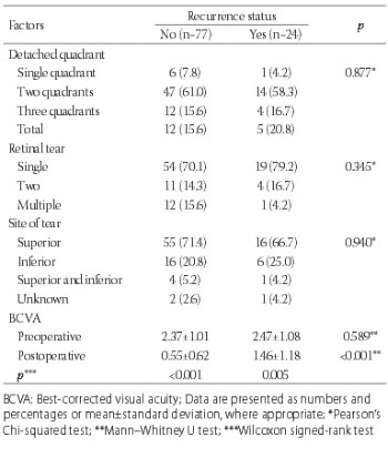
No statistically significant difference was determined between the recurrent and non-recurrent patients with regard to the number of detached quadrants, number and localization of breaks as well as with regard to preoperative BCVA. On contrary, a statistically significant difference was observed between the recurrent and non-recurrent patients in postoperative BCVA values (Table 4).

TABLE 4: Factors that may influence recurrence rate, preoperative, and postoperative BCVA values according to the recurrence status of patients

A statistically significant difference was found between the patients with and without macular detachment in terms of detachment time and number of detached quadrants (p < 0.05). The detachment time was longer and number of detached quadrants was higher in the patients with macular detachment. Although the mean pre-op BCVA was lower in the patients with macular detachment in comparison to the patients without macular detachment, the difference was not statistically significant (Table 5).

TABLE 5: Distribution of detachment time, number of detached quadrants, and preoperative and postoperative BCVA according to macular involvement

DISCUSSION

Various surgical methods have been used for the treatment of pseudophakic RD [11,12]. In 1997, Benson et al. [13] conducted a study to investigate the most preferred method in RD surgery and determined that 62% of vitreoretinal surgeons preferred SB surgery, 30% preferred PR, and 7% preferred PPV. The popularity of PPV in pseudophakic RD surgery has increased over time due to the advancements in vitrectomy techniques, as well as owing to the fact that peripheral retinal tears can be better observed and that the rate of PVR has declined. According to the results of the Preferences and Trends (PAT) survey (2013), conducted among the members of the American Society of Retina Specialists, the number of retina specialists that prefer treating RD with vitrectomy without SB has increased between 2005 and 2015. In the same case presentation of a 65-year-old patient with a pseudophakic RD, -3.00 D myopia, ½ clock-hour–size flap tear at 11:00 anterior to the equator, 45% detached, macula-on, with poor vision in the fellow eye, the preferred practice of retinal surgeons has dramatically shifted away from SB toward vitrectomy without SB. From 2005 to 2013, the number of those treating pseudophakic RD patients with vitrectomy without SB nearly doubled from 30 to 60% and, conversely, those using SB declined from 25 to 10%. The percentage of those preferring PR has remained stable at 25% [14]. In recent years, PR has become a less popular method for treating pseudophakic RD [15,16].

SB, which conventionally provides target-oriented retinal attachment, is one of two basic methods in the surgical treatment of RD. SB had the highest success rate and was the most frequently used method in the treatment of RD before the advancements in PPV technique [5-17]. The most significant advantage of PPV is that peripheral vitreous detachment can be visualized more clearly by eliminating vitreous and posterior hyaloid membrane. Furthermore, together with wide-angle imaging systems, it enables the microscopic visualization of peripheral fundus by scleral indentation and internal illumination. Thus, with clearer visualization of retinal tears in peripheral fundus and prompt intervention, it provides a high rate of anatomical success in treating RD. Ho et al. [18] detected retinal tears in 80% of the patients with pseudophakic RD using SB. In this study, we identified retinal breaks in 28 out of 30 patients (93.3%) that underwent SB. In the literature, the location of retinal breaks is detected in 94-100% patients undergoing PPV [19-21]. We found the location of retinal breaks in 70 out of 71 patients (98.5%) that underwent PPV.

In a case series of 225 patients with pseudophakic RRD, Campo et al. [22] achieved an initial success rate of 88% using PPV and final anatomical success of 96%. In our study, retinal reattachment was achieved after the initial surgery in 56 of 71 patients (78.8%) in PPV group, and 15 cases required a second surgery. After the second surgery, the retina was successfully reattached in all eyes (100%). Anatomical success after a single surgery varies between 84% and 94% in RD patients, whereas this rate reaches 96-100% after multiple surgeries [19-26]. The 100% final anatomical success observed in this study is related to the fact that the patient groups consisted of uncomplicated cases and the absence of vitreous or retinal pathologies. Other studies showed that the rate of retinal reattachment with SB in pseudophakic RD varies between 61% and 89% after a single surgery, and final anatomical success varies between 82% and 99% [18,27-30]. We achieved retinal reattachment after the initial surgery in 22 patients (73.3%) in SB group. Although 8 cases required a second surgery, the final anatomical success rate was 100%.

In general, 20/40 or better BCVA is achieved after PPV in 44% to 72% cases. Mendrinos et al. [31] reported 20/40 or better BCVA in 56% cases. In their study, 20/40 or better BCVA was more frequently reported in patients without macular involvement (69.6%) compared to patients with macular involvement (38.6%). In our study, the final BCVA was 0.83 ± 0.97 in PPV + Silicone group and 0.67 ± 0.70 in PPV + Gas group. A statistically significant increase in BCVA was observed in both groups compared to the preoperative BCVA. Mendrinos et al. reported 20/40 or better BCVA in 46.4% of patients in PPV group [31]. In the present study, the final BCVA of 20/40 or better was observed more frequently in patients without macular involvement at first presentation compared to the patients with macular involvement (54.5% and 45.0%, respectively). The difference in recovery of vision could be explained by the height of macular detachment and degeneration of photoreceptor cells in the macula. Awareness of the patient about this subject and early admission to the hospital as well as an early surgical intervention are of critical importance.

In a meta-analysis [32] covering the years from 1966 to 2004, a comparison of conventional SB and PPV revealed that PPV had better anatomical and visual outcomes in pseudophakic RD patients. Moreover, no difference was observed between PPV and combined technique (PPV + SB) in terms of early and final anatomical success. In the published randomized clinical studies, it was determined that PPV is at least as successful as SB [19,26]. Sharma et al. [26] determined that visual (functional) outcomes are better in PPV group when compared to SB group. However, a multicenter, prospective, randomized clinical trial in Europe, which recruited 416 phakic and 265 pseudophakic eyes with ‘medium severity’ RD, showed that visual outcomes were significantly better in patients treated with SB, while a single-operation success rate was significantly higher in pseudophakic patients treated with PPV [33]. Brazitikos et al. [19], however, determined no significant difference between the two methods in terms of final BCVA [19]. Another study reported a final posterior re-attachment rate of 77% in patients managed with PVR with SO and 79% in those treated with PVR with C3F8 gas tamponade [34]. In the Silicone Study, SO and C3F8 gas showed similar, long-term, efficacy in patients with retinal detachment and PVR [35]. Likewise, the present study determined no statistically significant difference between SO and C3F8 gas tamponade use in the PPV group, in terms of success rates, nor between the SB and PPV groups.

In their analysis of SB versus primary vitrectomy in RD patients, Feltgen et al. [36] showed that anatomical success was negatively correlated with multiple breaks in pseudophakic eyes [36]. In the present study, no statistically significant difference was determined between the recurrent and non-recurrent cases in terms of multiple breaks.

Although a number of previous studies investigated the efficacy of SB and PPV in pseudophakic RD patients [9,19,20,24-26], only a limited number of studies focused on the efficacy of gas and silicone oil tamponade use in pseudophakic RD patients. In the present study, we compared the efficacy of SB, PPV + Gas and PPV + Silicone Oil in pseudophakic RD patients and showed that no significant difference was determined between the groups regarding primary and final anatomical and visual success rates as well as recurrent retinal detachment rates.

There are several limitations in this study. First, the number of patients was limited only to 30-40 patients in each group. Second, this was a retrospective, comparative case series of a single center experience. Third, because one of the surgeons voluntarily chose to perform SB and the other surgeon performed PPV, these results may reflect their surgical expertise and it may be difficult to extrapolate these data to the general vitreoretinal surgery practice.

In conclusion, no difference was determined between the surgical procedures performed in pseudophakic RD patients in the case of early admission and rapid intervention, as well as when adequate technical facilities exist and the surgeries are performed by experienced surgeons. The surgeon can choose either SB or PPV safely by taking his or her own experience and the risk-benefit ratio into consideration, individually for each patient.

DECLARATION OF INTERESTS

The authors declare no conflict of interests.

REFERENCES

  1. , (). Update on a long-term, prospective study of capsulotomy and retinal detachment rates after cataract surgery. J Cataract Refract Surg. https://doi.org/10.1016/S0886-3350(00)00304-7
  2. , , (). National outcomes of cataract extraction. I. Retinal detachment after inpatient surgery. Ophthalmology. https://doi.org/10.1016/S0161-6420(91)32204-8
  3. , , , , (). Trends in vitreoretinal surgery at a tertiary referral center 1987 to 1996. Br J Ophthalmol. https://doi.org/10.1136/bjo.83.4.396
  4. , (). Pseudophakic retinal detachment. Surv Ophthalmol. https://doi.org/10.1016/S0039-6257(03)00083-3
  5. , , , , , (). Retinal detachment after cataract surgery. Surgical results. Ophthalmology. http://dx.doi.org/10.1016/S0161-6420(92)31952-9
  6. , , , , , (). Pseudophakic retinal detachments in the presence of various IOL types. Ophthalmology. https://doi.org/10.1016/S0161-6420(86)33596-6
  7. , , , , , (). Retinal detachment after cataract surgery. Predisposing factors. Ophthalmology. https://doi.org/10.1016/S0161-6420(92)31953-0
  8. , , (). Primary vitrectomy for pseudophakic retinal detachment. Br J Ophthalmol. https://doi.org/10.1136/bjo.80.4.346
  9. , (). Combined pars plana vitrectomy and scleral buckling for pseudophakic and aphakic retinal detachments in which a break is not seen preoperatively. Ophthalmic Surg Lasers.
  10. , , , (). Primary vitrectomy alone for repair of retinal detachments following cataract surgery. Retina. https://doi.org/10.1097/00006982-200005000-00005
  11. , (). Primary retinal detachment: Scleral buckle or pars plana vitrectomy?. Curr Opin Ophthalmol. https://doi.org/10.1097/01.icu.0000193097.28798.fc
  12. , , , , (). An evidence-based analysis of surgical interventions for uncomplicated rhegmatogenous retinal detachment. Acta Ophthalmol Scand. https://doi.org/10.1111/j.1600-0420.2006.00715.x
  13. , , , , , (). Current popularity of pneumatic retinopexy. Retina. https://doi.org/10.1097/00006982-199903000-00010
  14. , (). ASRS 2013 Preferences and Trends Membership Survey. American Society of Retina Specialists.
  15. , , , (). Reassessment of pneumatic retinopexy for primary treatment of rhegmatogenous retinal detachment. Clin Ophthalmol. DOI: 10.2147/OPTH.S91486
  16. , , , (). Pneumatic retinopexy for rhegmatogenous retinal detachment in pseudophakia. Semin Ophthalmol. Epub ahead of printhttps://doi.org/10.1080/08820538.2016.1190849
  17. , , , , (). Management of pseudophakic retinal detachment with various intraocular lens types. Ann Ophthalmol.
  18. , (). Pseudophakic retinal detachment. Surgical success rate with various types of IOLs. Ophthalmology. https://doi.org/10.1016/S0161-6420(84)34227-0
  19. , , , (). Primary pars plana vitrectomy versus scleral buckle surgery for the treatment of pseudophakic retinal detachment: A randomized clinical trial. Retina. https://doi.org/10.1097/00006982-200512000-00001
  20. , , , , , (). Pars plana vitrectomy versus combined pars plana vitrectomy-scleral buckle for primary repair of pseudophakic retinal detachment. Ophthalmology. https://doi.org/10.1016/j.ophtha.2006.05.038
  21. , , , (). Primary vitrectomy alone for repair of retinal detachments following cataract surgery. Retina. https://doi.org/10.1097/00006982-200005000-00005
  22. , , , , , (). Pars plana vitrectomy without scleral buckle for pseudophakic retinal detachments. Ophthalmology. https://doi.org/10.1016/S0161-6420(99)90353-6
  23. , , , , (). Pars plana vitrectomy, laser retinopexy, and aqueous tamponade for pseudophakic rhegmatogenous retinal detachment. Ophthalmology. https://doi.org/10.1016/j.ophtha.2006.07.037
  24. , , (). Surgical treatment of retinal detachment in pseudophakia: Comparison between vitrectomy and scleral buckling. Klin Monatsbl Augenheilkd. https://doi.org/10.1055/s-2008-1034893
  25. , , , , , (). Vitrectomy without scleral buckle versus ab-externo approach for pseudophakic retinal detachment: Comparative retrospective study. [Article in French]. J Fr Ophtalmol.
  26. , , , , , (). Functional and anatomic outcome of scleral buckling versus primary vitrectomy in pseudophakic retinal detachment. Acta Ophthalmol Scand. https://doi.org/10.1111/j.1600-0420.2005.00461.x
  27. , (). Pneumatic retinopexy. A multicenter randomized controlled clinical trial comparing pneumatic retinopexy with scleral buckling. The Retinal Detachment Study Group. Ophthalmology. https://doi.org/10.1016/S0161-6420(89)32820-X
  28. , , , , , (). Pseudophakic retinal detachments. Anatomic and visual results. Ophthalmology. https://doi.org/10.1016/S0161-6420(92)31983-9
  29. , , , , (). Comparison of pneumatic retinopexy and scleral buckling in the management of primary rhegmatogenous retinal detachment. Southern Wisconsin Pneumatic Retinopexy Study Group. Am J Ophthalmol. https://doi.org/10.1016/S0002-9394(98)00181-0
  30. , , , , (). Twenty-year follow-up for scleral buckling. Arch Ophthalmol. https://doi.org/10.1001/archopht.120.3.325
  31. , , , , (). Primary vitrectomy without scleral buckling for pseudophakic rhegmatogenous retinal detachment. Am J Ophthalmol. https://doi.org/10.1016/j.ajo.2008.01.018
  32. , , , , (). Surgical management of pseudophakic retinal detachments: A meta-analysis. Ophthalmology. https://doi.org/10.1016/j.ophtha.2006.05.044
  33. , , , , , (). Scleral buckling versus primary vitrectomy in rhegmatogenous retinal detachment: A prospective randomized multicenter clinical study. Ophthalmology. https://doi.org/10.1016/j.ophtha.2007.09.013
  34. , , , , , (). Vitrectomy with silicone oil or perfluoropropane gas in eyes with severe proliferative vitreoretinopathy: Results of a randomized clinical trial. Silicone Study Report 2. Arch Ophthalmol. DOI: 10.1001/archopht.1992.01080180052028
  35. , , , , , (). Vitrectomy with silicone oil or long-acting gas in eyes with severe proliferative vitreoretinopathy: Results of additional and long-term follow-up. Silicone Study report 11. Arch Ophthalmol. https://doi.org/10.1001/archopht.1997.01100150337005
  36. , , , , , (). Scleral buckling versus primary vitrectomy in rhegmatogenous retinal detachment (SPR study): Risk assessment of anatomical outcome. SPR study report. no. 7. Acta Ophthalmol. https://doi.org/10.1111/j.1755-3768.2011.02344.x