Potential inflammatory markers in obstructive sleep apnea-hypopnea syndrome

Authors

  • Dongmei Lu Postgraduate College of Xinjiang Medical University, Xinjiang Medical University, Urumqi, China; Department of Respiratory and Critical Care Medicine, People’s Hospital of Xinjiang Uygur Autonomous Region, Urumqi, China
  • Nanfang Li Hypertension Center, People’s Hospital of Xinjiang Uygur Autonomous Region, Hypertension Institute of Xinjiang, Urumqi, China
  • Xiaoguang Yao Hypertension Center, People’s Hospital of Xinjiang Uygur Autonomous Region, Hypertension Institute of Xinjiang, Urumqi, China
  • Ling Zhou Hypertension Center, People’s Hospital of Xinjiang Uygur Autonomous Region, Hypertension Institute of Xinjiang, Urumqi, China

DOI:

https://doi.org/10.17305/bjbms.2016.1579

Keywords:

Obstructive sleep apnea-hypopnea syndrome, hypoxia-inducible factor-1, nuclear factor-κB, surfactant protein

Abstract

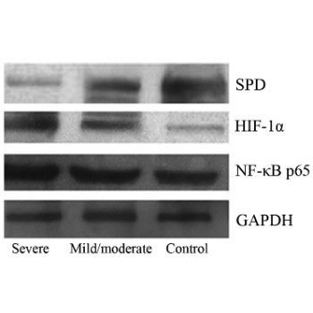
Obstructive sleep apnea-hypopnea syndrome (OSAHS) is a complex chronic inflammatory respiratory disease with multiple pathogenic factors and high morbidity and mortality. Serum levels of nuclear factor-κB (NF-κB), hypoxia-inducible factor-1 alpha (HIF-1α), and surfactant protein D (SPD) were investigated in OSAHS patients, to determine their clinical significance and correlation with the pathogenesis. Patients were classified into a mild and moderate OSAHS group (n = 25) and severe OSAHS group (n = 33). Twenty healthy patients served as a control group. Peripheral blood levels of NF-κB, HIF-1α, and SPD were determined by Western blot, and a correlation analysis was performed. Severe OSAHS patients received nasal continuous positive airway pressure (nCPAP) therapy and were followed up after 2 months. NF-κB p65, HIF-1α, and SPD expression levels were determined after valid nCPAP therapy. NF-κB p65 and HIF-1α expression was significantly higher in severe OSAHS group than in the other two groups (p < 0.01), and was positively correlated with the apnea-hypopnea index (AHI) (r = 0.696, p < 0.001; r = 0.634, p < 0.001). SPD expression was significantly lower in severe OSAHS group than in the control group (p < 0.01) and mild and moderate OSAHS group (p < 0.01), and was negatively correlated with AHI (r = −0.569, p < 0.001). OSAHS pathogenesis was associated with changes in NF-κB, HIF-1α, and SPD protein expression levels. nCPAP therapy could improve the clinical characteristics of the patients, lower serum NF-κB and HIF-1α levels, and increase serum SPD levels. We conclude that OSAHS is related to the expression of NF-κB, HIF-1, and SPD.

Downloads

Download data is not yet available.

Author Biographies

Dongmei Lu, Postgraduate College of Xinjiang Medical University, Xinjiang Medical University, Urumqi, China; Department of Respiratory and Critical Care Medicine, People’s Hospital of Xinjiang Uygur Autonomous Region, Urumqi, China

Department of Respiratory and Critical Care Medicine

Nanfang Li, Hypertension Center, People’s Hospital of Xinjiang Uygur Autonomous Region, Hypertension Institute of Xinjiang, Urumqi, China

Hypertension Center

Xiaoguang Yao, Hypertension Center, People’s Hospital of Xinjiang Uygur Autonomous Region, Hypertension Institute of Xinjiang, Urumqi, China

Hypertension Center

Ling Zhou, Hypertension Center, People’s Hospital of Xinjiang Uygur Autonomous Region, Hypertension Institute of Xinjiang, Urumqi, China

Hypertension Center

References

Daulatzai MA. Role of sensory stimulation in amelioration of obstructive sleep apnea. Sleep Disord 2011;2011:596879. http://dx.doi.org/10.1155/2011/596879.

Katz ES, D'Ambrosio CM. Pathophysiology of pediatric obstructive sleep apnea. Proc Am Thorac Soc 2008;5(2):253-62. http://dx.doi.org/10.1513/pats.200707-111MG.

Punjabi NM. The epidemiology of adult obstructive sleep apnea. Proc Am Thorac Soc 2008;5(2):136-43. http://dx.doi.org/10.1513/pats.200709-155MG.

Peppard PE, Young T, Barnet JH, Palta M, Hagen EW, Hla KM. Increased prevalence of sleep-disordered breathing in adults. Am J Epidemiol 2013;177(9):1006-14. http://dx.doi.org/10.1093/aje/kws342.

Ryan S, Taylor CT, McNicholas WT. Selective activation of inflammatory pathways by intermittent hypoxia in obstructive sleep apnea syndrome. Circulation 2005;112(17):2660-7. http://dx.doi.org/10.1161/CIRCULATIONAHA.105.556746.

Winnicki M, Shamsuzzaman A, Lanfranchi P, Accurso V, Olson E, Davison D, et al. Erythropoietin and obstructive sleep apnea. Am J Hypertens 2004;17(9):783-6. http://dx.doi.org/10.1016/j.amjhyper.2004.04.011.

van Uden P, Kenneth NS, Rocha S. Regulation of hypoxia-inducible factor-1alpha by NF-kappaB. Biochem J 2008;412(3):477-84. http://dx.doi.org/10.1042/BJ20080476.

Belaiba RS, Bonello S, Zähringer C, Schmidt S, Hess J, Kietzmann T, et al. Hypoxia up-regulates hypoxia-inducible factor-1alpha transcription by involving phosphatidylinositol 3-kinase and nuclear factor kappaB in pulmonary artery smooth muscle cells. Mol Biol Cell 2007;18(12):4691-7.

http://dx.doi.org/10.1091/mbc.E07-04-0391.

Bonello S, Zähringer C, BelAiba RS, Djordjevic T, Hess J, Michiels C, et al. Reactive oxygen species activate the HIF-1alpha promoter via a functional NFkappaB site. Arterioscler Thromb Vasc Biol 2007;27(4):755-61. http://dx.doi.org/10.1161/01.ATV.0000258979.92828.bc.

Arnardottir ES, Mackiewicz M, Gislason T, Teff KL, Pack AI. Molecular signatures of obstructive sleep apnea in adults: A review and perspective. Sleep 2009;32(4):447-70.

Cochrane CG, Spragg R, Revak SD. Pathogenesis of the adult respiratory distress syndrome. Evidence of oxidant activity in bronchoalveolar lavage fluid. J Clin Invest 1983;71(3):754-761. http://dx.doi.org/10.1172/JCI110823.

Kiefmann R, Rifkind JM, Nagababu E, Bhattacharya J. Red blood cells induce hypoxic lung inflammation. Blood 2008;111(10):5205-14. http://dx.doi.org/10.1182/blood-2007-09-113902.

Ito Y, Ahmad A, Kewley E, Mason RJ. Hypoxia-inducible factor regulates expression of surfactant protein in alveolar type II cells in vitro. Am J Respir Cell Mol Biol 2011;45(5):938-45. http://dx.doi.org/10.1165/rcmb.2011-0052OC.

Ghadiali SN, Gaver DP. Biomechanics of liquid-epithelium interactions in pulmonary airways. Respir Physiol Neurobiol 2008;163(1-3):232-43. DOI: 10.1016/j.resp.2008.04.008.

Tenner AJ, Robinson SL, Borchelt J, Wright JR. Human pulmonary surfactant protein (SP-A), a protein structurally homologous to Clq, can enhance FcR- and CR1-mediated phagocytosis. J Biol Chem 1989;264(23):13923-8.

van Iwaarden F, Welmers B, Verhoef J, Haagsman HP, van Golde LM. Pulmonary surfactant protein A enhances the host-defense mechanism of rat alveolar macrophages. Am J Respir Cell Mol Biol 1990;2(1):91-8. http://dx.doi.org/10.1165/ajrcmb/2.1.91.

Hohlfeld J, Fabel H, Hamm H. The role of pulmonary surfactant in obstructive airways disease. Eur Respir J 1997;10(2):482-91. http://dx.doi.org/10.1183/09031936.97.10020482.

Antal JM, Divis LT, Erzurum SC, Wiedemann HP, Thomassen MJ. Surfactant suppresses NF-kappa B activation in human monocytic cells. Am J Respir Cell Mol Biol 1996;14(4):374-9. http://dx.doi.org/10.1165/ajrcmb.14.4.8600942.

Phelps DS, Floros J. Localization of pulmonary surfactant proteins using immunohistochemistry and tissue in situ hybridization. Exp Lung Res 1991;17(6):985-95. http://dx.doi.org/10.3109/01902149109064330.

Takahashi H, Fujishima T, Koba H, Murakami S, Kurokawa K, Shibuya Y, et al. Serum surfactant proteins A and D as prognostic factors in idiopathic pulmonary fibrosis and their relationship to disease extent. Am J Respir Crit Care Med 2000;162(3 Pt 1):1109-14.

Stapleton RD, Dixon AE, Parsons PE, Ware LB, Suratt BT; NHLBI Acute Respiratory Distress Syndrome Network. The association between BMI and plasma cytokine levels in patients with acute lung injury. Chest 2010;138(3):568-77. http://dx.doi.org/10.1378/chest.10-0014.

Winkler C, Atochina-Vasserman EN, Holz O, Beers MF, Erpenbeck VJ, Krug N, et al. Comprehensive characterisation of pulmonary and serum surfactant protein D in COPD. Respir Res 2011;12:29. DOI: 10.1186/1465-9921-12-29.

He QY, Chen BY. Sleep related breathing disorders team, respiology branch, Chinese medical association, OSAHS diagnosis and treatment guideline (revision 2011) [Article in Chinese]. Chinese J Tuberc Respir Dis 2012;35(1):9-12.

Ciccone MM, Scicchitano P, Mitacchione G, Zito A, Gesualdo M, Caputo P, et al. Is there a correlation between OSAS duration/severity and carotid intima-media thickness? Respir Med 2012;106(5):740-6. http://dx.doi.org/10.1016/j.rmed.2011.12.016.

Atkeson A, Jelic S. Mechanisms of endothelial dysfunction in obstructive sleep apnea. Vasc Health Risk Manag 2008;4(6):1327-35.

https://dx.doi.org/10.2147/VHRM.S4078.

Damiani MF, Carratù P, Quaranta VN, Resta O. Influence of obstructive sleep apnea on endothelial function in obese patients. Chest 2012;141:1639; author reply 1640. http://dx.doi.org/10.1378/chest.12-0144.

Sarada SKS, Veeramohan PH, Mathew T, Saumya S, Chitharanjan M. Nifedipine inhibits hypoxia induced transvascular leakage through down regulation of NFkB. Respir Physiol Neurobiol 2012;183(1):26-34. http://dx.doi.org/10.1016/j.resp.2012.05.016.

Fitzpatrick SF, Tambuwala MM, Bruning U, Schaible B, Scholz CC, Byrne A, et al. An intact canonical NF-kB pathway is required for inflammatory gene expression in response to hypoxia. J Immunol 2011;186(2):1091-6. http://dx.doi.org/10.4049/jimmunol.1002256.

Htoo AK, Greenberg H, Tongia S, Chen G, Henderson T, Wilson D, et al. Activation of nuclear factor kappaB in obstructive sleep apnea: A pathway leading to systemic inflammation. Sleep Breath 2006;10(1):43-50.

http://dx.doi.org/10.1007/s11325-005-0046-6.

Ciccone MM, Scicchitano P, Zito A, Cortese F, Boninfante B, Falcone VA, et al. Correlation between inflammatory markers of atherosclerosis and carotid intima-media thickness in obstructive sleep apnea. Molecules 2014;19(2):1651-62. http://dx.doi.org/10.3390/molecules19021651.

Li RC, Haribabu B, Mathis SP, Kim J, Gozal D. Leukotriene B4 receptor-1 mediates intermittent hypoxia-induced atherogenesis. Am J Respir Crit Care Med 2011;184(1):124-31. http://dx.doi.org/10.1164/rccm.201012-2039OC.

Dyugovskaya L, Polyakov A, Ginsberg D, Lavie P, Lavie L. Molecular pathways of spontaneous and TNF-{alpha}-mediated neutrophil apoptosis under intermittent hypoxia. Am J Respir Cell Mol Biol 2011;45(1):154-62. http://dx.doi.org/10.1165/rcmb.2010-0025OC.

Greenberg H, Ye X, Wilson D, Htoo AK, Hendersen T, Liu SF. Chronic intermittent hypoxia activates nuclear factor-kappaB in cardiovascular tissues in vivo. Biochem Biophys Res Commun 2006;343(2):591-6. http://dx.doi.org/10.1016/j.bbrc.2006.03.015.

Carratù P, Zito A, Dragonieri S, Ciccone MM, Resta O. Effect of CPAP on cardiovascular risks in OSA patients. A 4-year follow-up, preliminary data. Sleep Breath 2016. [Epub ahead of print]. http://dx.doi.org/10.1007/s11325-016-1382-4.

Sin DD, Pahlavan PS, Man SF. Surfactant protein D: A lung specific biomarker in COPD? Ther Adv Respir Dis 2008;2(2):65-74. http://dx.doi.org/10.1177/1753465808088903.

Wirtz HR, Dobbs LG. Calcium mobilization and exocytosis after one mechanical stretch of lung epithelial cells. Science 1990;250(4985):1266-9. http://dx.doi.org/10.1126/science.2173861.

Wirtz H, Schmidt M. Ventilation and secretion of pulmonary surfactant. Clin Investig 1992;70(1):3-13. http://dx.doi.org/10.1007/BF00422930.

Massaro GD, Massaro D. Morphologic evidence that large inflations of the lung stimulate secretion of surfactant. Am Rev Respir Dis 1983;127(2):235-6.

Aderibigbe AO, Thomas RF, Mercer RR, Auten RL Jr. Brief exposure to 95% oxygen alters surfactant protein D and mRNA in adult rat alveolar and bronchiolar epithelium. Am J Respir Cell Mol Biol 1999;20(2):219-27. http://dx.doi.org/10.1165/ajrcmb.20.2.3348.

Liang S, Li N, Heizhati M, Yao X, Abdireim A, Wang Y, et al. What do changes in concentrations of serum surfactant proteins A and D in OSA mean? Sleep Breath 2015;19(3):955-62. http://dx.doi.org/10.1007/s11325-014-1106-6.

Potential inflammatory markers in obstructive sleep apnea-hypopnea syndrome

Downloads

Additional Files

Published

2017-02-21

How to Cite

1.
Lu D, Li N, Yao X, Zhou L. Potential inflammatory markers in obstructive sleep apnea-hypopnea syndrome. Biomol Biomed [Internet]. 2017Feb.21 [cited 2023Sep.25];17(1):47-53. Available from: https://www.bjbms.org/ojs/index.php/bjbms/article/view/1579

Issue

Section

Translational and Clinical Research

INTRODUCTION

Obstructive sleep apnea-hypopnea syndrome (OSAHS), which is characterized by repetitive episodes of airflow reduction (hypopnea) or cessation (apnea) due to upper airway collapse during sleep, often causes chronic intermittent hypoxia, repetitive waking, and sleep fragmentation. OSAHS increases all-cause mortality and the risk of death, and it results in multi-system and multi-organ damage [1,2]. As an independent risk factor for hypertension, coronary disease, left heart failure, pulmonary heart disease, myocardial infarction, and stroke, OSAHS is closely related to cardiac and cerebrovascular diseases. The prevalence of OSAHS has risen from 3 to 10% in adult population [3,4].

OSAHS is considered a complex, multi-factor and multi-gene disease, and its pathogenesis remains unexplored. Study of the pathological and physiological effects of intermittent hypoxia in OSAHS identified two molecular pathways: A nuclear factor-κB (NF-κB)-dependent inflammatory pathway, which produces inflammatory cytokines, and hypoxia-inducible factor-1 (HIF-1)-dependent adaptive pathway [5,6]. An active NF-κB binding site has been localized upstream of the hypoxia-inducible factor-1 alpha (HIF1A) promoter (at residues-197/188) [7], and the activation of NF-κB increases HIF1A messenger ribonucleic acid (mRNA) levels [8]. Mutations at these sites blocked this effect, indicating that the inflammatory pathways are significantly related to the hypoxic response pathway [8,9]. In intermittent hypoxia, the inflammatory signaling factors HIF-1α and NF-κB play essential roles in the transcriptional regulation of inflammatory cytokines. Although the two pathways can affect each other, the nature of their role in OSAHS pathogenesis and the exact relationship between them remain unclear. In OSAHS cases, repetitive hypoxia and reoxygenation occur during sleep, inducing an oxidative stress response similar to that induced by ischemia/reperfusion injury [10]. Oxidative stress not only damages endothelial cells in the peripheral circulation but also contributes to the damage of alveolar epithelial cells and endothelial cells in the lungs. This damage increases the permeability of the alveolar wall [11,12], which could decrease the function of pulmonary surfactants [13]. In addition, the repetitive airway collapse aggravates the damaged epithelial cells of the airway and lungs [14]. Surface-active materials regulate the functions of lung inflammatory cells [15,16]. The immune regulatory roles of surface-active materials include inhibiting cytokine secretion [17], activating transcriptional factors, and inhibiting NF-kB in human monocytes [18]. Surfactant protein D (SPD) is mainly synthesized and secreted by alveolar Type II cells (ATIIs), club (Clara) cells, and submucosal cells, and is found sparsely in the epithelium of conducting airways [19]. The restricted expression of SPD within the lung enables the analysis of specific markers in lung diseases; in fact, SPD levels in the systemic circulation have been measured and considered useful biomarkers for many lung diseases [20-22]. However, changes in these surface-active materials in OSAHS, and the relationship between such changes and the two pathways described above, remain to be elucidated.

In this study, the expression of NF-κB p65, HIF-1α, and SPD proteins in peripheral blood was determined in two OSAHS groups and in a control group. In addition, the activation of the anoxia pathway and pathogenesis were investigated.

MATERIALS AND METHODS

Patients

Random in- and out-patients, who underwent polysomnography (PSG) at the Sleep laboratory in Hypertension research center of the People’s Hospital of Xinjiang Uygur Autonomous Region between January 2015 and November 2015, were included.

The included patients were placed into either a mild and moderate OSAHS group (25 cases) or severe OSAHS group (33 cases). The inclusion criteria were as follows: 1) The patient met the diagnostic criteria for OSAHS, which were based on the adult OSAHS diagnosis and treatment guidelines (Draft), developed by the Respiratory Diseases Branch of the Chinese Medical Association in 2011 [23]; 2) The patient had no OSAHS treatment history; and 3) The patient provided written informed consent.

The exclusion criteria included the following: 1) History of neuromuscular disorders, infectious diseases, rheumatic diseases, immunological diseases, tumors, peripheral vascular diseases, coagulation disorders, liver or kidney diseases, severe psychogenic disorders, or acute or chronic kidney failure; 2) History of injury or surgery in the past 3 months; 3) Administration of drugs, hormones, immune suppressors, cytotoxins, or free radical scavengers; and 4) Other cases of chronic anoxia.

Twenty subjects who completed physical examinations during the same period were included as a control group. Among 33 patients in severe OSAHS group, 20 patients accepted the nasal continuous positive airway pressure (nCPAP) therapy with the Model Solo TMCPAP (Respironics, USA), and were considered as a treated group. The other 13 patients who did not receive the nCPAP therapy were considered as an untreated group. The 33 treated patients completed a 2-month follow-up visit, during which no antihypertensive therapy, antidiabetic therapy, or other OSAHS therapy was performed.

This study was registered as a clinical trial (No. ChiCTR-IOC-15002908). The protocols were approved by the Ethics Committee of the People’s Hospital of Xinjiang Uygur Autonomous Region, and informed consent was obtained from all participants.

Polysomnography

On the monitoring day, the patients were not permitted to nap during the afternoon, drink alcohol or coffee, or take sedatives. Beginning at 9:30 p.m., the patients’ sleep was monitored overnight (>7 hours) with the Poly Smith System (Neurotronics, Florida, USA), and indicators such as the apnea-hypopnea index (AHI), mean oxyhemoglobin saturation (mean SpO2), minimum SpO2 (min SpO2), and time points when the nocturnal pulse oximetric saturation was lower than 90% (tSpO2 <90%) were recorded.

Collection and evaluation of serum samples

The next morning at 7 a.m., on empty stomach, peripheral venous blood (5 mL) was collected from the patients into tubes without anticoagulation factors, and centrifuged at 1000 rpm for 10 minutes. Then, the supernatant was transferred into a cryogenic vial for storage at −80°C. Aseptic techniques were used to collect the blood, and the laboratory procedures were conducted blindly. Hemolysis was not observed in the samples, and the samples were not stored for more than 1 year. The samples were thawed only once.

NF-κB p65 and HIF-1α protein expression was determined using Western blot. Total proteins were extracted from 2 ml serum samples according to the manufacturer’s protocol (Merck, New Jersey, USA). A bicinchoninic acid assay was used to determine the protein concentration. After the proteins were denatured, protein samples (20 µg) were placed on polyacrylamide gels (8%) to conduct electrophoresis. After the proteins were transferred to membranes, the samples were blocked in a solution of 5% skim milk, incubated overnight with a primary antibody (Abcam, Massachusetts, USA, NF-κB p65 1:250, HIF-1α 1:400) at 4°C, incubated with fluorescein isothiocyanate-labeled goat anti-mouse immunoglobulin G (IgG) (Merck, New Jersey, USA, 1:10000) for 1 hour, and blots were developed by enhanced chemiluminescence after a 5 minutes exposure.

SPD protein expression was also determined by Western blot according to the procedures described in the manual. The protocol included the following steps: Polyacrylamide gel electrophoresis, membrane transfer, Ponceau staining, blocking step with protein powder, washing, incubation with a rabbit antihuman-SPD IgG polyclonal antibody (Santa Cruz Biotechnology, 1:500), incubation with a horseradish peroxidase-labeled goat anti-rabbit IgG (Santa Cruz Biotechnology, 1:2000), detection with a 3,3’-diaminobenzidine substrate system, image scanning, and analysis with the FluorChem Digital Imaging System V2.0 (Alpha Innotech, California, USA).

nCPAP therapy and follow-up visit

Instructions and educational materials were provided before the nCPAP therapy to promote patient cooperation. Based on the patient pressure titration data, the initial pressure was adjusted to a level that could eliminate 90-95% of the apnea. Then, the pressure was further adjusted, and the nocturnal SpO2 was monitored. The minimum pressure that could be endured by the patients, which generally prevented snoring and apnea and maintained the nocturnal SpO2 level above 90%, was defined as the target pressure for therapy. Telephone-based follow-ups were completed during the 1st, 2nd, and 4th weeks, and a clinical follow-up visit, that included laboratory assessments of NF-κB p65, HIF-1α, and SPD protein levels was completed during the 2nd month after the subjects were discharged from the hospital. In each follow-up visit, the number of hours that the noninvasive ventilator was used was confirmed.

Statistical analysis

The Statistical Package for the Social Sciences software version 17.0 (SPSS Inc., Chicago, IL, USA) was used for the data analysis. Data that demonstrated a normal distribution and had equal variance were presented as mean ± standard deviation (±s). One-way analysis of variance and subsequent pairwise comparisons were used to compare groups. The pairwise comparisons were tested using the least significant difference test or Mann–Whitney U test. The rank sum test was used to assess the heterogeneity of interclass variance. Enumeration data were presented as ratios, and the Chi-squared test was used for comparisons among groups. A pairwise t-test was used for pre- and post-therapy comparisons. The Pearson’s correlation was used to determine the relationship between two factors. A multivariate regression analysis was used to evaluate the role of confounding clinical factors on the final results. Values of p < 0.05 were considered statistically significant.

RESULTS

Baseline data

No significant differences in age or body mass index (BMI) were found between mild and moderate OSAHS group, severe OSAHS group, and control group. The serum levels of NF-κB p65 and HIF-1α increased with disease severity; the levels were the lowest in the control group and the highest in severe OSAHS group (Figure 1 and Table 1). In contrast, the serum SPD levels decreased with OSAHS severity; the lowest level was observed in severe OSAHS group (Table 1). The NF-κB p65 and HIF-1 expression levels in severe OSAHS group were significantly higher than those in control and mild and moderate groups (p < 0.05). No significant difference in NF-κB p65 expression was found between the control group and mild and moderate OSAHS group (p = 0.067 and p = 0.352). Similarly, the HIF-1α expression level in mild and moderate OSAHS group did not differ from that in the control group (p = 0.687). SPD expression in severe OSAHS group was higher than that in the control group and mild and moderate OSAHS group (p < 0.01); however, no significant difference was found between the control group and mild and moderate OSAHS group (p = 0.352). Furthermore, the expression levels of NF-κB p65 and HIF-1α were positively correlated (r = 0.726, p < 0.001). In severe OSAHS group, both NF-κB p65 (r = 0.513, 0.002) and HIF-1α (r = 0.343, p = 0.003) expression levels were negatively correlated with SPD expression (Table 1).

FIGURE 1: The expression levels of nuclear factor-κB (NF-κB) p65, hypoxia-inducible factor-1 alpha (HIF-1α), and surfactant protein D (SPD).
TABLE 1: Related clinical and physiological indexes

The results of the correlation analysis are summarized in Table 2. The serum levels of NF-κB p65, HIF-1α, and SPD were not related to the age or BMI. The NF-κB p65 level was positively correlated with the AHI (r = 0.696, p < 0.001) and with tSpO2 <90% (r=0.756, p < 0.001) but was negatively correlated with the mean SpO2 (r = −0.704, p < 0.001) and with the min SpO2 (r = −0.714, p < 0.001). The HIF-1α expression was positively correlated with the AHI (r = 0.634, p < 0.001) and with tSpO2 <90% (r = 0.632, p < 0.001) but was negatively correlated with the mean SpO2 (r = −0.565, p < 0.001) and with the min SpO2 (r = −0.596, p < 0.001). The SPD level was negatively correlated with the AHI and with tSpO2 <90% (r = −0.569, p < 0.001) but was positively correlated with the mean SpO2 and the min SpO2 (r = −0.609, p < 0.001). The multiple regression analysis showed that among the clinical indexes, the serum NF-κB p65, HIF-1α, and SPD levels were more strongly correlated with the AHI than with the other clinical indexes (Table 3).

TABLE 2: Correlation of serum NF-κB p65, HIF-1α, and SPD levels with the clinical indexes
TABLE 3: Evaluation of the correlation of NF-κB p65, HIF-1α, and SPD with clinical indexes based on the multivariate regression analysis

Follow-up and changes in the clinical and physiological indexes due to nCPAP therapy

Compared with the untreated patients, the patients who received the nCPAP therapy demonstrated a higher BMI (p = 0.003) and greater changes in OSAHS-related physical indexes, such as the AHI (p = 0.002), mean SpO2 (p = 0.001), and min SpO2 (p = 0.012). Out of the 58 patients, 33 completed the 2-month follow-up visit. Twenty of the 33 patients were treated by nCPAP, with 28 ± 1 days of more than 4 hours of ventilator use. Thirteen of the control subjects also completed the follow-up visit. In the group that was treated by nCPAP, the BMI did not change significantly (p = 0.967), whereas improvement was observed in the OSAHS-related indexes, including the AHI, mean SpO2, and min SpO2 (p < 0.001). NF-κB p65 and HIF-1α expression was down-regulated relative to the levels observed before nCPAP therapy, and SPD expression was significantly increased (p < 0.001). In the control group, the BMI (p = 0.865), OSAHS state (p = 0.595), and NF-κB p65 (p = 0.453), HIF-1α (p = 0.438), and SPD (p = 0.628) expressions did not change significantly. These results are summarized in Table 4.

TABLE 4: Therapy-induced changes in clinical and physiological indexes in OSAHS patients

DISCUSSION

Oxidative stress seems to be one of the primary causes of endothelial impairment in OSAHS patients [24]. Hypoxia is part of the natural course of OSAHS and causes damage to the endothelium [24]. Pathophysiologically, it is widely accepted that the role of OSAHS in the progression of endothelial damage is mediated by inflammation [25,26]. Intermittent hypoxemia causes anoxia and reoxygenation in OSAHS patients, which contributes to the production of oxygen radicals and elicits local and systemic inflammation. Previous studies have indicated that the NF-κB-related inflammatory pathway is associated with the pathogenesis of OSAHS. NF-κB is an essential initiator of inflammation and is associated with the development of OSAHS [5,27-29]. The upregulated NF-κB expression, that was observed in OSAHS cases, increased the expression of downstream inflammatory mediators and cytokines, such as tumor necrosis factor alpha (TNF-α), interleukin 6 and high-sensitivity C-reactive protein. This resulted in blood vessel endothelial damage and systemic inflammatory reactions, which are closely related to the progression of atherosclerosis and coronary artery disease [30]. Anoxia produces oxygen radicals and induces the separation of IκB from NF-κB, and then, the activated NF-κB translocates into the nucleus to enhance the transcription and cause damage [31-33]. The results of our study also indicated that NF-κB p65 protein expression was increased in the OSAHS patients, and that the expression level was positively correlated with the severity of the disease, which is consistent with the results of previous studies. These findings suggest that NF-κB contributes to the initiation of inflammation in OSAHS patients.

Hypoxia initiates the production of reactive oxygen species and subsequently induces inflammation, which is a good indicator of the level of hypoxia. In this study, the concurrent upregulation of NF-κB p65 protein and increase in the AHI provided evidence of a higher level of transcription and the occurrence of inflammatory reactions. In our study, 20 patients received nCPAP therapy for 2 months, and after this period the therapy relieved the patients’ symptoms and down-regulated NF-κB p65 expression. These were the results after only 2 months of nCPAP therapy; another study has shown that 4 years of CPAP therapy with good compliance improved endothelial function over time [34]. These findings endorse the use of CPAP at an early stage of disease to delay the occurrence of cardiovascular events. In contrast, the OSAHS patients who did not receive the therapy showed no symptom improvements and no changes in NF-κB p65 expression. Intermittent hypoxia could selectively activate the NF-κB-related inflammatory pathway through HIF-1α; this pathway could provide novel evidence of the involvement of the inflammatory pathway in the pathogenesis of human OSAHS.

The activation of the NF-κB-dependent inflammatory pathway increased TNF-α expression in OSAHS patients, and the activation of the HIF-1-dependent adaptive pathway increased the serum erythropoietin (EPO) levels in severe OSAHS cases [29], indicating a potential contribution of HIF-1 to the pathogenic effects of OSAHS. In our study, a significant increase in HIF-1α expression was found only in severe OSAHS cases, whereas no significant changes were found in mild and moderate OSAHS cases, which was consistent with the results from the study of Htoo et al. [29]. Hypoxia induces the expression and activation of HIF-1α, which then binds to HIF-1β. The two proteins then translocate into the nucleus, where they participate in the hypoxia-associated gene transcription that increases the expression of the downstream EPO molecules, inducible nitric oxide synthase, and vascular endothelial growth factor. Simultaneously, the degradation of HIF-1α helps maintain a stable HIF-1α level and oxygen balance [31]. Due to the changes induced by the disease state, namely, the longer periods of anoxia and shorter periods of reoxygenation, more HIF-1α is produced and accumulated. Although in mild and moderate cases, the necessary balance could be achieved through the downregulation of HIF-1α degradation, in severe cases, hypoxia becomes more critical. This condition can activate the mRNA expression of HIF-1α, which strengthens the compensatory role of HIF-1α and, ultimately, maintains HIF-1α expression at a high level. In this study, no significant differences in HIF-1α expression were observed between the control group and mild and moderate OSAHS group. The HIF-1α expression in severe OSAHS group was higher than in the control group and in mild and moderate OSAHS group. In patients with severe OSAHS, nCPAP therapy for 2 months could down-regulate the HIF-1α expression, whereas the HIF-1α expression did not change in the untreated patients. This study revealed the mutual impact of NF-κB and HIF-1α on the pathogenesis of OSAHS. Of particular note is that the activity of the HIF-1 pathway was more efficient in severe OSAHS cases and when anoxia lasted for longer time. Accumulating evidence indicates that lung surfactant proteins are related to respiratory diseases [6], and SPD has been found in multiple types of lung diseases. SPD has been considered as a potential biomarker of local and systemic inflammation in lung diseases, such as chronic obstructive pulmonary disease [35]. Mechanical stretching stimulates ATIIs to secrete surfactant proteins [36], and pulmonary ventilation is one of the essential stimulants of surfactant protein secretion [37]; therefore, high pulmonary ventilation [38] and high oxygen levels increase SPD secretion in alveolar cells and bronchial epithelial cells [39]. The fact that major portion of surfactant protein production can be attributed to pulmonary ventilation and mechanical stretching, suggests that low pulmonary ventilation could decrease local levels of surfactant proteins and cause a cycle that eventually results in airway obstruction. This sequence of events can enhance local inflammatory reactions when alveolar and bronchial functions are impaired due to infection or inflammatory stimuli and mediators, which could contribute to the pathogenesis of OSA. In this study, SPD protein expression was down-regulated in all of the OSAHS cases, although the decrease was significant only in severe OSAHS cases. Our results suggest that the decrease was associated with the disease severity, which is consistent with the findings of a previous study [40]. Frequent hypopnea and anoxia might inhibit the production and secretion of SPD, and in severe OSA cases, the increase in NF-κB and HIF-1α expression could inhibit the secretion of SPD from ATIIs. In this study, nCPAP therapy effectively increased the SPD level and down-regulated the expression of NF-κB and HIF-1α.

The findings of this study showed that OSAHS pathogenesis was associated with NF-κB, HIF-1α, and SPD, although the expression of these 3 molecules appears to be independently regulated. However, our study has several limitations. First, these results are based on a limited sample size, which does not allow us to draw a causal relationship between SPD and OSAHS. Second, as we did not directly assess the local expression levels of lung-specific proteins in the lungs of OSAHS patients, our results cannot explicitly establish the presence of lung inflammatory injury in OSA. Finally, our results are from a single center, increasing the likelihood that chance and selection bias may have influenced our results. As such, further detailed, large-scale research studies are required to confirm these findings.

CONCLUSION

OSAHS pathogenesis was associated with NF-κB, HIF-1α, and SPD expression, and nCPAP therapy not only improved the clinical characteristics of the patients but also lowered the serum NF-κB and HIF-1α levels and increased the serum SPD levels. Thus, the levels of NF-κB, HIF-1α, and SPD in the blood could serve as biomarkers of the severity of OSA; however, this possibility should be further investigated through long-term and prospective studies that measure the levels of inflammatory cytokines in the lungs and airway.

DECLARATION OF INTERESTS

The authors declare no conflicts of interests.

Acknowledgements

ACKNOWLEDGMENTS

This study was supported by the Natural Science Foundation of China (81160015).

REFERENCES

  1. (). Role of sensory stimulation in amelioration of obstructive sleep apnea. Sleep Disord. http://dx.doi.org/10.1155/2011/596879
  2. , (). Pathophysiology of pediatric obstructive sleep apnea. Proc Am Thorac Soc. http://dx.doi.org/10.1513/pats.200707-111MG
  3. (). The epidemiology of adult obstructive sleep apnea. Proc Am Thorac Soc. http://dx.doi.org/10.1513/pats.200709-155MG
  4. , , , , , (). Increased prevalence of sleep-disordered breathing in adults. Am J Epidemiol. http://dx.doi.org/10.1093/aje/kws342
  5. , , (). Selective activation of inflammatory pathways by intermittent hypoxia in obstructive sleep apnea syndrome. Circulation. http://dx.doi.org/10.1161/CIRCULATIONAHA.105.556746
  6. , , , , , (). Erythropoietin and obstructive sleep apnea. Am J Hypertens. http://dx.doi.org/10.1016/j.amjhyper.2004.04.011
  7. , , (). Regulation of hypoxia-inducible factor-1alpha by NF-kappaB. Biochem J. http://dx.doi.org/10.1042/BJ20080476
  8. , , , , , (). Hypoxia up-regulates hypoxia-inducible factor-1alpha transcription by involving phosphatidylinositol 3-kinase and nuclear factor kappaB in pulmonary artery smooth muscle cells. Mol Biol Cell. http://dx.doi.org/10.1091/mbc.E07-04-0391
  9. , , , , , (). Reactive oxygen species activate the HIF-1alpha promoter via a functional NFkappaB site. Arterioscler Thromb Vasc Biol. http://dx.doi.org/10.1161/01.ATV.0000258979.92828.bc
  10. , , , , (). Molecular signatures of obstructive sleep apnea in adults: A review and perspective. Sleep.
  11. , , (). Pathogenesis of the adult respiratory distress syndrome. Evidence of oxidant activity in bronchoalveolar lavage fluid. J Clin Invest. http://dx.doi.org/10.1172/JCI110823
  12. , , , (). Red blood cells induce hypoxic lung inflammation. Blood. http://dx.doi.org/10.1182/blood-2007-09-113902
  13. , , , (). Hypoxia-inducible factor regulates expression of surfactant protein in alveolar type II cells in vitro. Am J Respir Cell Mol Biol. http://dx.doi.org/10.1165/rcmb.2011-0052OC
  14. , (). Biomechanics of liquid-epithelium interactions in pulmonary airways. Respir Physiol Neurobiol. DOI: 10.1016/j.resp.2008.04.008
  15. , , , (). Human pulmonary surfactant protein (SP-A), a protein structurally homologous to Clq, can enhance FcR- and CR1-mediated phagocytosis. J Biol Chem.
  16. , , , , (). Pulmonary surfactant protein A enhances the host-defense mechanism of rat alveolar macrophages. Am J Respir Cell Mol Biol. http://dx.doi.org/10.1165/ajrcmb/2.1.91
  17. , , (). The role of pulmonary surfactant in obstructive airways disease. Eur Respir J. http://dx.doi.org/10.1183/09031936.97.10020482
  18. , , , , (). Surfactant suppresses NF-kappa B activation in human monocytic cells. Am J Respir Cell Mol Biol. http://dx.doi.org/10.1165/ajrcmb.14.4.8600942
  19. , (). Localization of pulmonary surfactant proteins using immunohistochemistry and tissue in situ hybridization. Exp Lung Res. http://dx.doi.org/10.3109/01902149109064330
  20. , , , , , (). Serum surfactant proteins A and D as prognostic factors in idiopathic pulmonary fibrosis and their relationship to disease extent. Am J Respir Crit Care Med.
  21. , , , , (). The association between BMI and plasma cytokine levels in patients with acute lung injury. Chest. http://dx.doi.org/10.1378/chest.10-0014
  22. , , , , , (). Comprehensive characterisation of pulmonary and serum surfactant protein D in COPD. Respir Res. DOI: 10.1186/1465-9921-12-29
  23. , (). Sleep related breathing disorders team, respiology branch, Chinese medical association, OSAHS diagnosis and treatment guideline (revision 2011) [Article in Chinese]. Chinese J Tuberc Respir Dis.
  24. , , , , , (). Is there a correlation between OSAS duration/severity and carotid intima-media thickness?. Respir Med. http://dx.doi.org/10.1016/j.rmed.2011.12.016
  25. , (). Mechanisms of endothelial dysfunction in obstructive sleep apnea. Vasc Health Risk Manag. https://dx.doi.org/10.2147/VHRM.S4078
  26. , , , (). Influence of obstructive sleep apnea on endothelial function in obese patients. Chest. author reply 1640http://dx.doi.org/10.1378/chest.12-0144
  27. , , , , (). Nifedipine inhibits hypoxia induced transvascular leakage through down regulation of NFkB. Respir Physiol Neurobiol. http://dx.doi.org/10.1016/j.resp.2012.05.016
  28. , , , , , (). An intact canonical NF-kB pathway is required for inflammatory gene expression in response to hypoxia. J Immunol. http://dx.doi.org/10.4049/jimmunol.1002256
  29. , , , , , (). Activation of nuclear factor kappaB in obstructive sleep apnea: A pathway leading to systemic inflammation. Sleep Breath. http://dx.doi.org/10.1007/s11325-005-0046-6
  30. , , , , , (). Correlation between inflammatory markers of atherosclerosis and carotid intima-media thickness in obstructive sleep apnea. Molecules. http://dx.doi.org/10.3390/molecules19021651
  31. , , , , (). Leukotriene B4 receptor-1 mediates intermittent hypoxia-induced atherogenesis. Am J Respir Crit Care Med. http://dx.doi.org/10.1164/rccm.201012-2039OC
  32. , , , , (). Molecular pathways of spontaneous and TNF-{alpha}-mediated neutrophil apoptosis under intermittent hypoxia. Am J Respir Cell Mol Biol. http://dx.doi.org/10.1165/rcmb.2010-0025OC
  33. , , , , , (). Chronic intermittent hypoxia activates nuclear factor-kappaB in cardiovascular tissues in vivo. Biochem Biophys Res Commun. http://dx.doi.org/10.1016/j.bbrc.2006.03.015
  34. , , , , (). Effect of CPAP on cardiovascular risks in OSA patients. A 4-year follow-up, preliminary data. Sleep Breath. Epub ahead of printhttp://dx.doi.org/10.1007/s11325-016-1382-4
  35. , , (). Surfactant protein D: A lung specific biomarker in COPD?. Ther Adv Respir Dis. http://dx.doi.org/10.1177/1753465808088903
  36. , (). Calcium mobilization and exocytosis after one mechanical stretch of lung epithelial cells. Science. http://dx.doi.org/10.1126/science.2173861
  37. , (). Ventilation and secretion of pulmonary surfactant. Clin Investig. http://dx.doi.org/10.1007/BF00422930
  38. , (). Morphologic evidence that large inflations of the lung stimulate secretion of surfactant. Am Rev Respir Dis.
  39. , , , (). Brief exposure to 95% oxygen alters surfactant protein D and mRNA in adult rat alveolar and bronchiolar epithelium. Am J Respir Cell Mol Biol. http://dx.doi.org/10.1165/ajrcmb.20.2.3348
  40. , , , , , (). What do changes in concentrations of serum surfactant proteins A and D in OSA mean?. Sleep Breath. http://dx.doi.org/10.1007/s11325-014-1106-6