Dose- and time-dependent effects of clodronate on orthodontic tooth movement

Authors

  • Enita Nakaš Department of Orthodontics, School of Dentistry, University of Sarajevo, Sarajevo, Bosnia and Herzegovina
  • Tomislav Lauc Department for Dental Medicine, Faculty of Medicine, University of Osijek, Osijek, Croatia; Department for Anthropology, Faculty for Social Studies and Humanities, University of Zagreb, Zagreb, Croatia http://orcid.org/0000-0002-4163-0856
  • Alisa Tiro Department of Orthodontics, School of Dentistry, University of Sarajevo, Sarajevo, Bosnia and Herzegovina
  • Vildana Džemidžić Department of Orthodontics, School of Dentistry, University of Sarajevo, Sarajevo, Bosnia and Herzegovina
  • Amila Zukanović Department of Preventive Dentistry, School of Dentistry, University of Sarajevo, Sarajevo, Bosnia and Herzegovina
  • Miljenko Franić Department of Orthopedics and Physical Rehabilitation, Faculty of Medicine, University of Osijek, Osijek, Croatia
  • Vladimir Ivković Laboratory for Neuroimaging and Integrative Physiology, Neural Systems Group, Department of Psychiatry, Harvard Medical School, Massachusetts General Hospital, Boston, USA

DOI:

https://doi.org/10.17305/bjbms.2017.1715

Keywords:

Histology, tooth movement, pharmacology, bone biology, clodronate, bisphosphonate

Abstract

Orthodontic tooth movement is the result of bone remodeling that occurs in periodontal ligament and alveolar bone tissue as a response to mechanical loading of the tooth. The aim of this study is to investigate the time- and dose-response effects of locally administered clodronate on tooth movement. Sixty Wistar rats were randomly assigned to 4 groups of 15 specimens: E1 - application of 10 mMol of clodronate in 3-day intervals; E2 - application of 2.5 mMol of clodronate in 3-day intervals; E3 - application of 10 mMol of clodronate in 7-day intervals; E4 - application of 2.5 mMol of clodronate in 7-day intervals. A 50 μL clodronate solution was injected into a subperiosteal area to the right maxillary incisor. The left maxillary incisor served as a control, with an injection of saline solution. In 3-day interval application regime, there was no effect of clodronate dosing on tooth movement. In 7-day interval application regime, decreased tooth movement was observed with 10 mMol compared with 2.5 mMol clodronate concentration. However, decreased tooth movement was also observed when 2.5 mMol of clodronate was applied in 7-versus 3-day intervals. Conversely, no difference was observed when 10 mMol concentration was applied in 3- versus 7-day intervals. When clodronate is applied subperiosteally in the root area, it decreases the tooth movement. Tooth movement is impeded by the higher clodronate dosing, as well as by shorter application interval even with lower dosing. The purpose of future trials should, therefore, be to determine a safe therapeutic dose/interval application of clodronate in humans and their potential side effects.

Downloads

Download data is not yet available.

Author Biographies

Enita Nakaš, Department of Orthodontics, School of Dentistry, University of Sarajevo, Sarajevo, Bosnia and Herzegovina

Department of Orthodontics

Tomislav Lauc, Department for Dental Medicine, Faculty of Medicine, University of Osijek, Osijek, Croatia; Department for Anthropology, Faculty for Social Studies and Humanities, University of Zagreb, Zagreb, Croatia

Department for Dental Medicine; Department for Anthropology

Alisa Tiro, Department of Orthodontics, School of Dentistry, University of Sarajevo, Sarajevo, Bosnia and Herzegovina

Department of Orthodontics

Vildana Džemidžić, Department of Orthodontics, School of Dentistry, University of Sarajevo, Sarajevo, Bosnia and Herzegovina

Department of Orthodontics

Amila Zukanović, Department of Preventive Dentistry, School of Dentistry, University of Sarajevo, Sarajevo, Bosnia and Herzegovina

Department of Preventive Dentistry

Miljenko Franić, Department of Orthopedics and Physical Rehabilitation, Faculty of Medicine, University of Osijek, Osijek, Croatia

Department of Orthopedics and Physical Rehabilitation

Vladimir Ivković, Laboratory for Neuroimaging and Integrative Physiology, Neural Systems Group, Department of Psychiatry, Harvard Medical School, Massachusetts General Hospital, Boston, USA

Department of Psychiatry

References

Garlet TP, Coelho U, Repeke CE, Silva JS, Cunha Fde Q, Garlet GP. Dif-ferential expression of osteoblast and osteoclast chemmoatractants in com-pression and tension sides during orthodontic movement. Cytokine 2008;42(3):330-5. https://doi.org/10.1016/j.cyto.2008.03.003.

Takano-Yamamoto T, Kawakami M, Kobayashi Y, Yamashiro T, Sakuda M. The effect of local application of 1,25-dihydroxycholecalciferol on os-teoclast numbers in orthodontically treated rats. J Dent Res 1992;71(1):53-9. https://doi.org/10.1177/00220345920710010901.

Mavragani M, Brudvik P, Selvig KA. Orthodontically induced root and alveolar bone resorption: Inhibitory effect of systemic doxycycline admin-istration in rats. Eur J Orthod 2005;27(3):215-25. https://doi.org/10.1093/ejo/cji015.

Baysal A, Uysal T, Ozdamar S, Kurt B, Kurt G, Gunhan O. Comparisons of the effects of systemic administration of L-thyroxine and doxycycline on orthodontically induced root resorption in rats. Eur J Orthod 2010;32(5):496-504. https://doi.org/10.1093/ejo/cjp124.

Russell RG. Bisphosphonates: The first 40 years. Bone 2011;49(1):2-19. https://doi.org/10.1016/j.bone.2011.04.022.

Igarashi K, Mitani H, Adachi H, Shinoda H. Anchorage and retentive ef-fects of a bisphosphonate (AHBuBP) on tooth movements in rats. Am J Orthod Dentofacial Orthop 1994;106(3):279-89. https://doi.org/10.1016/S0889-5406(94)70048-6.

Igarashi K, Adachi H, Mitani H, Shinoda H. Inhibitory effect of the topi-cal administration of a bisphosphonate (risedronate) on root resorption in-cident to orthodontic tooth movement in rats. J Dent Res 1996;75(9):1644-9. https://doi.org/10.1177/00220345960750090501.

Kim TW, Yoshida Y, Yokoya K, Sasaki T. An ultrastructural study of the effects of bisphosphonate administration on osteoclastic bone resorption during relapse of experimentally moved rat molars. Am J Orthod Dentofa-cial Orthop 1999;115(6):645-53.

https://doi.org/10.1016/S0889-5406(99)70290-8.

Rodrigues P, Hering F, Imperio M. Safety of I.V. nonnitrogen bisphos-phonates on the occurrence of osteonecrosis of the jaw: Long-term follow-up on prostate cancer patients. Clin Genitourin Cancer 2015;13(3):199-203. https://doi.org/10.1016/j.clgc.2014.10.001.

Frith JC, Mönkkönen J, Auriola S, Mönkkönen H, Rogers MJ. The mo-lecular mechanism of action of the antiresorptive and antiinflammatory drug clodronate: Evidence for the formation in vivo of a metabolite that inhibits bone resorption and causes osteoclast and macrophage apoptosis. Arthritis Rheum 2001;44(9):2201-10. https://doi.org/10.1002/1529-0131(200109)44:9<2201::AID-ART374>3.0.CO;2-E.

Liu L, Igarashi K, Haruyama N, Saeki S, Shinoda H, Mitani H. Effects of local administration of clodronate on orthodontic tooth movement and root resorption in rats. Eur J Orthod 2004;26(5):469-73. https://doi.org/10.1093/ejo/26.5.469.

Kilkenny C, Browne WJ, Cuthill IC, Emerson M, Altman DG. Improving bioscience research reporting: The ARRIVE guidelines for reporting animal research. PLoS Biol 2010;8(6):e1000412.

https://doi.org/10.1371/journal.pbio.1000412.

Arslan SG, Arslan H, Ketani A, Hamamci O. Effects of estrogen defi-ciency on tooth movement after force application: An experimental study in ovariectomized rats. Acta Odontol Scand 2007;65(6):319-23. https://doi.org/10.1080/00016350701678725.

Carvalho-Filho EP, Stabile AC, Ervolino E, Stuani MB, Iyomasa MM, Rocha MJ. Celecoxib treatment does not alter recruitment and activation of osteoclasts in the initial phase of experimental tooth movement. Eur J Hitochem 2012;56(4):e43. https://doi.org/10.4081/ejh.2012.e43.

Choi J, Baek SH, Lee JI, Chang YI. Effects of clodronate on early alveo-lar bone remodeling and root resorption related to orthodontic forces: A histomorphometric analysis. Am J Orthod Dentofacial Orthop 2010;138(5):548.e1-8. https://doi.org/10.1016/j.ajodo.2010.01.031.

Redlich M, Weinstock T, Abed Y, Schneor R, Holdstein Y, Fischer A. A new system for scanning, measuring and analyzing dental casts based on a 3D holographic sensor. Orthod Craniofac Res 2008;11(2):90-5. https://doi.org/10.1111/j.1601-6343.2007.00417.x.

Keating AP, Knox J, Bibb R, Zhurov AI. A comparison of plaster, digital and reconstructed study model accuracy. J Orthod 2008;35(3):191-201. https://doi.org/10.1179/146531207225022626.

Leifert MF, Leifert MM, Efstratiadis SS, Cangialosi TJ. Comparison of space analysis evaluations with digital models and plaster dental casts. Am J Orthod Dentofacial Orthop 2009;136(1):16.e1-4. DOI: 10.1016/j.ajodo.2008.11.019.

Fleisch H. New bisphosphonates in osteoporosis. Osteoporos Int 1993;3(S2):15-22. https://doi.org/10.1007/BF01623222.

Bagger YZ, Tankó LB, Alexandersen P, Ravn P, Christiansen C. Alen-dronate has a residual effect on bone mass in postmenopausal Danish women up to 7 years after treatment withdrawal. Bone 2003;33(3):301-7. https://doi.org/10.1016/S8756-3282(03)00112-1.

Rodan GA, Martin TJ. Therapeutic approaches to bone diseases. Science 2000;289(5484):1508-14. https://doi.org/10.1126/science.289.5484.1508.

Fleisch H. Development of bisphosphonates. Breast Cancer Res 2002;4(1):30-4. https://doi.org/10.1186/bcr414.

Sedghizadeh PP, Stanley K, Caligiuri M, Hofkes S, Lowry B, Shuler CF. Oral bisphosphonate use and the prevalence of osteonecrosis of the jaw: An institutional inquiry. J Am Dent Assoc 2009;140(1):61-6. https://doi.org/10.14219/jada.archive.2009.0019.

Mauri D, Valachis A, Polyzos IP, Polyzos NP, Kamposioras K, Pesce LL. Osteonecrosis of the jaw and use of bisphosphonates in adjuvant breast cancer treatment: A meta-analysis. Breast Cancer Res Treat 2009;116(3):433-9. https://doi.org/10.1007/s10549-009-0432-z.

Edwards BJ, Hellstein JW, Jacobsen PL, Kaltman S, Mariotti A, Migliorati CA, et al. Updated recommendations for managing the care of patients receiving oral bisphosphonate therapy: An advisory statement from the American Dental Association Council on Scientific Affairs. J Am Dent Assoc 2008;139(12):1674-7. https://doi.org/10.14219/jada.archive.2008.0110.
Dose- and time-dependent effects of clodronate on orthodontic tooth movement

Downloads

Additional Files

Published

2017-02-21

How to Cite

1.
Nakaš E, Lauc T, Tiro A, Džemidžić V, Zukanović A, Franić M, Ivković V. Dose- and time-dependent effects of clodronate on orthodontic tooth movement. Biomol Biomed [Internet]. 2017Feb.21 [cited 2023Mar.27];17(1):23-8. Available from: https://www.bjbms.org/ojs/index.php/bjbms/article/view/1715

Issue

Section

Pathology

INTRODUCTION

Orthodontic tooth movement is the result of bone remodeling that occurs in periodontal ligament and alveolar bone tissue as a response to mechanical loading of the tooth [1]. The force applied on the pressure side of the periodontal ligament alters the blood flow and results in a variety of regulatory factors. Finally, osteoclast- and osteoblast-like cells are activated. Because tooth movement is caused by osteoclastic activity, influencing this process can be an important factor in the pharmacological control of tooth movement.

The movement of anchorage teeth during orthodontic treatment is not desirable and is referred to as anchor loss. Anchor loss may, in turn, contribute to poor outcomes of orthodontic treatment, or relapse in the post-treatment period. The basis of both phenomena is alveolar bone resorption which occurs through osteoclastic activity. Therefore, several pharmacological agents that decrease cellular activity and neutralize the effect of mechanical force on tooth movement have been proposed [2-4]. Among chemical agents that inhibit bone resorption, bisphosphonates have been successfully used in the therapy of osteoporosis. However, their application in the control of orthodontic tooth movement is still under investigation. Many studies have shown that bisphosphonates can affect bone resorption by inhibition of osteoclast recruitment and differentiation, as well as by disruption of the osteoclast cytoskeleton and apoptosis [5]. The effects of different types, doses, and applications of bisphosphonates were also investigated in relation to tooth movement [6,7]. Igarashi et al. examined the effects of 4-amino-1-hydroxybutylidene-1,1-bisphosphonate (AHBuBP) and risedronate on the inhibition of tooth movement [6,7]. In both local and systemic applications, AHBuBP had a greater effect when administered subperiosteally [6]. Locally applied risedronate did not influence the growth of experimental animals but it did inhibit their tooth relapse [7]. Kim et al. analyzed the effect of pamidronate and emphasized that the single systemic application decreased the extent of initial relapse in experimentally moved rat molars [8]. This led to the conclusion that local application of bisphosphonate is helpful in anchoring and retaining teeth during and after orthodontic treatment.

The investigation of Rodrigues et al. indicates that the main adverse event of bisphosphonates, the osteonecrosis of the jaw, seems to be exclusively related to nitrogen-containing bisphosphonates [9]. Clodronate is non-nitrogen containing bisphosphonate which contains two chlorine atoms in the side chains and possesses anti-resorptive as well as anti-inflammatory effects [10]. Liu et al. confirmed that local administration of clodronate every 3rd day causes significant and dose-dependent reduction in tooth movement, and inhibits root resorption related to tooth movement in rats [11]. Presently, research evidence on the effects of bisphosphonate dosage and dosing frequency remain scarce [5]. Therefore, the purpose of this study was to investigate the time- and dose-response effects of locally administered clodronate on tooth movement.

MATERIALS AND METHODS

Teaching and Science Research Council of the School of Dental Medicine, University of Sarajevo approved the protocol for this experiment. The study was conducted at the Institute of Pharmacology, Toxicology and Clinical Pharmacology, University of Sarajevo, Faculty of Medicine. Experiments were conducted according to the Guide for the Care and Use of Laboratory Animals, Institute for Laboratory Animal Research, Division on Earth and Life Studies, National Research Council, USA and Animal Research: Reporting of In Vivo Experiments (ARRIVE) guidelines for reporting animal research [12]. All invasive procedures have been conducted under ether anesthesia.

Samples

For the experimental procedures, we used 60 adult Wistar rats. All animals were 8 to 9 weeks old and in the same developmental stage. The mean weight was 343 g (min = 315, max = 370) in a group of 14 males and 233 g (min = 210, max = 250) in a group of 46 females. The weight did not change after the experiment. The animals were kept in a vivarium (3-4 per cage) at a room temperature of T = 25°C. They were exposed to a standard 12-hour cycle of light and darkness and were given pelleted food and water ad libitum. Throughout the study, the weight and general conditions of all animals were evaluated.

The animals were randomly assigned to 4 groups of 15 specimens: Experimental (E1) - application of 10 mMol of clodronate in 3-day intervals; E2 - application of 2.5 mMol of clodronate in 3-day intervals; E3 - application of 10 mMol of clodronate in 7-day intervals; E4 - application of 2.5 mMol of clodronate in 7-day intervals. The impressions (Express™ STD, 3M ESPE, U.S.A.) of the upper jaw of all animals were taken with an individual acrylic impression tray on the 1st day of the experiment. The previous studies have used the incisors in rats as an experimental model for exploring the effect of pharmaco-therapeutic agents on orthodontic tooth movement [13,14]. After the impression had been taken, an elastic band (elastic separator Ø 1.5 mm Dentalastics® Separators, Dentaurum, Germany) was inserted between the incisors. To align with the vestibular and palatal surface of the incisors, the excess of the separator was removed with scissors. To allow for continuous movement of the teeth, elastics were changed every 3 days.

During the research, 10 animals were excluded: Two due to eye injuries as a result of an internal conflict; one died; and seven lost the elastic separator during the experiment. After the research, seven animals were excluded due to errors during the dissection of the upper jaw. Consequently, the total number of animals from which data was collected was 43.

At the end of the 3-week experimental period, the animals were sacrificed by diethyl ether anesthesia.

Clodronate administration

Based on findings in previous studies [6,11,15], the short application interval was set to 3 days. For clinical reasons, we also investigated the effects of application in longer, 7-day intervals.

The clodronate solution was prepared by dissolving bisphosphonate clodronate Bonefos® (disodium clodronate 60 mg/ml, 5 ml ampoules, molecular weight 360.92, Schering AG, Germany) in saline solution (0.9% NaCl, pH 7.4). The “high” dose of 10 mMol of clodronate solution was prepared by mixing one clodronate ampoule with 90 ml of saline solution in a sterile container. The “low” dose of 2.5 mMol-solution was prepared by mixing one clodronate ampoule with 400 ml of saline solution.

Drug administration began 3 days after the insertion of the elastic band. The application was performed with a disposable insulin syringe. Fifty µL of clodronate solution with a high (10 mMol) or low (2.5 mMol) concentration of clodronate was injected into the subperiosteal area adjacent to the right maxillary incisor. The application of clodronate was distally from the right incisor to eliminate the effect of clodronate on the left incisor. The left maxillary incisor served as a control, with an injection of 50 µL of saline solution into the corresponding area. Information on study groups, clodronate dosage and application intervals is presented in Table 1.

TABLE 1: Information on study groups, clodronate dosage, and application intervals

Measurement of tooth movement

On the last day of the experiment, the impressions were taken and the plaster model of the upper jaw was prepared and sent to Ortholab Laboratory (Czestochowa, Poland) where they were scanned. Digital measurements of incisor movements were performed using O3DM software package (Ortholab, Czestochowa, Poland) as suggested as a very reliable and precise method for this type of research [16-18]. The maxillary incisor movements were evaluated according to the fixed position of molars. The superimposition of models before and after tooth movement confirmed fixed molar position.

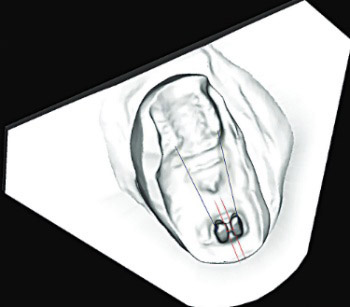
Teeth movements were calculated by measuring the distance between incisors and molars in the treated and control side (Figure 1), as follows:

FIGURE 1: Measurements of tooth movements. Tooth movements were calculated by measuring the distance between incisors (the middle of the distoproximal surface of incisors 2 mm from the gingiva) and molars (the middle of the mesioproximal ridge of the first molar) in the treated and control side. The measured distance between determined points on the incisor and molars represents bilaterally tooth movement in millimeters.

  • Point 1 - The middle of the distoproximal surface of incisors on the right side 2 mm from the gingiva

  • Point 1’ - The middle of the mesioproximal ridge of the first molar on the right side

  • Point 2 - The middle of the distoproximal surface of incisors on the left side 2 mm from the gingiva

  • Point 2’ - The middle of the mesioproximal ridge of the first molar on the left side.

The measured distance between points 1 and 1’ and 2 and 2’ was used to represent tooth movement in millimeters. A greater space between the incisor and the molar was interpreted as a decreased incisor movement.

The reliability of the measurements was calculated as correlations between the two measurements on 30 randomly selected plaster models by a single investigator. The assessment of the error of measurements showed that the coefficients of reliability were 0.998.

Histological examination

The upper jaws were dissected and the separation of the right and left maxilla was performed with a diamond separator (Diamantscheibe 30 mm Ø, Bredent, Germany). The same region was excised in all rats, along the upper left central incisor root toward the apex of the alveolar bone on the mesial and distal sides, 3 mm above the apex towards the crown, as per previously described method [13].

Bones with corresponding teeth were decalcified in 10% formic acid and routinely processed. They were embedded in paraffin, cut at 5 micrometers, and stained with hematoxylin-eosin (HE). For immunohistochemical (IH) staining the tissue slices were applied onto glass slides and coated with 3-aminopropyltriethoxy silane (APES, Sigma, St. Luis, MO, USA). The slices were air-dried and stored at 4°C until being processed for indirect immunoperoxidase staining. The tissue slices were briefly deparaffinized in xylene and rehydrated in ethanol. Endogenous peroxidase and nonspecific binding were blocked by incubation in 0.3% H2O in methanol and 5% non-immune serum. The sections were incubated for 60 minutes at room temperature with primary anti-calcitonin antibody (clone CAL-3-F5, dilution 1 [DAKO, Glostrup, Denmark]).

All tissue samples were analyzed using an Olympus BX50 light microscope with a 40 × 10 magnification. Osteoclasts were counted using the method described by Carvalho-Filho et al. [14]. Each of the five histologic sections showing the alveolar bone surface on the compression side adjacent to the entire root was examined. The number of osteoclasts on the alveolar bone surface of the pressure side was counted in each animal in four areas of the section. Cells were considered osteoclasts if they were multinucleated and located on or close to bone surfaces (Figure 2).

FIGURE 2: Osteoclasts in alveolar bone (immunohistochemistry, anti-calcitonin antibody, clone CAL-3-F5, ×400). Cells were considered osteoclasts if they were multinucleated and located on, or close to bone surfaces.

Statistical analysis

All data were subjected to Shapiro–Wilk test (p < 0.05) to test the probability distribution of the sample. One-way analysis of variance (ANOVA) was used to test the differences in tooth movement depending on different time and clodronate concentration regimes.

RESULTS

Data on study groups, clodronate dosage and application intervals are presented in Table 1.

No serious or nonserious adverse events were registered during the investigation.

Table 2 shows the effects of short and long interval application (3 days and 7 days) of different clodronate concentrations (10 mM and 2.5 mM) on tooth movement, compared to control side (0 mM).

TABLE 2: The effects of short and long interval application using 10 mMol and 2.5 mMol clodronate bisphosphonate concentrations

In the 3-day interval clodronate application groups (E1 and E2), there was no difference in the tooth movement between 10 and 2.5 mMol concentrations (F = 0.748, p > 0.05).

In the 7-day interval clodronate application groups (E3 and E4), 10 mMol clodronate bisphosphonate concentration showed a greater effect on tooth movement than 2.5 mMol concentration (F = 4.592, p < 0.05; Table 3).

TABLE 3: The effects of 10 mMol and 2.5 mMol clodronate bisphosphonate concentrations in short-term and long-term application intervals

The duration of clodronate application interval had no effect on tooth movement when 10 mMol clodronate bisphosphonate concentration was used (F = 0.677, p > 0.05). Conversely, application of 2.5 mMol clodronate bisphosphonate concentration showed a greater effect on tooth movement in the 3-day interval group (E2) compared to 7-day interval group [E4] (F = 7.084, p < 0.05).

The obtained values are shown as percentage of the distance between incisors and molars [11]. The value of 100% signifies equal movement on both sides, while the value less of 100% indicates a decreased tooth movement on the experimental side in relation to the control side.

Statistical analysis of osteoclast cell numbers was not possible, due to a limited number of detected osteoclast cells in the tissue sample (max = 6; min = 0).

There was no difference in the number of detected osteoclast cells between HE and IH staining methods.

DISCUSSION

In this study, we investigated the effects of local application of 10 mMol and 2.5 mMol clodronate bisphosphonate concentrations, over 3- and 7-day intervals on tooth movement.

The research model was adjusted so that both doses could be tested depending on the frequency of application throughout a period of 21 days. During the first 3 days of the procedure, no application took place to allow for the initial tooth movement and creation of osteoclasts. The results indicate that a higher dose of clodronate results in a substantially decreased tooth movement if applied over 7-day application intervals (Table 2). This finding is in accordance with the results reported by Liu et al. [11], suggesting a dose- and time-dependent effect of clodronate on tooth movement. Previous studies have shown a similar dose-dependent reduction of tooth movement with different bisphosphonates [6-8].

Second, we tested the time-dependent effect of clodronate. It is well documented that bisphosphonates accumulate in the body and can have a long-lasting, cumulative effect in reducing bone turnover [19,20]. This effect can be enhanced and extended by other types of bisphosphonates [5].

Based on the reported cumulative effects of bisphosphonate application, the same rate of bone resorption inhibition would be expected if bisphosphonates were administered more frequently in smaller doses than if they were administered in larger doses but less frequently.

We, therefore, investigated the effects of two doses of clodronate and two application intervals. The obtained results suggest that higher clodronate doses permit a longer interval between consecutive applications. The dose of 2.5 mMol of clodronate is effective when applied every 3 days but less effective when applied every 7 days. This indicates that bisphosphonate clodronate dose should be selected in accordance with the type of tooth movement to be prevented, e.g. orthodontic anchor loss or relapse, periodontal problems, etc. When partial pharmacological control of tooth movement is sufficient, a higher dose can be used in a 7-day interval or lower dose in 3-day interval. To prevent possible side effects, we recommend the dose of clodronate to be selected according to clinical needs; hence in cases when total control of tooth movement is in question, a higher dose in a shorter interval can be used and when clodronate is an additional factor in tooth movement control we can decrease the dose as well as intervals between the applications.

Despite the fact that clodronate can reduce tooth movement and even reduce root resorption, there are some concerns regarding the use of bisphosphonate in orthodontics. It is well known that high doses administered systematically inhibit dentinogenesis and cementogenesis, however, the low doses may be ineffective [21,22]. Nevertheless, if we use high doses of bisphosphonates, the possible side-effects can be irreversible.

The second concern is so-called “bisphosphonate-related osteonecrosis of the jaw” [23]. It has been shown that osteonecrosis usually occurs in adult patients who have received intravenous as well as oral bisphosphonate treatment. However, meta-analytic studies suggest a greater impact of amino-bisphosphonate on osteonecrosis [23,24]. The investigation of Rodrigues et al. indicate that the main adverse event of bisphosphonates, the osteonecrosis of the jaw, is exclusively related to nitrogen-containing bisphosphonates, at least in the patients with prostate cancer [9]. With respect to the use of bisphosphonates in children, there are currently no cases of “bisphosphonate-related osteonecrosis of the jaw” during and after orthodontic tooth movement [25].

CONCLUSION

For decreasing the tooth movement, low-risk bisphosphonate clodronate can be applied in the root area. Local subperiosteal injection of clodronate shows time- and dose-dependent effect on the tooth movement. To decrease the dose concentration, the interval between applications should be shorter. Future studies should evaluate the safety, efficacy, and minimum effective dose for clinical use of clodronate against tooth movement in humans.

DECLARATION OF INTERESTS

The authors declare no conflict of interests.

Acknowledgements

ACKNOWLEDGMENTS

The authors want to acknowledge Dr. Samir Prohić, Professor Nedžad Mulabegović and Professor Svjetlana Radović, for their assistance in experimental protocols and support in research.

REFERENCES

  1. , , , , , (). Differential expression of osteoblast and osteoclast chemmoatractants in compression and tension sides during orthodontic movement. Cytokine. https://doi.org/10.1016/j.cyto.2008.03.003
  2. , , , , (). The effect of local application of 1,25-dihydroxycholecalciferol on osteoclast numbers in orthodontically treated rats. J Dent Res. https://doi.org/10.1177/00220345920710010901
  3. , , (). Orthodontically induced root and alveolar bone resorption: inhibitory effect of systemic doxycycline administration in rats. Eur J Orthod. https://doi.org/10.1093/ejo/cji015
  4. , , , , , (). Comparisons of the effects of systemic administration of L-thyroxine and doxycycline on orthodontically induced root resorption in rats. Eur J Orthod. https://doi.org/10.1093/ejo/cjp124
  5. (). Bisphosphonates: the first 40 years. Bone. https://doi.org/10.1016/j.bone.2011.04.022
  6. , , , (). Anchorage and retentive effects of a bisphosphonate (AHBuBP) on tooth movements in rats. Am J Orthod Dentofacial Orthop. https://doi.org/10.1016/S0889-5406(94)70048-6
  7. , , , (). Effects of topical administration of a bisphosphonate (risedronate) on orthodontic tooth movements in rats. J Dent Res.
  8. , , , (). An ultrastructural study of the effects of bisphosphonate administration on osteoclastic bone resorption during relapse of experimentally moved rat molars. Am J Orthod Dentofacial Orthop. https://doi.org/10.1016/S0889-5406(99)70290-8
  9. , , (). Safety of I. V. nonnitrogen bisphosphonates on the occurrence of osteonecrosis of the jaw: long-term follow-up on prostate cancer patients. Clin Genitourin Cancer. https://doi.org/10.1016/j.clgc.2014.10.001
  10. , , , , (). The molecular mechanism of action of the antiresorptive and antiinflammatory drug clodronate: Evidence for the formation in vivo of a metabolite that inhibits bone resorption and causes osteoclast and macrophage apoptosis. Arthritis Rheum. https://doi.org/10.1002/1529-0131(200109)44:9<2201:AID - ART 374>3.0.CO;2-E
  11. , , , , , (). Effects of local administration of clodronate on orthodontic tooth movement and root resorption in rats. Eur J Orthod. https://doi.org/10.1093/ejo/26.5.469
  12. , , , , (). Improving bioscience research reporting: The ARRIVE guidelines for reporting animal research. PLoS Biol. https://doi.org/10.1371/journal.pbio.1000412
  13. , , , (). Effects of estrogen deficiency on tooth movement after force application: an experimental study in ovariectomized rats. Acta Odontol Scand. https://doi.org/10.1080/00016350701678725
  14. , , , , , (). Celecoxib treatment does not alter recruitment and activation of osteoclasts in the initial phase of experimental tooth movement. Eur J Hitochem. https://doi.org/10.4081/ejh.2012.e43
  15. , , , (). Effects of clodronate on early alveolar bone remodeling and root resorption related to orthodontic forces: a histomorphometric analysis. Am J Orthod Dentofacial Orthop. https://doi.org/10.1016/j.ajodo.2010.01.031
  16. , , , , , (). A new system for scanning, measuring and analyzing dental casts based on a 3D holographic sensor. Orthod Craniofac Res. https://doi.org/10.1111/j.1601-6343.2007.00417.x
  17. , , , (). A comparison of plaster, digital and reconstructed study model accuracy. J Orthod. https://doi.org/10.1179/146531207225022626
  18. , , , (). Comparison of space analysis evaluations with digital models and plaster dental casts. Am J Orthod Dentofacial Orthop. DOI: 10.1016/j.ajodo.2008.11.019
  19. (). New bisphosphonates in osteoporosis. Osteoporos Int. https://doi.org/10.1007/BF01623222
  20. , , , , (). Alendronate has a residual effect on bone mass in postmenopausal Danish women up to 7 years after treatment withdrawal. Bone. https://doi.org/10.1016/S8756-3282(03)00112-1
  21. , (). Therapeutic approaches to bone diseases. Science. https://doi.org/10.1126/science.289.5484.1508
  22. (). Development of bisphosphonates. Breast Cancer Res. https://doi.org/10.1186/bcr414
  23. , , , , , (). Oral bisphosphonate use and the prevalence of osteonecrosis of the jaw: an institutional inquiry. J Am Dent Assoc. https://doi.org/10.14219/jada.archive.2009.0019
  24. , , , , , (). Osteonecrosis of the jaw and use of bisphosphonates in adjuvant breast cancer treatment: a meta-analysis. Breast Cancer Res Treat. https://doi.org/10.1007/s10549-009-0432-z
  25. , , , , , (). Updated recommendations for managing the care of patients receiving oral bisphosphonate therapy: an advisory statement from the American Dental Association Council on Scientific Affairs. J Am Dent Assoc. https://doi.org/10.14219/jada.archive.2008.0110