K-RAS and N-RAS mutations in testicular germ cell tumors

Authors

  • Bekir Muhammet Hacioglu Department of Medical Oncology, Faculty of Medicine, Trakya University, Edirne, Turkey
  • Hilmi Kodaz Department of Medical Oncology, Acıbadem Hospital, Eskişehir, Turkey
  • Bulent Erdogan Department of Medical Oncology, Faculty of Medicine, Trakya University, Edirne, Turkey
  • Ahmet Cinkaya Department of Radiation Oncology, Dumlupınar University Evliya Celebi Training and Research Hospital, Kutahya, Turkey
  • Ebru Tastekin Department of Pathology, Faculty of Medicine, Trakya University, Edirne, Turkey
  • Ilhan Hacibekiroglu Department of Medical Oncology, Sakarya University, Training and Research Hospital, Adapazarı, Turkey
  • Esma Turkmen Department of Medical Oncology, Kocaeli Derince Training and Research Hospital, Kocaeli, Turkey
  • Osman Kostek Department of Medical Oncology, Faculty of Medicine, Trakya University, Edirne, Turkey
  • Ezgi Genc Department of Pathology, Faculty of Medicine, Trakya University, Edirne, Turkey
  • Sernaz Uzunoglu Department of Medical Oncology, Faculty of Medicine, Trakya University, Edirne, Turkey
  • Irfan Cicin Department of Medical Oncology, Faculty of Medicine, Trakya University, Edirne, Turkey

DOI:

https://doi.org/10.17305/bjbms.2017.1764

Keywords:

Testicular germ cell tumors, seminoma, non-seminoma, K-RAS mutation, N-RAS mutation, TGCT

Abstract

Testicular cancer is a relatively rare tumor type, accounting for approximately 1% of all cancers in men. However, among men aged between 15 and 40 years, testicular cancer is the most commonly diagnosed malignancy. Testicular germ cell tumors (TGCTs) are classified as seminoma and non-seminoma. The RAS oncogene controls several cellular functions, including cell proliferation, apoptosis, migration, and differentiation. Thus, RAS signaling is important for normal germ cell development. Mutations of the Kirsten RAS (K-RAS) gene are present in over 20% of all cancers. RAS gene mutations have also been reported in TGCTs. We investigated K-RAS and N-RAS mutations in seminoma and non-seminoma TGCT patients. A total of 24 (55%) pure seminoma cases and 19 (45%) non-seminoma cases were included in the study. K-RAS and N-RAS analyses were performed in our molecular pathology laboratory, using K-RAS and N-RAS Pyro Kit 24 V1 (Qiagen). In total, a RAS mutation was present in 12 patients (27%): 7 seminoma (29%) and 5 non-seminoma cases (26%) [p = 0.55]. A K-RAS mutation was present in 4 pure seminoma tumors (16%) and 3 non-seminoma tumors (15%) [p = 0.63], and an N-RAS mutation was observed in 4 seminoma tumors (16%) and 3 non-seminoma tumors (15%) [p = 0.63]. Both, K-RAS and N-RAS mutations were present in two patients: one with seminoma tumor and the other with non-seminoma tumor. To date, no approved targeted therapy is available for the treatment of TGCTs. The analysis of K-RAS and N-RAS mutations in these tumors may provide more treatment options, especially in platinum-resistant tumors.

Downloads

Download data is not yet available.

Author Biographies

Bekir Muhammet Hacioglu, Department of Medical Oncology, Faculty of Medicine, Trakya University, Edirne, Turkey

Department of Medical Oncology

Hilmi Kodaz, Department of Medical Oncology, Acıbadem Hospital, Eskişehir, Turkey

Department of Medical Oncology

Bulent Erdogan, Department of Medical Oncology, Faculty of Medicine, Trakya University, Edirne, Turkey

Department of Medical Oncology

Ahmet Cinkaya, Department of Radiation Oncology, Dumlupınar University Evliya Celebi Training and Research Hospital, Kutahya, Turkey

Department of Radiation Oncology

Ebru Tastekin, Department of Pathology, Faculty of Medicine, Trakya University, Edirne, Turkey

Department of Pathology

Ilhan Hacibekiroglu, Department of Medical Oncology, Sakarya University, Training and Research Hospital, Adapazarı, Turkey

Department of Medical Oncology

Esma Turkmen, Department of Medical Oncology, Kocaeli Derince Training and Research Hospital, Kocaeli, Turkey

Department of Medical Oncology

Osman Kostek, Department of Medical Oncology, Faculty of Medicine, Trakya University, Edirne, Turkey

Department of Medical Oncology

Ezgi Genc, Department of Pathology, Faculty of Medicine, Trakya University, Edirne, Turkey

Department of Pathology

Sernaz Uzunoglu, Department of Medical Oncology, Faculty of Medicine, Trakya University, Edirne, Turkey

Department of Medical Oncology

Irfan Cicin, Department of Medical Oncology, Faculty of Medicine, Trakya University, Edirne, Turkey

Department of Medical Oncology

References

Shanmugalingam T, Soultati A, Chowdhury S, Rudman S, Van Hemelrijck M. Global incidence and outcome of testicular cancer. Clin Epidemiol 2013;5(1):417-27. https://doi.org/10.2147/CLEP.S34430.

Chia VM, Quraishi SM, Devesa SS, Purdue MP, Cook MB, McGlynn KA. International trends in the incidence of testicular cancer, 1973-2002. Cancer Epidemiol Biomarkers Prev 2010;19(5):1151-9. https://doi.org/10.1158/1055-9965.EPI-10-0031.

International Germ Cell Consensus Classification: A prognostic factor-based staging system for metastatic germ cell cancers. International Germ Cell Cancer Collaborative Group. J Clin Oncol 1997;15(2):594-603.

Raghavan D. Testicular cancer: Maintaining the high cure rate. Oncology (Williston Park) 2003;17(2):218-28.

Li J, Xia F, Li WX. Coactivation of STAT and Ras is required for germ cell proliferation and invasive migration in Drosophila. Dev Cell 2003;5(5):787-98. https://doi.org/10.1016/S1534-5807(03)00328-9.

Bamford S, Dawson E, Forbes S, Clements J, Pettett R, Dogan A, et al. The COSMIC (Catalogue of Somatic Mutations in Cancer) database and website. Br J Cancer 2004;91(2):355-8. https://doi.org/10.1038/sj.bjc.6601894.

Moul JW, Theune SM, Chang EH. Detection of RAS mutations in archival testicular germ cell tumors by polymerase chain reaction and oligonucleotide hybridization. Genes Chromosomes Cancer 1992;5(2):109-18. https://doi.org/10.1002/gcc.2870050204.

Chaganti RS, Houldsworth J. Genetics and biology of adult human male germ cell tumors. Cancer Res 2000;60(6):1475-82.

Reuter VE. Origins and molecular biology of testicular germ cell tumors. Mod Pathol 2005;18(Suppl 2):S51-60. https://doi.org/10.1038/modpathol.3800309.

Colicelli J. Human RAS superfamily proteins and related GTPases. Sci STKE 2004;2004(250):RE13. https://doi.org/10.1126/stke.2502004re13.

Lin JS, Webber EM, Senger CA, Holmes RS, Whitlock EP. Systematic review of pharmacogenetic testing for predicting clinical benefit to anti-EGFR therapy in metastatic colorectal cancer. Am J Cancer Res 2011;1(5):650-62.

Sommerer F, Hengge UR, Markwarth A, Vomschloss S, Stolzenburg JU, Wittekind C, et al. Mutations of BRAF and RAS are rare events in germ cell tumours. Int J Cancer 2005;113(2):329-35. https://doi.org/10.1002/ijc.20567.

Ganguly S, Murty VV, Samaniego F, Reuter VE, Bosl GJ, Chaganti RS. Detection of preferential NRAS mutations in human male germ cell tumors by the polymerase chain reaction. Genes Chromosomes Cancer 1990;1(3):228-32. https://doi.org/10.1002/gcc.2870010307.

Price TJ, Bruhn MA, Lee CK, Hardingham JE, Townsend AR, Mann KP, et al. Correlation of extended RAS and PIK3CA gene mutation status with outcomes from the phase III AGITG MAX STUDY involving capecitabine alone or in combination with bevacizumab plus or minus mitomycin C in advanced colorectal cancer. Br J Cancer 2015;112(6):963-70. https://doi.org/10.1038/bjc.2015.37.

Amado RG, Wolf M, Peeters M, Van Cutsem E, Siena S, Freeman DJ, et al. Wild-type KRAS is required for panitumumab efficacy in patients with metastatic colorectal cancer. J Clin Oncol 2008;26(10):1626-34. https://doi.org/10.1200/JCO.2007.14.7116.

Boublikova L, Bakardjieva-Mihaylova V, Skvarova Kramarzova K, Kuzilkova D, Dobiasova A, Fiser K, et al. Wilms tumor gene 1 (WT1), TP53, RAS/BRAF and KIT aberrations in testicular germ cell tumors. Cancer Lett 2016;376(2):367-76. https://doi.org/10.1016/j.canlet.2016.04.016.

Goddard NC, McIntyre A, Summersgill B, Gilbert D, Kitazawa S, Shipley J. KIT and RAS signalling pathways in testicular germ cell tumours: New data and a review of the literature. Int J Androl 2007;30(4):337-48.

https://doi.org/10.1111/j.1365-2605.2007.00769.x.

Skotheim RI, Monni O, Mousses S, Fosså SD, Kallioniemi OP, Lothe RA, et al. New insights into testicular germ cell tumorigenesis from gene expression profiling. Cancer Res 2002;62(8):2359-64.

Feldman DR, Iyer G, Van Alstine L, Patil S, Al-Ahmadie H, Reuter VE, et al. Presence of somatic mutations within PIK3CA, AKT, RAS, and FGFR3 but not BRAF in cisplatin-resistant germ cell tumors. Clin Cancer Res 2014;20(14):3712-20. https://doi.org/10.1158/1078-0432.CCR-13-2868.

Honecker F, Wermann H, Mayer F, Gillis AJ, Stoop H, van Gurp RJ, et al. Microsatellite instability, mismatch repair deficiency, and BRAF mutation in treatment-resistant germ cell tumors. J Clin Oncol 2009;27(13):2129-36. https://doi.org/10.1200/JCO.2008.18.8623.
K-RAS and N-RAS mutations in testicular germ cell tumors

Downloads

Additional Files

Published

2017-05-20

How to Cite

1.
Hacioglu BM, Kodaz H, Erdogan B, Cinkaya A, Tastekin E, Hacibekiroglu I, Turkmen E, Kostek O, Genc E, Uzunoglu S, Cicin I. K-RAS and N-RAS mutations in testicular germ cell tumors. Biomol Biomed [Internet]. 2017May20 [cited 2023Sep.23];17(2):159-63. Available from: https://www.bjbms.org/ojs/index.php/bjbms/article/view/1764

Issue

Section

Translational and Clinical Research

INTRODUCTION

Testicular cancer is a relatively rare tumor type, accounting for approximately 1% of all cancers in men. However, among men aged between 15 and 40 years, testicular cancer is the most commonly diagnosed malignancy [1]. Furthermore, roughly 95% of all malignant tumors of the testis are germ cell tumors. Although these tumors can also arise in extragonadal primary sites, they are still managed in the same way as testicular germ cell tumors (TGCTs) [2]. TGCTs are classified into two groups: Seminoma and non-seminoma tumors. Non-seminoma tumors include several subtypes of cancers, including embryonal cell carcinoma, choriocarcinoma, yolk sac tumor, and teratoma. Serum tumor markers such as alpha-fetoprotein (AFP), lactate dehydrogenase (LDH), and beta-human chorionic gonadotropin (beta-hCG) are critical in diagnosing TGCTs, determining prognosis, and assessing treatment outcomes [3].

It has been shown that TGCTs are highly sensitive to radiation and chemotherapy, and consequently, the cure rate is high in these cases. However, approximately 5% of patients develop resistance to the treatment [4].

The RAS oncogene controls several cellular functions, including cell proliferation, apoptosis, migration, and differentiation. Thus, RAS signaling is important for normal germ cell development [5]. Kirsten RAS (K-RAS) mutations are present in over 20% of all cancers [6]. Although RAS gene mutations have also been reported in TGCTs [7], the role of RAS pathway alterations in the treatment response is still not clear. In addition, seminomas and non-seminomas may also occur as mixed germ cell tumors, but in clinical management, they are categorized as non-seminomas. In this study, we investigated K-RAS and N-RAS mutations in seminoma and non-seminoma TGCT patients, aiming to determine the relationship between these mutations and pathologic factors, prognostic factors, and response to therapy.

MATERIALS AND METHODS

Patients and samples

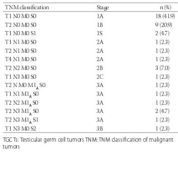
We retrospectively reviewed the records of patients diagnosed with TGCT between 2007 and 2015 at Trakya University Medical Oncology Department. Paraffin-embedded tissue blocks from these patients were collected from the archive of our pathology department. Twenty-four pure seminoma cases and 19 non-seminoma cases were included in the study. All tumoral slides were reevaluated and the pathologic diagnoses were confirmed by a pathologist. Cancer staging was performed according to the TNM Classification of Malignant Tumors (TNM staging) in the Seventh Edition American Joint Committee on Cancer (AJCC) Cancer Staging Manual. The study was approved by the Institutional Ethics Board. Informed consent was obtained from all individual participants or their legal representatives.

Mutation analysis

K-RAS and N-RAS analyses were performed in our molecular pathology laboratory, and the following mutations were analyzed: CAA>CTA Q61L, CAA>CAT Q61H, CAA>CGA Q61R, GGT>GTT G12V, GGC>GAC G13D, GGT>GAT G12D, GGT>TGT G12C, GGT>AGT G12S, GGT>GCT G12A, GGT>CGT G12R, and GGT>c.34_35GG>TT G12F. DNA isolation was performed from the primary tumor tissue samples using QIAamp DNA FFPE Tissue Kit (Qiagen, Hilden, Germany). The Pyro Kit 24 V1 (Qiagen, Hilden, Germany) was used for K-RAS and N-RAS analyses, and K-RAS and N-RAS point mutations were analyzed with the PyroMark Q24 Software System (Qiagen, Hilden, Germany).

Statistical analysis

We used IBM Statistical Package for Social Sciences (SPSS, Chicago, IL, USA) version 18.0 to perform statistical analyses in this study. Overall, patient survival was calculated from the date of diagnosis to date of death from the disease or to the patient’s last follow-up. The relationship between nonparametric variables was studied using Chi-square test, and parametric variables were compared using an independent samples t-test. The value of p < 0.05 was considered statistically significant. Survival estimates were calculated using the Kaplan-Meier method.

RESULTS

Clinical-pathological features and TNM staging of the cases are shown in Tables 1 and 2.

TABLE 1: Comparison of clinical and tumor pathological characteristics according to RAS mutations
TABLE 2: TNM classification of patients with TGCT

Overall, 24 patients (55%) were diagnosed with pure seminoma tumors and 19 patients (45%) were diagnosed with non-seminoma tumors. The median age of participants was 30 years (min: 19 - max: 63). In total, a RAS mutation was present in 12 tumors (27%): 7 pure seminoma cases (29%) and 5 non-seminoma cases (26%) (p = 0.55). A K-RAS mutation was present in 4 pure seminoma tumors (16%) and 3 non-seminoma tumors (15%) (p = 0.63); an N-RAS mutation was also present in 4 pure seminoma tumors (16%) and 3 non-seminoma tumors (15%) (p = 0.63). While all of the N-RAS mutations were 181 C>A (Q61K) in the codon 61 of the exon 3, K-RAS mutations were detected in different codons. The most common K-RAS mutation was 436G>A (A146T) in the codon 146 of the exon 4 (in 3 cases). In addition, two patients had GGT>AGT (G12S) mutation in the codon 12 of the exon 2, one patient had GGC>GAC (G13D) mutation in the codon 13 of the exon 2, and another had CAA>CTA (Q61L) mutation in the codon 61 of the exon 3.

Furthermore, a total of 19 patients (44%) had lymphovascular invasion (LVI) at the time of diagnosis. There was no significant difference between RAS mutant and wild-type patients in the rate of LVI. Moreover, involvement of tunica albuginea was observed in 12 patients (28%) and this was not associated with the presence of RAS mutations. Other pathologic features of the testes (i.e., tunica vaginalis, spermatic cord, and scrotal invasion) were not statistically different between the RAS wild-type and mutant patients (p = 0.61).

It should be noted that two patients involved in the study died before the time of this analysis. Both patients had RAS wild-type tumors. One tumor was seminoma and the other was non-seminoma. The patient who had seminoma tumor did not receive adjuvant chemotherapy but did receive adjuvant radiotherapy. The overall survival was 14 months for this patient. The patient with non-seminoma died 31 months after the diagnosis. Aside from these two cases, the progression was observed in four patients. Three of these patients had seminoma tumor and the fourth had non-seminoma tumor. Only in one of these patients CAA>CTA (Q61L) mutation was observed in the K-RAS codon 61. This patient received a first-line combination chemotherapy containing bleomycin, etoposide, and cisplatin (BEP). Following the progression, he received cisplatin, etoposide, and ifosfamide as the second-line chemotherapy. The other patients received BEP therapy at the time of progression. After the second-line chemotherapy, recurrence or metastasis did not occur in these patients. They were still alive at the time of writing this manuscript. In our study, there was a limited number of events for the overall and progression-free survival analyses.

DISCUSSION

A distinguishing characteristic of TGCTs is a mutation on the short arm of chromosome 12 (12p), identified in almost all invasive TGCTs, as well as in intratubular embryonal carcinoma and intratubular seminoma [8,9]. K-RAS and N-RAS are components of the Ras-Raf-MEK-ERK pathway. K-RAS and N-RAS mutations in the codons 12, 13, and 61 cause inappropriate activation of the pathway independent of epidermal growth factor-epidermal growth factor receptor (EGF-EGFR) binding. The constitutively active Ras-Raf-MEK-ERK pathway initiates carcinogenesis by promoting cellular proliferation, gene expression, differentiation, mitosis, and cell survival. Patients with lung and colorectal cancers who have these mutations showed unfavorable response to anti-EGFR-targeted or receptor tyrosine kinase inhibitor (TKI) therapy [10,11].

In the COSMIC study, RAS mutations (K-RAS [13/214] and N-RAS [8/145]) were observed in approximately 6% of seminomas; both types of mutations were absent in non-seminomas in this study (K-RAS [0/138] and N-RAS [0/71]) [6]. In another study, Sommerer et al. [12] identified K-RAS mutations in 7% of seminomas (2/30) and 9% of non-seminoma TGCTs (3/32) [12]. Furthermore, Ganguly et al. [13] found that 59% of seminomas (13/22) and 78% of non-seminomas (7/9) had N-RAS gene mutations [13]. In these studies, none of the patients had both K-RAS and N-RAS mutations. In previous studies, the incidence of K-RAS and N-RAS mutations in TGCTs was 6-7%; however, it was 27.9% in our study. In the present study, a K-RAS mutation was present in 4 pure seminoma tumors (16%) and 3 non-seminomas (15%) and an N-RAS mutation was present in 4 seminomas (16%) and in 3 non-seminomas (15%). All of the N-RAS mutations [181C>A (Q61K)] were detected in the codon 61 of the exon 3, while the K-RAS mutations were detected in different codons. The most common K-RAS mutation [436G>A (A146T)] was in the codon 146 of the exon 4, detected in three patients. In addition, two patients had GGT>AGT (G12S) mutation in the codon 12 of the exon 2, one patient had GGC>GAC (G13D) mutation in the codon 13 of the exon 2, and another had CAA>CTA (Q61L) mutation in the codon 61 of the exon 3. In colorectal cancer, mutations in the codons 12 and 13 of the exon 2 represent the most common K-RAS and N-RAS mutations [14]. Approximately 40% of K-RAS gene mutations in colorectal cancer are observed in the codons 12 and 13 of the exon 2 [15]. Our results showed that the RAS mutations in TGCT patients are present in different codons compared to the patients with colorectal cancer.

It is important to note that the rate of K-RAS and N-RAS mutations was higher in our study compared with the other studies. This may be due to the fact that we performed extended RAS mutation testing.

In two patients, both K-RAS and N-RAS mutations were present; one patient had seminoma tumor and the other had non-seminoma tumor. The patient with seminoma had GGT>AGT (G12S) K-RAS mutation in the codon 12 and 181C>A (Q61K) N-RAS mutation in the codon 61. The patient with non-seminoma had 436G>A (A146T) K-RAS mutation in the codon 146 and 181C>A (Q61K) N-RAS mutation in the codon 61. One of these patients was treated with one cycle of carboplatin and the other with four cycles of BEP as the adjuvant therapy. No recurrence was observed in either of the two patients after the adjuvant chemotherapy.

A study by Boublikova et al. [16] revealed that there was no significant difference in the frequency of BRAF and RAS mutations between seminoma and non-seminoma patients, even though BRAF mutations were more common in seminomas and RAS variants were more frequent in non-seminomas [16]. Goddard et al. [17] found that testicular seminomas are associated with c-Kit mutations [17]. Skotheim et al. [18] showed gene expression differences between seminoma and non-seminoma TGCTs [18]. Feldman et al. [19] showed 4.2% rate of K-RAS mutations in cisplatin-resistant germ cell tumors [19]. A recent study by Honecker et al. [20] assessed 100 control (50 seminomas and 50 nonseminomatous germ cell tumors [NSGCT]) and 35 cisplatin-resistant cases of TGCTs (3 seminomas and 32 NSGCTs). A K-RAS mutation was not observed in the cisplatin-resistant group and was present in two cases (one seminoma and one non-seminoma; 2%) in the control group [20]. In our study, two cases were cisplatin-resistant, and these patients died before the time of analysis. Neither K-RAS nor N-RAS mutations were detected in these two cases. In addition, four recurrences were observed after orchiectomy and adjuvant chemotherapy; a K-RAS mutation was detected in only one of these cases (nonseminomatous type), and none of the RAS mutations were observed. The major limitation of our study was the small number of samples. However, this might be explained by the fact that TGCTs are uncommon tumors.

CONCLUSION

TGCTs include a heterogeneous group of tumors. To date, no approved targeted therapy is available for the treatment of TGCTs. However, analysis of K-RAS and N-RAS mutations in these tumors may provide more treatment options with a lower toxicity profile compared to the current therapies. Therefore, further studies investigating the genetic profile of TGCTs are warranted.

DECLARATION OF INTERESTS

The authors declare no conflict of interests.

REFERENCES

  1. , , , , (). Global incidence and outcome of testicular cancer. Clin Epidemiol. https://doi.org/10.2147/CLEP.S34430
  2. , , , , , (). International trends in the incidence of testicular cancer 1973-2002. Cancer Epidemiol Biomarkers Prev. https://doi.org/10.1158/1055-9965.EPI-10-0031
  3. (). International Germ Cell Cancer Collaborative Group. J Clin Oncol.
  4. (). Testicular cancer: Maintaining the high cure rate. Oncology (Williston Park).
  5. , , (). Coactivation of STAT and Ras is required for germ cell proliferation and invasive migration in Drosophila . Dev Cell. https://doi.org/10.1016/S1534-5807(03)00328-9
  6. , , , , , (). The COSMIC (Catalogue of Somatic Mutations in Cancer) database and website. Br J Cancer. https://doi.org/10.1038/sj.bjc.6601894
  7. , , (). Detection of RAS mutations in archival testicular germ cell tumors by polymerase chain reaction and oligonucleotide hybridization. Genes Chromosomes Cancer. https://doi.org/10.1002/gcc.2870050204
  8. , (). Genetics and biology of adult human male germ cell tumors. Cancer Res.
  9. (). Origins and molecular biology of testicular germ cell tumors. Mod Pathol. https://doi.org/10.1038/modpathol.3800309
  10. (). Human RAS superfamily proteins and related GTPases. Sci STKE. https://doi.org/10.1126/stke.2502004rne13
  11. , , , , (). Systematic review of pharmacogenetic testing for predicting clinical benefit to anti-EGFR therapy in metastatic colorectal cancer. Am J Cancer Res.
  12. , , , , , (). Mutations of BRAF and RAS are rare events in germ cell tumours. Int J Cancer. https://doi.org/10.1002/ijc.20567
  13. , , , , , (). Detection of preferential NRAS mutations in human male germ cell tumors by the polymerase chain reaction. Genes Chromosomes Cancer. https://doi.org/10.1002/gcc.2870010307
  14. , , , , , (). Correlation of extended RAS and PIK3CA gene mutation status with outcomes from the phase III AGITG MAX STUDY involving capecitabine alone or in combination with bevacizumab plus or minus mitomycin C in advanced colorectal cancer. Br J Cancer. https://doi.org/10.1038/bjc.2015.37
  15. , , , , , (). Wild-type KRAS is required for panitumumab efficacy in patients with metastatic colorectal cancer. J Clin Oncol. https://doi.org/10.1200/JCO.2007.14.7116
  16. , , , , , (). Wilms tumor gene 1 (WT1), TP53, RAS/BRAF and KIT aberrations in testicular germ cell tumors. Cancer Lett. https://doi.org/10.1016/j.canlet.2016.04.016
  17. , , , , , (). KIT and RAS signalling pathways in testicular germ cell tumours: New data and a review of the literature. Int J Androl. https://doi.org/10.1111/j.1365-2605.2007.00769.x
  18. , , , , , (). New insights into testicular germ cell tumorigenesis from gene expression profiling. Cancer Res.
  19. , , , , , (). Presence of somatic mutations within PIK3CA, AKT, RAS, and FGFR3 but not BRAF in cisplatin-resistant germ cell tumors. Clin Cancer Res. https://doi.org/10.1158/1078-0432.CCR-13-2868
  20. , , , , , (). Microsatellite instability, mismatch repair deficiency, and BRAF mutation in treatment-resistant germ cell tumors. J Clin Oncol. https://doi.org/10.1200/JCO.2008.18.8623