Salvia miltiorrhiza extract dihydrotanshinone induces apoptosis and inhibits proliferation of glioma cells

Authors

  • Yong Cao Department of Neurosurgery, Henan Province Hospital of TCM, Zhengzhou, China
  • Bin Huang Department of Neurosurgery, Tongchuan Mineral Bureau Center Hospital in Shaanxi Province, Tongchuan, China
  • Changqing Gao Department of Neurosurgery II, The First People’s Hospital of Xianyang City, Xianyang, China

DOI:

https://doi.org/10.17305/bjbms.2017.1800

Keywords:

Dihydrotanshinone, glioma, SHG-44 cells, cell apoptosis, cell proliferation, MTT assay

Abstract

Dihydrotanshinone, a functional food in China, is an effective anti-cardiovascular disease substance isolated from Salvia miltiorrhiza (S. miltiorrhiza). Glioma is considered to be fatal due to its poor prognosis and few effective therapeutic options. In this study, we investigated the anticancer effects of S. miltiorrhiza extract dihydrotanshinone on human glioma SHG-44 cells, by using 3-(4, 5-dimethyl-2-thiazolyl)-2, 5-diphenyl-2H-tetrazolium bromide assay, Hoechst 33258 nuclear staining, Annexin V/propidium iodide double staining, as well as western blot analysis. The results showed that dihydrotanshinone effectively suppressed SHG-44 cells proliferation and induced apoptosis in both dose- and time-dependent manner. Moreover, we demonstrated that dihydrotanshinone increased the activation of caspases (caspase-3 and caspase-9) and the release of cytochrome c in SHG-44 cells. Overall, dihydrotanshinone could induce apoptosis and inhibit proliferation of glioma cells by regulating caspases and cytochrome c. This study suggests that dihydrotanshinone may serve as a potential treatment option for patients with glioma.

Downloads

Download data is not yet available.

Author Biographies

Yong Cao, Department of Neurosurgery, Henan Province Hospital of TCM, Zhengzhou, China

Department of Neurosurgery

Bin Huang, Department of Neurosurgery, Tongchuan Mineral Bureau Center Hospital in Shaanxi Province, Tongchuan, China

Department of Neurosurgery

Changqing Gao, Department of Neurosurgery II, The First People’s Hospital of Xianyang City, Xianyang, China

Department of Neurosurgery II

References

Tzen JT, Jinn TR, Chen YC, Li FY, Cheng FC, Shi LS, et al. Magnesium lithospermate B possesses inhibitory activity on Na+, K+ - ATPase and neuroprotective effects against ischemic stroke. Acta Pharmacol Sin 2007;28(5):609-15.

https://doi.org/10.1111/j.1745-7254.2007.00544.x.

Wang X, Morris-Natschke SL, Lee KH. New developments in the chemistry and biology of the bioactive constituents of Tanshen. Med Res Rev 2007;27(1):133-48. https://doi.org/10.1002/med.20077.

Tian XH, Wu JH. Tanshinone derivatives: A patent review (January 2006 - September 2012). Expert Opin Ther Pat 2013;23(1):19-29. https://doi.org/10.1517/13543776.2013.736494.

Franek KJ, Zhou Z, Zhang WD, Chen WY. In vitro studies of baicalin alone or in combination with Salvia miltiorrhiza extract as a potential anti-cancer agent. Int J Oncol 2005;26(1):217-24. https://doi.org/10.3892/ijo.26.1.217.

Chang JY, Chang CY, Kuo CC, Chen LT, Wein YS, Kuo YH. Salvinal, a novel microtubule inhibitor isolated from Salvia miltiorrhizae Bunge (Danshen), with antimitotic activity in multidrug-sensitive and -resistant human tumor cells. Mol Pharmacol 2004;65(1):77-84. https://doi.org/10.1124/mol.65.1.77.

Chen W, Lu Y, Chen G, Huang S. Molecular evidence of cryptotanshinone for treatment and prevention of human cancer. Anticancer Agents Med Chem 2013;13(7):979-87. https://doi.org/10.2174/18715206113139990115.

Bian W, Chen F, Bai L, Zhang P, Qin W. Dihydrotanshinone I inhibits angiogenesis both in vitro and in vivo. Acta Biochim Biophys Sin (Shanghai) 2008;40(1):1-6. https://doi.org/10.1111/j.1745-7270.2008.00370.x.

D'Agostino VG, Lal P, Mantelli B, Tiedje C, Zucal C, Thongon N, et al. Dihydrotanshinone-I interferes with the RNA-binding activity of HuR affecting its post-transcriptional function. Sci Rep 2015;5:16478. https://doi.org/10.1038/srep16478.

Lee DS, Lee SH. Biological activity of dihydrotanshinone I: Effect on apoptosis. J Biosci Bioeng 2000;89(3):292-3. https://doi.org/10.1016/S1389-1723(00)88838-6.

Chen MH, Jenh YJ, Wu SK, Chen YS, Hanagata N, Lin FH. Non-invasive photodynamic therapy in brain cancer by use of Tb3 - Doped LaF3 nanoparticles in combination with photosensitizer through X-ray irradiation: A proof-of-concept study. Nanoscale Res Lett 2017;12(1):62. https://doi.org/10.1186/s11671-017-1840-3.

Perry NS, Houghton PJ, Theobald A, Jenner P, Perry EK. In-vitro inhibition of human erythrocyte acetylcholinesterase by salvia lavandulaefolia essential oil and constituent terpenes. J Pharm Pharmacol 2000;52(7):895-902. https://doi.org/10.1211/0022357001774598.

Zhang JH, Xu M. DNA fragmentation in apoptosis. Cell Res 2000;10(3):205-11. https://doi.org/10.1038/sj.cr.7290049.

Kajstura M, Halicka HD, Pryjma J, Darzynkiewicz Z. Discontinuous fragmentation of nuclear DNA during apoptosis revealed by discrete "sub-G1" peaks on DNA content histograms. Cytometry A 2007;71(3):125-31. https://doi.org/10.1002/cyto.a.20357.

Barciszewska AM. MicroRNAs as efficient biomarkers in high-grade gliomas. Folia Neuropathol 2016;54(4):369-74. https://doi.org/10.5114/fn.2016.64812.

Hamza MA, Gilbert M. Targeted therapy in gliomas. Curr Oncol Rep 2014;16(4):379. https://doi.org/10.1007/s11912-014-0379-z.

Kaufmann JK, Chiocca EA. Glioma virus therapies between bench and bedside. Neuro Oncol 2014;16(3):334-51. https://doi.org/10.1093/neuonc/not310.

Zhuo Z, Hu J, Yang X, Chen M, Lei X, Deng L, et al. Ailanthone inhibits Huh7 cancer cell growth via cell cycle arrest and apoptosis in vitro and in vivo. Sci Rep 2015;5:16185. https://doi.org/10.1038/srep16185.

Liu JJ, Wu HH, Chen TH, Leung W, Liang YC. 15,16-dihydrotanshinone I from the functional food Salvia miltiorrhiza exhibits anticancer activity in human HL-60 leukemia cells: In vitro and in vivo studies. Int J Mol Sci 2015;16(8):19387-400. https://doi.org/10.3390/ijms160819387.

Saraste A, Pulkki K. Morphologic and biochemical hallmarks of apoptosis. Cardiovasc Res 2000;45(3):528-37. https://doi.org/10.1016/S0008-6363(99)00384-3.

Huang WW, Chiu YJ, Fan MJ, Lu HF, Yeh HF, Li KH, et al. Kaempferol induced apoptosis via endoplasmic reticulum stress and mitochondria-dependent pathway in human osteosarcoma U-2 OS cells. Mol Nutr Food Res 2010;54(11):1585-95. https://doi.org/10.1002/mnfr.201000005.

Alnemri ES, Livingston DJ, Nicholson DW, Salvesen G, Thornberry NA, Wong WW, et al. Human ICE/CED-3 protease nomenclature. Cell 1996;87(2):171. https://doi.org/10.1016/S0092-8674(00)81334-3.

Zou H, Henzel WJ, Liu X, Lutschg A, Wang X. Apaf-1, a human protein homologous to C. Elegans CED-4, participates in cytochrome c-dependent activation of caspase-3. Cell 1997;90(3):405-13. https://doi.org/10.1016/S0092-8674(00)80501-2.

Wolf BB, Green DR. Suicidal tendencies: Apoptotic cell death by caspase family proteinases. J Biol Chem 1999;274(29):20049-52. https://doi.org/10.1074/jbc.274.29.20049.

Tsai SL, Suk FM, Wang CI, Liu DZ, Hou WC, Lin PJ, et al. Anti-tumor potential of 15, 16-dihydrotanshinone I against breast adenocarcinoma through inducing G1 arrest and apoptosis. Biochem Pharmacol 2007;74(11):1575-86. https://doi.org/10.1016/j.bcp.2007.08.009.

Wang L, Hu T, Shen J, Zhang L, Chan RL, Lu L, et al. Dihydrotanshinone I induced apoptosis and autophagy through caspase dependent pathway in colon cancer. Phytomedicine 2015;22(12):1079-87. https://doi.org/10.1016/j.phymed.2015.08.009.

Lee WY, Liu KW, Yeung JH. Reactive oxygen species-mediated kinase activation by dihydrotanshinone in tanshinones-induced apoptosis in HepG2 cells. Cancer Lett 2009;285(1):46-57. https://doi.org/10.1016/j.canlet.2009.04.040.
Salvia miltiorrhiza extract dihydrotanshinone induces apoptosis and inhibits proliferation of glioma cells

Downloads

Additional Files

Published

2017-08-20

How to Cite

1.
Cao Y, Huang B, Gao C. Salvia miltiorrhiza extract dihydrotanshinone induces apoptosis and inhibits proliferation of glioma cells. Biomol Biomed [Internet]. 2017Aug.20 [cited 2023Nov.30];17(3):235-40. Available from: https://www.bjbms.org/ojs/index.php/bjbms/article/view/1800

Issue

Section

Molecular Biology

INTRODUCTION

Native in China and Japan, Salvia miltiorrhiza Bunge, also called Chinese sage or danshen, is a perennial plant in the genus Salvia and has been considered as a functional food in Asia. In traditional Chinese herbal medicine, dried root of S. miltiorrhiza is famous for its high medicinal value and is widely used for promoting blood circulation, especially in angina pectoris and myocardial infarction [1,2]. Modern pharmacological studies demonstrated that Salvia has some antitumor effect, and it has been used as an auxiliary anticancer drug [3,4]. For example, it was demonstrated that Salvia alcohol extract significantly inhibited the growth of human colorectal cancer cells (HRT-18), colon cancer cells (HT-29), and liver cancer cells (HepG2) in a cell-culture setting [5,6]. The major ingredients isolated from Salvia ethanol extract include cryptotanshinone, dihydrotanshinone I, tanshinone IIA, and tanshinone IIB among others. While tanshinone IIA, IIB and cryptotanshinone have been reported for their effective antitumor activity [2,6], the role of dihydrotanshinone in cancer treatment remains relatively unclear.

Cytotoxic effects of dihydrotanshinone have been reported in a variety of tumor cells [7]. For instance, dihydrotanshinone could interfere with the RNA-binding activity of HuR and thus inhibit viability, proliferation, and chemotaxis of breast cancer cells [8]. Dihydrotanshinone induced cell growth arrest during the S phase of the cell cycle and, subsequently, apoptosis following its application to human leukemia multidrug resistant K562/ADR cells [9]. However, the role of dihydrotanshinone in brain cancer remains completely unexplored.

Brain cancer has very limited treatment options due to the presence of the blood–brain barrier (BBB), which protects infiltrating glioma cells from the effect of chemotherapeutic agents [10]. It has been previously reported that tanshinones have the potential to penetrate the BBB [11], and based on this, anti-glioma effects of dihydrotanshinone have been proposed.

In this study, 0-100 μg/L of dihydrotanshinone was applied to human glioblastoma SHG-44 cells and the antitumor properties of dihydrotanshinone were observed. Specifically, dihydrotanshinone could remarkably inhibit SHG-44 cell growth and proliferation, as well as induce cellular apoptosis. Dihydrotanshinone increased the activation of caspase-3 and caspase-9, as well as promoted the release of cytochrome c. This study may shed lights on the application of dihydrotanshinone in treating gliomas.

MATERIALS AND METHODS

Cell culture

Human glioma cell line SHG-44 was purchased from Shanghai Institute of Cell Bank (Shanghai, China) and grown in RPMI-1640 medium (Gibco, Carlsbad, CA, USA) containing 10% heat-inactivated fetal bovine serum (Gibco) and 100 U/mL of penicillin-streptomycin (Gibco). The cells were incubated in a humidified incubator at 37°C with 5% CO2. All cells used in the assays were in the logarithmic growth phase (adherent density of about 60-80%).

Treatment with dihydrotanshinone

Dihydrotanshinone, with purity of more than 98%, was purchased from Baoji Herbest Bio-Tech Co., Ltd. (Baoji, China). In all experiments, indicated drugs were dissolved in dimethyl sulfoxide (DMSO, Sigma-Aldrich, St. Louis, MO), and all original stock solutions were maintained at 100 mg/L and stored at 4°C. A series of different dihydrotanshinone concentrations (0, 10, 20, 40, 50, 60, 80, and 100 μg/L) were added upon attachment of the cells, to detect the effects of dihydrotanshinone on SHG-44 cells.

3-(4, 5-dimethyl-2-thiazolyl)-2, 5-diphenyl-2H-tetrazolium bromide (MTT) assay

SHG-44 cells were seeded in 96-well plates with 1 × 10⁴ cells/100 μL. The following groups were included: background group (blank), control group, and the experimental group in which increasing concentrations of dihydrotanshinone were used. Various concentrations (0, 10, 20, 40, 50, 60, 80, and 100 μg/L) of dihydrotanshinone were added upon attachment of the cells to the dishes, and four parallel wells were set up for each dihydrotanshinone concentration. After 24, 48 and 72 hours of incubation, 30 μL of MTT (5 mg/mL; Sigma-Aldrich) was added until formazan was formed. These 96-well plates were then placed upside down and dried, and 0.1 mL DMSO was added to dissolve formazan. Absorbance or optical density (OD) values of each well at λ = 595 nm were measured using a multi-plate reader, and the cell growth inhibition rate was calculated according to the formula as follows:

% inhibition of cell proliferation rate = (1−experimental group mean OD value/control group mean OD value) × 100%.

Flow cytometry analysis

SHG-44 cells were seeded in 6-well plates at 5 × 10⁴ cells per well. After 24 hours, 10, 50 and 100 μg/L of dihydrotanshinone, and DMSO as the control, were added to the cells for 24 hours. The cells were collected and gently washed 2-3 times in phosphate-buffered saline (PBS), then fixed with 75% ethanol (4°C pre-cooled) overnight. After the cells were collected by centrifugation, 0.5 mL of PBS containing propidium iodide (PI) (0.02 mg/mL; Sigma-Aldrich) and RNase A (0.1 mg/mL; Sigma-Aldrich) were added and mixed gently. The samples were then incubated at 37°C in the dark for 30 minutes. Cell sieves with 8.47 μm pore size were used to prepare single cell suspension for measuring DNA content by Epics XL flow cytometry system (Beckman-Coulter, USA).

Hoechst 33258 staining

SHG-44 cells were seeded in 6-well plates at 5 × 10⁴ cells per well. After 24 hours, 10, 50 and 100 μg/L of dihydrotanshinone, and DMSO as the control, were added to the cells for 24 hours. The cells were collected and gently washed 2-3 times in PBS, then stained with 1 mL of PBS solution containing 10 mg/mL of Hoechst 33258 (Sigma-Aldrich) and incubated at 37°C in the dark for 30 minutes. The cells were then washed 3 times with PBS and imaged using fluorescence microscopy at an excitation wavelength of 350 nm and an emission wavelength at 460 nm.

Annexin V-FITC/PI double staining

SHG-44 cells were seeded in 6-well plates at 5 × 10⁴ cells per well. After 24 hours, 10, 50 and 100 μg/L of dihydrotanshinone, and DMSO as the control, were added to the cells for 24 hours. The cells were collected and gently washed 2-3 times in PBS, then resuspended in 0.5 mL of PBS containing 5 μL of Annexin V-FITC (Pharmingen, San Diego, CA) and 10 μL of PI (Sigma-Aldrich). The reactions were incubated at 37°C in the dark for 30 minutes. Cell sieves with 8.47 μm pore size were used to prepare single cell suspension for Epics XL flow cytometry analyses of cell apoptosis.

Western blot analysis

SHG-44 cells were seeded in 6-well plates at 5 × 10⁴ cells per well. After 24 hours, 10, 50 and 100 μg/L of dihydrotanshinone, and DMSO as the control, were added to the cells for 24 hours. The cells were collected and lysed in RIPA lysis buffer (Beyotime, Beijing, China), and protein concentrations in whole cell lysates were measured by the bicinchoninic acid assay. Forty milligrams of total protein were loaded on 30% sodium dodecyl sulfate polyacrylamide gel electrophoresis and transferred to polyvinylidene difluoride (PVDF) membrane (Millipore Corp, Bedford, MA, USA). After blocking with 5% non-fat milk, indicated primary antibodies (1:1000, Cell Signaling Technology, USA) and secondary antibodies (1:5000, Cell Signaling Technology, USA) were incubated with PVDF membrane, and the signals were visualized using the enhanced chemiluminescence reagent (GE Healthcare, Little Chalfont, UK).

Statistics

Data were presented as mean ± standard deviation (SD). GraphPad Prism 5.0 statistical software (GraphPad, San Diego, CA) and Student’s t-test (two-tailed) were used to analyze the significant difference between the groups. Values of p < 0.05 were considered statistically significant.

RESULTS

Dihydrotanshinone significantly inhibits SHG-44 cell growth in vitro

To investigate whether dihydrotanshinone can inhibit cell proliferation in human glioma, SHG-44 cells were exposed to various doses of dihydrotanshinone for 24-72 hours. As shown in Figure 1, dihydrotanshinone inhibited SHG-44 cell growth in a dose- and time-dependent manner, with an IC50 value of 50.32 ± 2.49 μg/L at 24 hours, 42.35 ± 2.25 μg/L at 48 hours, and 31.25 ± 2.82 μg/L at 72 hours. These data confirm a possible role of dihydrotanshinone in suppression of SHG-44 cell growth.

FIGURE 1: Dihydrotanshinone suppresses SHG-44 cell growth in time- and dose-dependent manner. SHG-44 cells were treated with various concentrations (0, 10, 20, 40, 50, 60, 80, and 100 μg/L) of dihydrotanshinone for 24, 48 and 72 hours, and analyzed with 3-(4, 5-dimethyl-2-thiazolyl)-2, 5-diphenyl-2H-tetrazolium bromide (MTT) assay.

Dihydrotanshinone treatment induces DNA fragmentation in SHG-44 cells

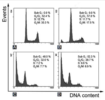
Next, we examined the possible mechanism(s) responsible for dihydrotanshinone-mediated suppression of SHG-44 cell growth. Because the cleavage of chromosomal DNA by the DNA fragment factor is associated with cellular ­apoptosis [12], we first examined whether dihydrotanshinone treatment leads to DNA fragmentation in SHG-44 cells. Notably, we observed that the treatments with increased doses of dihydrotanshinone led to correspondingly increased amount of sub-G1 DNA content (<2N location apoptotic peaks in Figure 2A-D), a marker of cellular apoptosis [13]. Specifically, the number of cells with sub-G1 DNA content indicated that the apoptosis levels were 9.0%, 48.0%, and 52.3% for dihydrotanshinone concentrations at 10, 50, and 100 μg/L, respectively. This suggests that dihydrotanshinone treatment may induce cell apoptosis in SHG-44 cells in a dose-dependent manner.

FIGURE 2: Dihydrotanshinone induces DNA fragmentation. SHG-44 cells were treated with dimethyl sulfoxide (A), and 10 μg/L (B), 50 μg/L (C) and 100 μg/L (D) of dihydrotanshinone for 24 hours, followed by propidium iodide staining and flow cytometry analysis.

Dihydrotanshinone treatment leads to nuclear condensation in SHG-44 cells

To further confirm the pro-apoptosis role of dihydrotanshinone in SHG-44 cells, Hoechst 33258 staining was applied to examine the nucleus morphology. After the staining, a blue fluorescent signal in normal cells is usually weak and evenly colored, while in apoptotic cells, fluorescent signals are unevenly distributed with increased fluorescence intensity. After the treatment of SHG-44 cells with different concentrations of dihydrotanshinone for 24 hours, the nuclei exhibited condensed and fragmented chromatin with increased staining intensity (Figure 3A-D), which is another feature of cellular apoptosis [13]. Moreover, the increase in staining intensity of condensed nuclei was positively correlated with the increasing doses of dihydrotanshinone used in the treatments. Taken together, these results suggest that dihydrotanshinone treatment results in nuclei condensation and fragmentation in SHG-44 cells.

FIGURE 3: Dihydrotanshinone induces nuclear condensation. SHG-44 cells were treated with dimethyl sulfoxide (A), and 10 μg/L (B), 50 μg/L (C) and 100 μg/L (D) of dihydrotanshinone for 24 hours. The nuclei were stained with Hoechst 33258, and analyzed using a fluorescent microscopy. The representative images are shown. The arrowheads indicate Hoechst 33258 stained nucleus.

Dihydrotanshinone-induced cellular apoptosis in SHG-44 cells

As both DNA fragmentation and nuclear condensation were observed in dihydrotanshinone-treated SHG-44 cells, we further examined whether these phenotypes were the result of the dihydrotanshinone-induced cellular apoptosis. We used the Annexin V-FITC/PI double staining method to check whether the cells undergo apoptosis. The cells stained only with Annexin V are shown in the lower right quadrant of the FACS histogram, and the cells stained with both dyes are presented in the upper right quadrant of the FACS histogram (Figure 4A-D). Notably, the early apoptotic population was positively increased with the increasing doses of dihydrotanshinone, from 2.6% in the control group gradually to 83.1% in the group treated with 100 μg/L of dihydrotanshinone (Figure 4A-D). However, the cell population in the late apoptosis stages (stained by both PI and Annexin V) did not change dramatically with the increasing doses of dihydrotanshinone (Figure 4B-D). These data suggest that dihydrotanshinone induces apoptosis in SHG-44 cells, and consequently triggers DNA fragmentation and nuclear condensation.

FIGURE 4: Dihydrotanshinone induces cell apoptosis in a dose-dependent manner. SHG-44 cells were treated with dimethyl sulfoxide (A), and 10 μg/L (B), 50 μg/L (C) and 100 μg/L (D) of dihydrotanshinone for 24 hours, followed by Annexin V/propidium iodide (PI) staining and flow cytometry analysis.

Dihydrotanshinone-induced cellular apoptosis is mediated by increased activation of caspases and release of cytochrome c

To investigate the possible molecular mechanism of dihydrotanshinone in suppressing SHG-44 cell growth through apoptosis, we performed western blot analyses using the dihydrotanshinone-treated cells. There was no significant change in the levels of caspase-8-mediated death receptor pathway, while the cleavage of caspase-9, downstream of the mitochondrial pathway, was markedly increased, as well as the levels of cleaved caspase-3 and poly ADP-ribose polymerase (PARP) that are signs of apoptosis (Figure 5).

FIGURE 5: Dihydrotanshinone promotes caspase activation. SHG-44 glioma cells were treated with dimethyl sulfoxide (A), and 10 μg/L (B), 50 μg/L (C) and 100 μg/L (D) dihydrotanshinone for 24 hours, followed by cell lysis and western blot analyses with indicated antibodies.

Because the cleavage-mediated activation of caspase-3 requires cytochrome c, we examined whether dihydrotanshinone also influences the cytoplasmic levels of cytochrome c. After the treatment with 10 μg/L and 50 μg/L of dihydrotanshinone, the cytoplasmic, but not the total levels of cytochrome c were markedly increased (Figure 6), further indicating the role of dihydrotanshinone in facilitating cytochrome c release, which activates caspase-3 and promotes apoptosis.

FIGURE 6: Dihydrotanshinone promotes cytochrome c release from mitochondria. SHG-44 cells were treated with dimethyl sulfoxide (A), and 10 μg/L (B) and 50 μg/L (C) of dihydrotanshinone for 24 hours, followed by cell lysis and western blot analyses with indicated antibodies.

DISCUSSION

Human glioma is one of the most frequent malignant neoplasms, accounting for over 70% of all primary brain tumors in adults [14]. Studies have reported that in more than 60% of adults over the age of 50 glioma relapsed, due to the absence of timely diagnosis and treatment [15,16]. The current treatment options for glioma patients include surgery, radiotherapy, chemotherapy and targeted therapy, most of which have profound side effects. Moreover, many anticancer chemotherapies kill a significant portion of healthy cells in addition to glioma cancer cells. On the other hand, the traditional Chinese herbal medicine has drawn more and more attention, due to its high efficiency and low toxicity effect. One effective way is to search for or isolate active ingredients from the plants that display antitumor activity, which would be helpful in discovery of new drugs but could also provide a molecular basis for the design of active lead compounds [17].

Salvia is one of the Chinese traditional medicinal plants that has been broadly studied. The active, fat-soluble ingredients such as quinone derivatives (i.e., tanshinone I, cryptotanshinone, isotanshinone I and others), have demonstrated broad antitumor activity [6]. Previous studies reported the cytotoxic effect of dihydrotanshinone from Salvia against various types of tumor cells [18]. To our knowledge, this is the first report of dihydrotanshinone antitumor activity in glioma cells. In this study, dihydrotanshinone showed a significant antiproliferative effect in the human SHG-44 metastatic glioma cells, in both time- and dose-dependent manner, with IC50 values of 50.32 ± 2.49 μg/L, 42.35 ± 2.25 μg/L, and 31.25 ± 2.82 μg/L at 24, 48, and 72 hours post treatment, respectively. More importantly, we further demonstrated that dihydrotanshinone triggers cellular apoptosis by releasing cytochrome c from mitochondria and thus activating caspases.

During cell apoptosis, changes in the cell morphology are observed including cell shrinkage, cytoplasmic vacuole formation, swelling of mitochondria, nuclear membrane shrinkage, chromatin aggregation, and appearance of apoptotic bodies [19]. In this study, dihydrotanshinone triggered cell shrinkage, nuclear chromatin aggregation and the movement of chromatin closer to the nuclear membrane, suggesting that dihydrotanshinone might suppress SHG-44 cell growth by inducing cell apoptosis. Furthermore, cellular debris was identified by flow cytometry upon the treatment with increasing concentrations of dihydrotanshinone, which also suggests the role of dihydrotanshinone in inducing cell apoptosis. Finally, we performed Annexin V-FITC/PI double staining analysis to confirm that dihydrotanshinone can induce apoptosis in dihydrotanshinone dose-dependent manner. Cumulatively, our results confirm that dihydrotanshinone suppresses SHG-44 cell proliferation by inducing cell apoptosis.

There are two major apoptotic pathways, i.e., intracellular and extracellular apoptotic pathway [20]. The intracellular pathway, also known as mitochondrial pathway, is the major apoptotic signaling pathway in the cell. Apoptosis is the result of complex interactions between a variety of genes and proteins in cells where, in particular, caspase protein family plays an important role [21]. The proteins from caspase family are activated by cytochrome c, released from mitochondria, and by the apoptotic protease activating factor 1 upon apoptotic signals, through the formation of apoptotic bodies that ultimately induce apoptosis [22]. Once capase-3 and -9 are activated, they can cause PARP cleavage [23]. Our western blot analyses demonstrated that, upon the dihydrotanshinone treatment, the cytoplasmic level of cytochrome c was increased which activated caspase-9 and caspase-3, as well as PARP cleavage, and triggered the cell apoptosis. These findings are consistent with the results from previous studies [24,25].

CONCLUSION

Overall, our results suggest that dihydrotanshinone can effectively inhibit the proliferation of SHG-44 cells possibly through the activation of the mitochondrial apoptosis pathway. These results shed new light on the application of dihydrotanshinone as a new class of anticancer drugs in treating patients with glioma. As reactive oxygen species (ROS)-mediated p38 mitogen-activated protein kinases (MAPK) activation has been shown to play a key role in dihydrotanshinone-induced apoptosis in HepG2 cells [26], it warrants further studies to investigate molecular signaling pathways that are altered upon dihydrotanshinone treatment, to search for additional drug targets, or to overcome possible drug resistance triggered by dihydrotanshinone administration.

DECLARATION OF INTERESTS

The authors declare no conflict of interests.

Acknowledgements

ACKNOWLEDGMENTS

We thank all the Gao Lab members for helpful discussions.

REFERENCES

  1. , , , , , (). Magnesium lithospermate B possesses inhibitory activity on Na+, K+- ATPase and neuroprotective effects against ischemic stroke. Acta Pharmacol Sin. https://doi.org/10.1111/j.1745-7254.2007.00544.x
  2. , , (). New developments in the chemistry and biology of the bioactive constituents of Tanshen. Med Res Rev. https://doi.org/10.1002/med.20077
  3. , (). Tanshinone derivatives: A patent review (January 2006 - September 2012). Expert Opin Ther Pat. https://doi.org/10.1517/13543776.2013.736494
  4. , , , (). In vitro studies of baicalin alone or in combination with Salvia miltiorrhiza extract as a potential anti-cancer agent. Int J Oncol. https://doi.org/10.3892/ijo.26.1.217
  5. , , , , , (). Salvinal, a novel microtubule inhibitor isolated from Salvia miltiorrhizae Bunge (Danshen), with antimitotic activity in multidrug-sensitive and -resistant human tumor cells. Mol Pharmacol. https://doi.org/10.1124/mol.65.1.77
  6. , , , (). Molecular evidence of cryptotanshinone for treatment and prevention of human cancer. Anticancer Agents Med Chem. https://doi.org/10.2174/18715206113139990115
  7. , , , , (). Dihydrotanshinone I inhibits angiogenesis both in vitro and in vivo. Acta Biochim Biophys Sin (Shanghai). https://doi.org/10.1111/j.1745-7270.2008.00370.x
  8. , , , , , (). Dihydrotanshinone-I interferes with the RNA-binding activity of HuR affecting its post-transcriptional function. Sci Rep. https://doi.org/10.1038/srep16478
  9. , (). Biological activity of dihydrotanshinone I: Effect on apoptosis. J Biosci Bioeng. https://doi.org/10.1016/S1389-1723(00)88838-6
  10. , , , , , (). Non-invasive photodynamic therapy in brain cancer by use of Tb3 - Doped LaF3 nanoparticles in combination with photosensitizer through X-ray irradiation: A proof-of-concept study. Nanoscale Res Lett. https://doi.org/10.1186/s11671-017-1840-3
  11. , , , , (). In-vitro inhibition of human erythrocyte acetylcholinesterase by salvia lavandulaefolia essential oil and constituent terpenes. J Pharm Pharmacol. https://doi.org/10.1211/0022357001774598
  12. , (). DNA fragmentation in apoptosis. Cell Res. https://doi.org/10.1038/sj.cr.7290049
  13. , , , (). Discontinuous fragmentation of nuclear DNA during apoptosis revealed by discrete “sub-G1” peaks on DNA content histograms. Cytometry A. https://doi.org/10.1002/cyto.a.20357
  14. (). MicroRNAs as efficient biomarkers in high-grade gliomas. Folia Neuropathol. https://doi.org/10.5114/fn.2016.64812
  15. , (). Targeted therapy in gliomas. Curr Oncol Rep. https://doi.org/10.1007/s11912-014-0379-z
  16. , (). Glioma virus therapies between bench and bedside. Neuro Oncol. https://doi.org/10.1093/neuonc/not310
  17. , , , , , (). Ailanthone inhibits Huh7 cancer cell growth via cell cycle arrest and apoptosis in vitro and in vivo. Sci Rep. https://doi.org/10.1038/srep16185
  18. , , , , (). 15,16-dihydrotanshinone I from the functional food Salvia miltiorrhiza exhibits anticancer activity in human HL-60 leukemia cells In vitro and in vivo studies. Int J Mol Sci. https://doi.org/10.3390/ijms160819387
  19. , (). Morphologic and biochemical hallmarks of apoptosis. Cardiovasc Res. https://doi.org/10.1016/S0008-6363(99)00384-3
  20. , , , , , (). Kaempferol induced apoptosis via endoplasmic reticulum stress and mitochondria-dependent pathway in human osteosarcoma U-2 OS cells. Mol Nutr Food Res. https://doi.org/10.1002/mnfr.201000005
  21. , , , , , (). Human ICE/CED-3 protease nomenclature. Cell. https://doi.org/10.1016/S0092-8674(00)81334-3
  22. , , , , (). Apaf-1, a human protein homologous to C. Elegans CED-4, participates in cytochrome c-dependent activation of caspase-3. Cell. https://doi.org/10.1016/S0092-8674(00)80501-2
  23. , (). Suicidal tendencies: Apoptotic cell death by caspase family proteinases. J Biol Chem. https://doi.org/10.1074/jbc.274.29.20049
  24. , , , , , (). Anti-tumor potential of 15, 16-dihydrotanshinone I against breast adenocarcinoma through inducing G1 arrest and apoptosis. Biochem Pharmacol. https://doi.org/10.1016/j.bcp.2007.08.009
  25. , , , , , (). Dihydrotanshinone I induced apoptosis and autophagy through caspase dependent pathway in colon cancer. Phytomedicine. https://doi.org/10.1016/j.phymed.2015.08.009
  26. , , (). Reactive oxygen species-mediated kinase activation by dihydrotanshinone in tanshinones-induced apoptosis in HepG2 cells. Cancer Lett. https://doi.org/10.1016/j.canlet.2009.04.040