Apocrine carcinoma of the breast: A brief update on the molecular features and targetable biomarkers

Authors

  • Semir Vranic Department of Pathology, University of Sarajevo Clinical Center
  • Rebecca Feldman Levine Cancer Institute, Charlotte, NC
  • Zoran Gatalica Caris Life Sciences, Phoenix, AZ

DOI:

https://doi.org/10.17305/bjbms.2016.1811

Keywords:

breast cancer, special types, apocrine carcinoma, androgen receptor, biomarkers, targeted therapy

Abstract

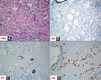
Apocrine carcinoma of the breast is a rare, primary breast cancer characterized by the apocrine morphology, estrogen receptor-negative and androgen receptor-positive profile with a frequent overexpression of Her-2/neu protein (~30%). Apart from the Her-2/neu target, advanced and/or metastatic apocrine carcinomas have limited treatment options. In this review, we briefly describe and discuss the molecular features and new theranostic biomarkers for this rare mammary malignancy. The importance of comprehensive profiling is highlighted due to synergistic and potentially antagonistic molecular events in the individual patients.

Downloads

Download data is not yet available.

References

Eusebi V, Millis R, Cattani M, Bussolati G, Azzopardi J. Apocrine carcinoma of the breast. A morphologic and immunocytochemical study. Am J Pathol 1986;123(3): 532-41.

Gatalica Z. Immunohistochemical analysis of apocrine breast lesions. Pathol Res Pract 1997;193(11-12):753-8.

Vranic S, Tawfik O, Palazzo J, Bilalovic N, Eyzaguirre E, Lee LM, et al. EGFR and HER-2/neu expression in invasive apocrine carcinoma of the breast. Mod Pathol 2010;23(5):644-53.

Vranic S, Schmitt F, Sapino A, Costa JL, Reddy S, Castro M, et al. Apocrine carcinoma of the breast: A comprehensive review. Histol Histopathol 2013;28(11):1393-1409.

Vranic S, Marchiò C, Castellano I, Botta C, Scalzo MS, Bender RP, et al. Immunohistochemical and molecular profiling of histologically defined apocrine carcinomas of the breast. Hum Pathol 2015;46(9):1350-9.

Mills AM, E Gottlieb C, M Wendroth S, M Brenin C, Atkins KA. Pure Apocrine Carcinomas Represent a Clinicopathologically Distinct Androgen Receptor-Positive Subset of Triple-Negative Breast Cancers. Am J Surg Pathol 2016;40(8):1109-16.

Bratthauer GL, Lininger RA, Man YG, Tavassoli FA. Androgen and estrogen receptor mRNA status in apocrine carcinomas. Diagn Mol Pathol 2002;11(2):113-8.

Vranic S, Gatalica Z, Deng H, Frkovic-Grazio S, Lee LM, Gurjeva O, et al. ER-α36 a novel isoform of ER-α66 is commonly over-expressed in apocrine and adenoid cystic carcinoma of the breast. J Clin Pathol 2011;64(1):54-7.

Costa LJ, Justino A, Gomes M, Alvarenga CA, Gerhard R, Vranic S, et al. Comprehensive genetic characterization of apocrine lesions of the breast. Cancer Res 2013;73(8):2013.

Naderi A, Meyer M, Dowhan DH. Cross-regulation between FOXA1 and ErbB2 signaling in estrogen receptor-negative breast cancer. Neoplasia 2012;14(4):283-96.

Naderi A, Hughes-Davies L. A functionally significant cross-talk between androgen receptor and ErbB2 pathways in estrogen receptor negative breast cancer. Neoplasia 2008;10(6):542-8.

Naderi A, Meyer M. Prolactin-induced protein mediates cell invasion and regulates integrin signaling in estrogen receptor-negative breast cancer. Breast Cancer Res 2012;14(4):R111.

Lehmann-Che J, Hamy AS, Porcher R, Barritault M, Bouhidel F, Habuellelah H, et al. Molecular apocrine breast cancers are aggressive estrogen receptor negative tumors overexpressing either HER2 or GCDFP15. Breast Cancer Res 2013;15(3):R37.

Weisman PS, Ng CK, Brogi E, Eisenberg RE, Won HH, Piscuoglio S, et al. Genetic alterations of triple negative breast cancer by targeted next-generation sequencing and correlation with tumor morphology. Mod Pathol 2016;29(5):476-88.

Arce-Salinas C, Riesco-Martinez MC, Hanna W, Bedard P, Warner E. Complete Response of Metastatic Androgen Receptor-Positive Breast Cancer to Bicalutamide: Case Report and Review of the Literature. J Clin Oncol 2016;34(4):e21-4.

Bonnefoi H, Grellety T, Tredan O, Saghatchian M, Dalenc F, Mailliez A, et al. A phase II trial of abiraterone acetate plus prednisone in patients with triple-negative androgen receptor positive locally advanced or metastatic breast cancer (UCBG 12-1). Ann Oncol 2016;27(5):812-8.

Gucalp A, Tolaney S, Isakoff SJ, Ingle JN, Liu MC, Carey LA, et al. Phase II trial of bicalutamide in patients with androgen receptor-positive, estrogen receptor-negative metastatic Breast Cancer. Clin Cancer Res 2013;19(19):5505-12.

Lehmann BD, Bauer JA, Schafer JM, Pendleton CS, Tang L, Johnson KC, et al. PIK3CA mutations in androgen receptor-positive triple negative breast cancer confer sensitivity to the combination of PI3K and androgen receptor inhibitors. Breast Cancer Res 2014;16(4):406. doi: 10.1186/s13058-014-0406-x.

Shi W, Jiang T, Nuciforo P, Hatzis C, Holmes E, Harbeck N, et al. Pathway level alterations rather than mutations in single genes predict response to HER2-targeted therapies in the neo-ALTTO trial. Ann Oncol 2016 Sep 29. pii: mdw434.

Gatalica Z, Snyder C, Maney T, Ghazalpour A, Holterman DA, Xiao N, et al. Programmed cell death (PD-1) and its ligand (PD-L1) expression in common cancers and their correlation with molecular cancer type. Cancer Epidemiol Biomarkers Prev 2014;23(12):2965-70.

Joneja U, Vranic S, Swensen J, Feldman R, Chen W, Kimbrough J, et al. Comprehensive profiling of metaplastic breast carcinomas reveals frequent overexpression of PD-L1. J Clin Pathol. 2016 Aug 16. pii: jclinpath-2016-203874. doi: 10.1136/jclinpath-2016-203874. [Epub ahead of print]

Apocrine carcinoma of the breast: A brief update on the molecular features and targetable biomarkers

Downloads

Additional Files

Published

2017-02-21

How to Cite

1.
Vranic S, Feldman R, Gatalica Z. Apocrine carcinoma of the breast: A brief update on the molecular features and targetable biomarkers. Biomol Biomed [Internet]. 2017Feb.21 [cited 2023Sep.22];17(1):9-11. Available from: https://www.bjbms.org/ojs/index.php/bjbms/article/view/1811

Issue

Section

Reviews

INTRODUCTION

Invasive apocrine carcinoma of the breast is a rare primary breast cancer characterized by the apocrine cells with abundant eosinophilic and granular cytoplasm, centrally to eccentrically located nuclei with prominent nucleoli and distinctive cell borders [1] (Figure 1A). Mammary apocrine epithelium has a characteristic steroid receptor profile that is negative for full length estrogen receptor-alpha (ER) and progesterone receptor (PR) and is androgen receptor (AR) positive [2,3]. The presence of apocrine cells in more than 90% of the tumor population defines invasive apocrine carcinoma, but it is necessary to have the characteristic immunohistochemical (IHC) profile of steroid receptors (ER-/PR-/AR+) to qualify as “pure” apocrine carcinoma (PAC) [3-5] (Figures 1B-D). Such strictly defined PAC constitutes < 1% of all breast cancers [6].

FIGURE 1: (A) Hematoxylin and Eosin slide of a case of invasive mammary carcinoma with apocrine morphology; The apocrine cells are negative for estrogen receptor [ER] (B) and progesterone receptor [PR] (C) but positive for androgen receptor [AR] (D); note the presence of ER and PR expression in adjacent normal ducts.

At the molecular level, PAC is characterized by detectable mRNA transcript of estrogen receptor, but only the alternatively spliced variant ER-36alpha (ER-36α) can be detected at the protein level (IHC) [7,8]. Progression of neoplastic disease from apocrine hyperplasia, apocrine ductal carcinoma in situ (DCIS) to invasive apocrine carcinoma had been proposed and investigated for genetic changes using a microdissection approach [9]. Array comparative genomic hybridization (aCGH) and next-generation sequencing (NGS) revealed accumulation of chromosomal losses and gains (particularly along the chromosome 17q) as well as tumor suppressor/oncogene mutations during the phases of progression [5,9].

Activation of the AR signaling pathway is a prominent feature present in apocrine lesions of the breast including apocrine DCIS and invasive apocrine carcinoma [2]. AR upregulation is mediated through forkhead box A1 (FoxA1) and human epidermal growth factor receptor 2 (HER2) activities [10]. Prolactin-induced protein (or GCDFP-15) is also actively regulated by the AR/extracellular signal-regulated kinase (ERK) feedback loop in apocrine cells [11,12]. Based on these data, targeting of the androgen receptor with androgen-targeting therapies, and potential combination strategies based on additional genomic alterations have been under investigation in clinical trials for triple-negative breast carcinomas, including PAC.

“Molecular apocrine” carcinomas exhibit prognostically poor gene signature (using validated qRT-PCR signature) with a high-risk recurrence score and a poor prognosis [13]. Comprehensive cancer profiling of PAC reveals numerous, yet individualized therapeutic options. PIK3CA/PTEN/AKT and TP53 alterations are most common in apocrine carcinoma [5,9,13,14]. Deregulation of the mitogen-activated protein kinase (MAPK) pathway components (KRAS, NRAS, BRAF) is less frequent [5,9] (Table 1). AR expression presents an opportunity for the treatment with androgen targeting therapies seemingly in all cases [6,15-17]. The presence of PIK3CA mutations and androgen overexpression may confer sensitivity to the combination of phosphatidylinositol-4,5-bisphosphate 3-kinase catalytic subunit alpha (PIK3CA) and AR inhibitors [18]. HER2 amplification and over-expression are seen in ~30% of tumors, while EGFR gene is amplified in a small (6%) percentage of cases despite the common epidermal growth factor receptor (EGFR) protein expression [3,5,6,13]. These two receptors may be targeted with monoclonal antibodies; however the high frequency of PIK3CA mutation may contribute to attenuated responses [19], and a comprehensive sample profiling (e.g. IHC for receptors and sequencing for mutations) is needed to provide optimal personalized therapy.

TABLE 1: A summary of the biomarker profiling using a multiplatform approach (immunohistochemistry, in situ hybridization and next-generation sequencing) [data from the Caris Life Science commercial laboratory, September 2016]

Biomarkers of classic chemotherapy that are variably expressed in individual cases of PAC (and the agents used to target them) include topoisomerase 2 alpha (TOPO2A) [anthracyclines], excision repair cross-complementation group 1 protein (ERCC1) [platinum drugs], ribonucleotide reductase catalytic subunit M1 (RRM1) [gemcitabine], transducin like enhancer of split 3 (TLE3) [taxanes], and thymidylate synthase (TS) [antifolates] (Table 1). Recently described immune therapies based on immune checkpoint inhibitors have not been described specifically for apocrine carcinomas, which in our limited experience do not express programmed death-ligand 1 (PD-L1) (0/8; using antibody clone SP142), in contrast to classic triple-negative breast cancers, which are more frequently PD-L1 positive (9-59%) [20,21] (Table 1).

In summary, apocrine carcinoma is a rare, distinct subtype of breast cancer with consistent AR overexpression/full length ER-α loss, frequent deregulation of the PIK3CA/phosphatase and tensin homolog (PTEN) pathway and HER2 activation, which along with the biomarkers of conventional chemotherapy may tailor optimal treatment modalities for the patients with advanced and/or metastatic apocrine carcinoma. Targeted therapy based on immune check point inhibitors needs additional investigations as preliminary results show lack of PD-L1 expression in this cancer type.

DECLARATION OF INTERESTS

Zoran Gatalica is an employee of Caris Life Sciences; Semir Vranic and Rebecca Feldman declare no conflict of interest.

Acknowledgements

ACKNOWLEDGMENTS

The results provided here were in part presented at the XXXI International Congress of the International Academy of Pathology and the 28th Congress of the European Society of Pathology that was held in Cologne, Germany, September 25-29, 2016 (The Breast Cancer Working Group Long Course entitled “Breast Pathology: Histology-based prognostic markers besides grade and stage (Session I) & Special types of breast cancers with clinical impact (Session II)”.

REFERENCES

  1. , , , , (). Apocrine carcinoma of the breast. A morphologic and immunocytochemical study. Am J Pathol.
  2. (). Immunohistochemical analysis of apocrine breast lesions. Pathol Res Pract. https://doi.org/10.1016/S0344-0338(97)80053-2
  3. , , , , , (). EGFR and HER-2/neu expression in invasive apocrine carcinoma of the breast. Mod Pathol. https://doi.org/10.1038/modpathol.2010.50
  4. , , , , , (). Apocrine carcinoma of the breast: A comprehensive review. Histol Histopathol.
  5. , , , , , (). Immunohistochemical and molecular profiling of histologically defined apocrine carcinomas of the breast. Hum Pathol. https://doi.org/10.1016/j.humpath.2015.05.017
  6. , , , , (). Pure apocrine carcinomas represent a clinicopathologically distinct androgen receptor-positive subset of triple-negative breast cancers. Am J Surg Pathol. https://doi.org/10.1097/PAS.0000000000000671
  7. , , , (). Androgen and estrogen receptor mRNA status in apocrine carcinomas. Diagn Mol Pathol. https://doi.org/10.1097/00019606-200206000-00008
  8. , , , , , (). ER-α36 a novel isoform of ER-α66 is commonly over-expressed in apocrine and adenoid cystic carcinoma of the breast. J Clin Pathol. https://doi.org/10.1136/jcp.2010.082776
  9. , , , , , (). Comprehensive genetic characterization of apocrine lesions of the breast. Cancer Res. https://doi.org/10.1158/1538-7445.AM2013-2013
  10. , , (). Cross-regulation between FOXA1 and ErbB2 signaling in estrogen receptor-negative breast cancer. Neoplasia. https://doi.org/10.1593/neo.12294
  11. , (). A functionally significant cross-talk between androgen receptor and ErbB2 pathways in estrogen receptor negative breast cancer. Neoplasia. https://doi.org/10.1593/neo.08274
  12. , (). Prolactin-induced protein mediates cell invasion and regulates integrin signaling in estrogen receptor-negative breast cancer. Breast Cancer Res. https://doi.org/10.1186/bcr3232
  13. , , , , , (). Molecular apocrine breast cancers are aggressive estrogen receptor negative tumors overexpressing either HER2 or GCDFP15. Breast Cancer Res. https://doi.org/10.1186/bcr3421
  14. , , , , , (). Genetic alterations of triple negative breast cancer by targeted next-generation sequencing and correlation with tumor morphology. Mod Pathol. https://doi.org/10.1038/modpathol.2016.39
  15. , , , , (). Complete response of metastatic androgen receptor-positive breast cancer to bicalutamide: Case report and review of the literature. J Clin Oncol. https://doi.org/10.1200/JCO.2013.49.8899
  16. , , , , , (). A phase II trial of abiraterone acetate plus prednisone in patients with triple-negative androgen receptor positive locally advanced or metastatic breast cancer (UCBG 12-1). Ann Oncol. https://doi.org/10.1093/annonc/mdw067
  17. , , , , , (). Phase II trial of bicalutamide in patients with androgen receptor-positive, estrogen receptor-negative metastatic breast cancer. Clin Cancer Res. https://doi.org/10.1158/1078-0432.CCR-12-3327
  18. , , , , , (). PIK3CA mutations in androgen receptor-positive triple negative breast cancer confer sensitivity to the combination of PI3K and androgen receptor inhibitors. Breast Cancer Res. https://doi.org/10.1186/s13058-014-0406-x
  19. , , , , , (). Pathway level alterations rather than mutations in single genes predict response to HER2-targeted therapies in the neo-ALTTO trial. Ann Oncol. pii: mdw434https://doi.org/10.1093/annonc/mdw434
  20. , , , , , (). Programmed cell death (PD-1) and its ligand (PD-L1) expression in common cancers and their correlation with molecular cancer type. Cancer Epidemiol Biomarkers Prev. https://doi.org/10.1158/1055-9965.EPI-14-0654
  21. , , , , , (). Comprehensive profiling of metaplastic breast carcinomas reveals frequent overexpression of PD-L1. J Clin Pathol. pii: jclinpath-2016-203874. DOI: 10.1136/jclinpath-2016-203874. [Epub ahead of print]