Soluble transferrin receptor as a marker of erythropoiesis in patients undergoing high-flux hemodialysis

Authors

  • Pei Yin Department of Nephrology, First Affiliated Hospital of Chinese PLA General Hospital, Beijing, China
  • Yan Song Department of Nephrology, First Affiliated Hospital of Chinese PLA General Hospital, Beijing, China
  • Jijun Li Department of Nephrology, First Affiliated Hospital of Chinese PLA General Hospital, Beijing, China

DOI:

https://doi.org/10.17305/bjbms.2017.1972

Keywords:

Hemodialysis, renal anemia, high-flux dialysis, soluble transferrin receptor, HFD, erythropoiesis, sTfR, iron status

Abstract

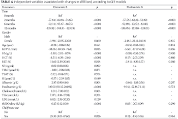
Anemia is a common complication in chronic kidney disease (CKD) patients receiving hemodialysis. The effect of high-flux dialysis (HFD) on anemia remains unclear. This prospective study aimed to evaluate the effect of HFD on anemia, and the potential of soluble transferrin receptor (sTfR) as a marker of iron status and erythropoiesis in CKD patients on hemodialysis. Forty patients, who switched from conventional low-flux dialysis to HFD for 12 months, were enrolled in this study. The levels of sTfR, hemoglobin (Hb), iron, and nutritional markers, as well as the dose of recombinant human erythropoietin (rhEPO) and use of chalybeate were determined at 0, 2, 6, and 12 months after starting HFD. HFD significantly increased the hemoglobin level and reduced sTfR level in CKD patients (p < 0.05). In addition, significant decreasing linear trends were observed for rhEPO dosage and chalybeate use (p < 0.05). The level of sTfR was positively correlated with the percentage of reticulocytes (RET%), rhEPO dose, and chalybeate use, while it was negatively correlated with Hb levels and total iron-binding capacity results (all p < 0.05). A univariate generalized estimating equation (GEE) model showed that the Hb level, RET%, rhEPO dose, and chalybeate use were the variables associated with sTfR levels. A multivariate GEE model showed that the time points when hemodialysis was performed were the variables associated significantly with sTfR levels. Overall, our findings suggest that HFD can effectively improve renal anemia in hemodialysis patients, and sTfR could be used as a marker of erythropoiesis in HFD patients.

Downloads

Download data is not yet available.

Author Biographies

Pei Yin, Department of Nephrology, First Affiliated Hospital of Chinese PLA General Hospital, Beijing, China

Department of Nephrology

Yan Song, Department of Nephrology, First Affiliated Hospital of Chinese PLA General Hospital, Beijing, China

Department of Nephrology

Jijun Li, Department of Nephrology, First Affiliated Hospital of Chinese PLA General Hospital, Beijing, China

Department of Nephrology

References

Locatelli F, Bárány P, Covic A, De Francisco A, Del Vecchio L, Goldsmith D, et al. Kidney disease: Improving global outcomes guidelines on anaemia management in chronic kidney disease: A European Renal Best Practice position statement. Nephrol Dial Transplant 2013;28(6):1346-59. https://doi.org/10.1093/ndt/gft033.

Locatelli F, Aljama P, Canaud B, Covic A, De Francisco A, Macdougall IC, et al. Target haemoglobin to aim for with erythropoiesis-stimulating agents: A position statement by ERBP following publication of the trial to reduce cardiovascular events with Aranesp therapy (TREAT) study. Nephrol Dial Transplant 2010;25(9):2846-50. https://doi.org/10.1093/ndt/gfq336.

Ofsthun N, Labrecque J, Lacson E, Keen M, Lazarus JM. The effects of higher hemoglobin levels on mortality and hospitalization in hemodialysis patients. Kidney Int 2003;63(5):1908-14. https://doi.org/10.1046/j.1523-1755.2003.00937.x.

Babitt JL, Lin HY. Mechanisms of anemia in CKD. J Am Soc Nephrol 2012;23(10):1631-4. https://doi.org/10.1681/ASN.2011111078.

Zadrazil J, Horak P. Pathophysiology of anemia in chronic kidney diseases: A review. Biomed Pap Med Fac Univ Palacky Olomouc Czech Repub 2015;159(2):197-202. DOI: 10.5507/bp.2013.093.

Tsagalis G. Renal anemia: A nephrologist’s view. Hippokratia 2011;15(Suppl 1):39-43.

Merello Godino JI, Rentero R, Orlandini G, Marcelli D, Ronco C. Results from EuCliD (European Clinical Dialysis Database): Impact of shifting treatment modality. Int J Artif Organs 2002;25(11):1049-60.

Ayli D, Ayli M, Azak A, Yüksel C, Kosmaz GP, Atilgan G, et al. The effect of high-flux hemodialysis on renal anemia. J Nephrol 2004;17(5):701-6.

Locatelli F, Andrulli S, Pecchini F, Pedrini L, Agliata S, Lucchi L, et al. Effect of high-flux dialysis on the anaemia of haemodialysis patients. Nephrol Dial Transplant 2000;15(9):1399-409. https://doi.org/10.1093/ndt/15.9.1399.

Schneider A, Drechsler C, Krane V, Krieter DH, Scharnagl H, Schneider MP, et al. The effect of high-flux hemodialysis on hemoglobin concentrations in patients with CKD: Results of the MINOXIS study. Clin J Am Soc Nephrol 2012;7(1):52-9. https://doi.org/10.2215/CJN.02710311.

Kliger AS, Foley RN, Goldfarb DS, Goldstein SL, Johansen K, Singh A, et al. KDOQI US commentary on the 2012 KDIGO Clinical Practice Guideline for Anemia in CKD. Am J Kidney Dis 2013;62(5):849-59. https://doi.org/10.1053/j.ajkd.2013.06.008.

Cameron JS. European best practice guidelines for the management of anaemia in patients with chronic renal failure. Nephrol Dial Transplant 1999;14(Suppl 2):61-5. https://doi.org/10.1093/ndt/14.suppl_2.61.

van Zyl-Smit R. Diagnosis and management of iron deficiency in chronic dialysis patients. Curr Opin Nephrol Hypertens 2000;9(6):669-74.

https://doi.org/10.1097/00041552-200011000-00013.

IV. NKF-K/DOQI clinical practice guidelines for anemia of chronic kidney disease: Update 2000. Am J Kidney Dis 2001;37(1 Suppl 1):S182-238. https://doi.org/10.1016/S0272-6386(01)70008-X.

Weiss G, Goodnough LT. Anemia of chronic disease. N Engl J Med 2005;352(10):1011-23. https://doi.org/10.1056/NEJMra041809.

Tarng DC, Hung SC, Huang TP. Effect of intravenous ascorbic acid medication on serum levels of soluble transferrin receptor in hemodialysis patients. J Am Soc Nephrol 2004;15(9):2486-93. https://doi.org/10.1097/01.ASN.0000137884.59308.E7.

Tarng DC, Huang TP. Determinants of circulating soluble transferrin receptor level in chronic haemodialysis patients. Nephrol Dial Transplant 2002;17(6):1063-9. https://doi.org/10.1093/ndt/17.6.1063.

Braga F, Infusino I, Dolci A, Panteghini M. Soluble transferrin receptor in complicated anemia. Clin Chim Acta 2014;431:143-7. https://doi.org/10.1016/j.cca.2014.02.005.

Chiang WC, Tsai TJ, Chen YM, Lin SL, Hsieh BS. Serum soluble transferrin receptor reflects erythropoiesis but not iron availability in erythropoietin-treated chronic hemodialysis patients. Clin Nephrol 2002;58(5):363-9. https://doi.org/10.5414/CNP58363.

Lorenzo JD, Rodríguez MM, Martín SS, Romo JM. Assessment of erythropoiesis activity during hemodialysis therapy by soluble transferrin receptor levels and ferrokinetic measurements. Am J Kidney Dis 2001;37(3):550-6. https://doi.org/10.1053/ajkd.2001.22079.

World Health Organization. Iron deficiency anaemia: Assessment, prevention, and control: A guide for programme managers. Geneva, Switzerland: World Health Organization; 2001.

Tonbul HZ, Kaya H, Selçuk NY, Tekin SB, San A, Akçay F, et al. The importance of serum transferrin receptor level in the diagnosis of functional iron deficiency due to recombinant human erythropoietin treatment in haemodialysis patients. Int Urol Nephrol 1998;30(5):645-51. https://doi.org/10.1007/BF02550560.

Gupta S, Uppal B, Pawar B. Is soluble transferrin receptor a good marker of iron deficiency anemia in chronic kidney disease patients? Indian J Nephrol 2009;19(3):96-100. https://doi.org/10.4103/0971-4065.57105.

Yoon SH, Kim DS, Yu ST, Shin SR, Choi du Y. The usefulness of soluble transferrin receptor in the diagnosis and treatment of iron deficiency anemia in children. Korean J Pediatr 2015;58(1):15-9. https://doi.org/10.3345/kjp.2015.58.1.15.

Choi JW, Pai SH. Reticulocyte subpopulations and reticulocyte maturity index (RMI) rise as body iron status falls. Am J Hematol 2001;67(2):130-5. https://doi.org/10.1002/ajh.1091.

Eleftheriadis T, Kartsios C, Liakopoulos V, Antoniadi G, Ditsa M, Papadopoulos C, et al. Does hepcidin affect erythropoiesis in hemodialysis patients? Acta Haematol 2006;116(4):238-44. https://doi.org/10.1159/000095873.

Soluble transferrin receptor as a marker of erythropoiesis in patients undergoing high-flux hemodialysis

Downloads

Additional Files

Published

2017-11-20

How to Cite

1.
Yin P, Song Y, Li J. Soluble transferrin receptor as a marker of erythropoiesis in patients undergoing high-flux hemodialysis. Biomol Biomed [Internet]. 2017Nov.20 [cited 2023Jun.6];17(4):333-8. Available from: https://www.bjbms.org/ojs/index.php/bjbms/article/view/1972

Issue

Section

Translational and Clinical Research

INTRODUCTION

Anemia is a common complication of chronic kidney disease (CKD), especially in patients on hemodialysis for end-stage renal disease (ESRD); it results in reduced quality of life and is associated with considerable morbidity and mortality [1-3]. The main cause of renal anemia is decreased production of erythropoietin (EPO), a glycoprotein hormone that is produced by the kidneys and is involved in the regulation of erythropoiesis [4]. Deficiency or limited availability of iron for erythropoiesis is another important cause of renal anemia [5]. Moreover, in hemodialysis patients, poor iron absorption, and the loss of blood and iron associated with hemodialysis also contribute to renal anemia [4,6].

High-flux dialysis (HFD) was developed to improve the efficiency of dialysis and to decrease the adverse effects. However, the effects of HFD on anemia remain unclear. A European multicenter controlled study by Merello et al. [7] showed that, during 6 months of HFD, there was a significant increase in hemoglobin (Hb) levels in these patients. Similarly, Ayli et al. [8] reported that 6-month HFD treatment significantly increased Hb levels and reduced the dose of recombinant human EPO (rhEPO) used in patients with ESRD. Contradictory to these results, multicenter controlled studies by Locatelli et al. [9] and Schneider et al. [10] reported that HFD treatment lasting for 3 months and 1 year, respectively, had no superior effect on Hb levels. Thus, any advantages of HFD on renal anemia remain yet to be determined.

At present, rhEPO is frequently used to treat renal anemia. The clinical practice guidelines for anemia in CKD patients developed by The National Kidney Foundation Kidney Disease Outcomes Quality Initiative (NKF KDOQI)™ [11] and European Best Practice Guidelines for the management of anemia in CKD [12], both indicate that iron status should be evaluated and iron deficiency should be corrected in patients with renal anemia receiving rhEPO therapy. This is due to the fact that optimal iron stores are necessary for rhEPO to be maximally effective [13]. The standard biochemical markers of iron status, also widely used in hemodialysis patients, include serum ferritin (SF) and transferrin saturation (TSAT) [14]. However, these standard tests may not always accurately reflect the iron status of patients due to interference from disease, nutritional status, or inflammatory conditions.

The soluble transferrin receptor (sTfR) is an extracellular portion of the transferrin receptor, a transmembrane protein responsible for iron transport. It is truncated from the cell surface and released into the serum [15]. sTfR has been used as a marker for the iron status in long-term hemodialysis patients [16,17]. Furthermore, low iron stores caused elevated levels of sTfR, and this was not affected by age, sex, tissue injury, or acute phase inflammation [18]. In addition, sTfR is a marker of erythropoiesis [17-20]. So far, no study has investigated sTfR levels in patients treated with HFD.

The purpose of this study was to prospectively investigate whether HFD improves renal anemia and to evaluate sTfR as a marker of iron status and erythropoiesis in patients with CKD treated with HFD.

MATERIALS AND METHODS

Patients

A total of 40 patients undergoing maintenance hemodialysis at the Blood Purification Center of the First Affiliated Hospital of Chinese PLA General Hospital, from June 2012 to June 2013, were enrolled in this study. The inclusion criteria were 1) 18-75 years old; 2) treated with low-flux hemodialysis for at least 6 months; 3) receiving low-flux hemodialysis 3 times/week, 4 hours each session, and a single-pool Kt/V (spKt/V) index ≥1.2; 4) had a permanent vascular access; 5) receiving rhEPO or iron therapy for at least 1 month. The exclusion criteria were 1) Vitamin B12 or B9 (folate) deficiency; 2) severe heart failure; 3) active or concealed bleeding; 4) received a blood transfusion or antibiotic therapy in the month prior to HFD; 5) malignancy, end-stage liver disease, or other serious comorbidities. The study was performed in accordance with the ethical principles of the Declaration of Helsinki and approved by the Institutional Review Board of Our Hospital. Written informed consent was obtained from each participant.

Hemodialysis

Hemodialysis was performed with a Fresenius FX-class dialyzer (Fresenius Medical Care, Germany) using a high-flux membrane (FX-60, surface area of 1.4 m2, ultrafiltration coefficient of 46 mL/h/mm Hg, Fresenius Medical Care) through an autologous arteriovenous fistula, for 4 hours and 3 times a week. The blood flow rate was 250-300 mL/min, and the dialysate flow rate was 500 mL/min. Low-molecular heparin was used for anticoagulation.

Anemia therapy

During the study period, the Hb level of patients was maintained within a range of 10-13 g/dL by EPO therapy. When the Hb level was outside of the desired range, rhEPO was administered according to the following criteria: 1) Hb <10 g/dL: rhEPO dose increased by 25%; 2) Hb >13 g/dL: rhEPO dose reduced by 25%; 3) Hb >14 g/dL: rhEPO administration stopped immediately. In the patients with iron deficiency (SF <100 µg/L or TSAT <20%), iron replacement therapy was conducted in accordance with the standard practice at our center. rhEPO and chalybeate doses were adjusted weekly based on the laboratory test results of each patient.

Data collection

At 0, 2, 6, and 12 months after beginning of HFD, fasting blood samples were collected at the first dialysis session of the week. The following commercial kits, reagents, or devices were used for biochemical analysis: enzyme-linked immunosorbent assay (sTfR ELISA kit, Santa Cruz, USA) for serum sTfR; COULTER LH 780 hematology analyzer (Beckman Coulter, USA) for Hb, hematocrit (HCT), and percentage of reticulocytes (RET%); Ferritin Reagent (Beckman Coulter) and a UniCel DxI 800 Immunoassay System (Beckman Coulter) for SF; Quick auto neo Fe 7170 kit (SHINO-TEST Co., Japan) for total iron-binding capacity (TIBC), TSAT, and serum iron (SI); Bromocresol green albumin assay kit (SLB010, Beijing Leadman Biochemistry, China) for albumin; an assay kit from Desai Diagnostic System Co. (China) for cholesterol; Triglycerides (TGs) glycerol phosphate oxidase (GPO)-peroxidase (POD) assay kit (Proline, Indonesia) for TG; Low-density lipoprotein (LDL) test kit (GS141Z, Beijing Strong Biotechnologies, China) for LDL; TIBC, TSAT, SI, albumin, cholesterol, TG, and LDL were analyzed on an automatic biochemistry analyzer (Hitachi 7600-120E, Tokyo, Japan). All assays were performed according to the respective manufacturer’s instructions. rhEPO and chalybeate doses were recorded weekly. The rate of chalybeate use was defined as the percentage of patients using chalybeate in the current month.

Statistical analysis

Continuous variables were reported as mean ± standard deviation (SD) and compared with one-way repeated measures analysis of variance (ANOVA) at different time points (i.e. 0, 2, 6, and 12 months after beginning of HFD). Fisher’s Least Significant Difference test was used for post hoc comparisons. Categorical variables were presented as number and percentage (%) and compared using Chi-square test. The linear trend across the 4 time points was analyzed by the linearity test. The correlation between variables was determined by the Pearson correlation coefficient. Generalized estimating equations (GEE) models were applied to determine independent variables associated with sTfR, as there were four repeated measurements for all patients. The significance level for all tests was set at a two-tailed p < 0.05. Statistical analyses were performed using IBM SPSS Version 20 (SPSS Statistics V20, IBM Corporation, Somers, New York, USA).

RESULTS

Patients

A total of 40 patients treated with conventional low-flux hemodialysis were enrolled in the study, and switched to HFD for 12 months. The sample included 13 (32.5%) males and 27 (67.5%) females, with a mean age of 55.83 ± 14.54 years and a mean hemodialysis duration of 5 years (range: 2-7 years). The list of reasons why patients required hemodialysis is shown in Table 1, and no significant difference was found between the male and female patients (p = 0.284).

TABLE 1: The main reasons for hemodialysis among chronic kidney disease patients

Biochemical parameters of patients after HFD

To evaluate the effect of HFD on the biochemical status of patients, markers of anemia, iron, and nutrition were assessed at 0, 2, 6, and 12 months after the onset of HFD. The changes across time points and linear trends were analyzed. As shown in Table 2, the sTfR, Hb, HCT, albumin, prealbumin levels, rhEPO dose, and rate of chalybeate use were significantly different across the 4 time points (all p < 0.05, one-way repeated measures ANOVA), with a significant linear trend (all p < 0.05, linearity test). The TIBC, SF, and LDL levels were also significantly different across the 4 time points (all p < 0.05), but no linear trend was observed (all p > 0.05). We did not observed significant differences in the SI, TSAT, RET%, Kt/V, cholesterol, and TG level across the 4 time points (all p > 0.05).

TABLE 2: Clinical characteristics of chronic kidney disease patients

Independent variables associated with sTfR

To evaluate sTfR as a marker of iron status and erythropoiesis, we identified independent variables associated with sTfR, by correlation analysis and regression models. As shown in Table 3, the Hb, HCT, and TIBC were negatively correlated with sTfR (all p < 0.05), while the RET%, albumin, prealbumin, rhEPO dose, and rate of chalybeate use were positively correlated with sTfR (all p < 0.05). Next, univariate and multivariate GEE models were used to investigate the variables associated with sTfR. As shown in Table 4, the time points when hemodialysis was performed, HCT, Hb, RET%, Kt/V, albumin, prealbumin, rhEPO dose, and rate of chalybeate use were the variables significantly associated with sTfR in the univariate GEE model (all p < 0.05). However, only the time points when hemodialysis was performed were significantly associated with sTfR, in the multivariate model adjusted for other clinical parameters and patient sex and age (p < 0.001). These data suggest that the level of sTfR gradually decreased after the onset of HFD

TABLE 3: Correlation analysis between sTfR and independent variables
TABLE 4: Independent variables associated with changes in sTfR level, according to GEE models

DISCUSSION

In this study, we investigated the effect of HFD on renal anemia and the potential of sTfR as a marker of iron status and erythropoiesis, in 40 patients who switched from conventional low-flux dialysis to HFD for 12 months. The results showed that HFD significantly increased the Hb and HCT, and reduced serum sTfR level, rhEPO dose, and rate of chalybeate use in the CKD patients, with significant linear trends. sTfR was positively correlated with the RET%, albumin, prealbumin, rhEPO dose, and rate of chalybeate use, while it was negatively correlated with the Hb, HCT, and TIBC. Furthermore, the univariate GEE model revealed that the time points when hemodialysis was conducted, HCT, Hb, RET%, rhEPO dose, and rate of chalybeate use were the variables associated with sTfR. However, in the multivariate GEE model, only the time points when hemodialysis was performed were significantly associated with sTfR. Taken together, these data suggest that HFD could improve renal anemia in hemodialysis patients, and that sTfR might be used as a marker of erythropoiesis in HFD patients. To the best of our knowledge, this is the first study to report changes of sTfR level in patients treated with HFD.

In our study, both Hb and HCT were significantly increased at 2 months after the onset of HFD, and this trend continued during the next 10 months, indicating that HFD can reduce the severity of renal anemia. Furthermore, the significantly reduced rhEPO dose and rate of chalybeate use by HFD, also support the positive effects of HFD on renal anemia. In addition, our findings are consistent with previous reports [7,8]. By contrast, changes in the four conventional markers of iron status (i.e. SF, TIBC, TSAT, and SI) did not show a linear trend during the 12 months of HFD treatment. Among these markers, measurement of SF is the most specific biochemical test for determining relative total body iron store [21]. However, SF level can be affected by infectious or inflammatory conditions [21]. Our results showed that SF levels were significantly different across the 4 time points, but a linear trend was not observed for these changes. For example, the SF levels were increased at 6 months, but markedly decreased at 12 months, which is inconsistent with the linear trends observed for the Hb, HCT, rhEPO dose, and rate of chalybeate use. These observations may indicate that SF level is easily affected by other factors and, thus, is not a suitable marker for iron status. However, we did not assess inflammatory markers in these patients; therefore, it is unclear if any inflammatory process contributed to the changes in SF levels.

In this study, sTfR level was significantly reduced after the onset of HFD. In addition, the multivariate GEE model confirmed that sTfR level had gradually declined over time. These changes of sTfR showed a significant decreasing linear trend, consistent with the trends observed for rhEPO dose and rate of chalybeate use. Contrary to that, Hb and HCT levels showed a significant increasing linear trend. In addition, sTfR was significantly correlated with the Hb, HCT, RET%, rhEPO dose, and rate of chalybeate use, indicating that sTfR could be used as a marker of erythropoiesis, which is also consistent with previous studies [17-20, 22-25]. Only one (TIBC) out of the four iron parameters was negatively correlated with sTfR, indicating that sTfR could not accurately reflect the iron status of HFD patients in our study.

Our study has several limitations. First, the small sample size was included. Second, we did not enrolled patients undergoing low-flux dialysis as a control group. Finally, we did not collect the data of the same patient before switching to HFD. A well-designed study with a large sample size is necessary to validate our findings. Furthermore, we did not assess inflammatory status nor hepcidin levels of the patients, during HFD. Nevertheless, a study on hemodialysis patients reported that sTfR was positively associated with rhEPO dose, and no correlation between sTfR and C-reactive protein (an inflammatory marker) was observed, suggesting that sTfR level is not affected by inflammatory processes [26].

CONCLUSION

In summary, our study demonstrated that HFD can improve renal anemia in hemodialysis patients, and that sTfR could be used as a potential marker of erythropoiesis in HFD patients.

Acknowledgements

ACKNOWLEDGMENTS

This study was supported by the grant from the Capital Health Development Research Fund (2011-5002-04).

DECLARATION OF INTERESTS

The authors declare no conflict of interests.

REFERENCES

  1. , , , , , (). Kidney disease: Improving global outcomes guidelines on anaemia management in chronic kidney disease: A European Renal Best Practice position statement. Nephrol Dial Transplant. https://doi.org/10.1093/ndt/gft033
  2. , , , , , (). Target haemoglobin to aim for with erythropoiesis-stimulating agents: A position statement by ERBP following publication of the trial to reduce cardiovascular events with Aranesp therapy (TREAT) study. Nephrol Dial Transplant. https://doi.org/10.1093/ndt/gfq336
  3. , , , , (). The effects of higher hemoglobin levels on mortality and hospitalization in hemodialysis patients. Kidney Int. https://doi.org/10.1046/j.1523-1755.2003.00937.x
  4. , (). Mechanisms of anemia in CKD. J Am Soc Nephrol. https://doi.org/10.1681/ASN.2011111078
  5. , (). Pathophysiology of anemia in chronic kidney diseases: A review. Biomed Pap Med Fac Univ Palacky Olomouc Czech Repub. DOI: 10.5507/bp.2013.093
  6. (). Renal anemia: A nephrologist’s view. Hippokratia.
  7. , , , , (). Results from EuCliD (European Clinical Dialysis Database): Impact of shifting treatment modality. Int J Artif Organs.
  8. , , , , , (). The effect of high-flux hemodialysis on renal anemia. J Nephrol.
  9. , , , , , (). Effect of high-flux dialysis on the anaemia of haemodialysis patients. Nephrol Dial Transplant. https://doi.org/10.1093/ndt/15.9.1399
  10. , , , , , (). The effect of high-flux hemodialysis on hemoglobin concentrations in patients with CKD: Results of the MINOXIS study. Clin J Am Soc Nephrol. https://doi.org/10.2215/CJN.02710311
  11. , , , , , (). KDOQI US commentary on the 2012 KDIGO Clinical Practice Guideline for Anemia in CKD. Am J Kidney Dis. https://doi.org/10.1053/j.ajkd.2013.06.008
  12. (). European best practice guidelines for the management of anaemia in patients with chronic renal failure. Nephrol Dial Transplant. https://doi.org/10.1093/ndt/14.suppl_2.61
  13. (). Diagnosis and management of iron deficiency in chronic dialysis patients. Curr Opin Nephrol Hypertens. https://doi.org/10.1097/00041552-200011000-00013
  14. (). IV. NKF-K/DOQI clinical practice guidelines for anemia of chronic kidney disease: Update 2000. Am J Kidney Dis. https://doi.org/10.1016/S0272-6386(01)70008-X
  15. , (). Anemia of chronic disease. N Engl J Med. https://doi.org/10.1056/NEJMra041809
  16. , , (). Effect of intravenous ascorbic acid medication on serum levels of soluble transferrin receptor in hemodialysis patients. J Am Soc Nephrol. https://doi.org/10.1097/01.ASN.0000137884.59308.E7
  17. , (). Determinants of circulating soluble transferrin receptor level in chronic haemodialysis patients. Nephrol Dial Transplant. https://doi.org/10.1093/ndt/17.6.1063
  18. , , , (). Soluble transferrin receptor in complicated anemia. Clin Chim Acta. https://doi.org/10.1016/j.cca.2014.02.005
  19. , , , , (). Serum soluble transferrin receptor reflects erythropoiesis but not iron availability in erythropoietin-treated chronic hemodialysis patients. Clin Nephrol. https://doi.org/10.5414/CNP58363
  20. , , , (). Assessment of erythropoiesis activity during hemodialysis therapy by soluble transferrin receptor levels and ferrokinetic measurements. Am J Kidney Dis. https://doi.org/10.1053/ajkd.2001.22079
  21. (). . Iron deficiency anaemia: Assessment, prevention, and control: A guide for programme managers.
  22. , , , , , (). The importance of serum transferrin receptor level in the diagnosis of functional iron deficiency due to recombinant human erythropoietin treatment in haemodialysis patients. Int Urol Nephrol. https://doi.org/10.1007/BF02550560
  23. , , (). Is soluble transferrin receptor a good marker of iron deficiency anemia in chronic kidney disease patients?. Indian J Nephrol. https://doi.org/10.4103/0971-4065.57105
  24. , , , , (). The usefulness of soluble transferrin receptor in the diagnosis and treatment of iron deficiency anemia in children. Korean J Pediatr. https://doi.org/10.3345/kjp.2015.58.1.15
  25. , (). Reticulocyte subpopulations and reticulocyte maturity index (RMI) rise as body iron status falls. Am J Hematol. https://doi.org/10.1002/ajh.1091
  26. , , , , , (). Does hepcidin affect erythropoiesis in hemodialysis patients?. Acta Haematol. https://doi.org/10.1159/000095873