Differential diagnosis of hepatopulmonary syndrome (HPS): Portopulmonary hypertension (PPH) and hereditary hemorrhagic telangiectasia (HHT)

Authors

  • Inna Krynytska Functional and Laboratory Diagnostics Department, I. Horbachevsky Ternopil State Medical University, Ternopil, Ukraine
  • Mariya Marushchak Functional and Laboratory Diagnostics Department, I. Horbachevsky Ternopil State Medical University, Ternopil, Ukraine
  • Anna Mikolenko Functional and Laboratory Diagnostics Department, I. Horbachevsky Ternopil State Medical University, Ternopil, Ukraine
  • Anzhela Bob Functional and Laboratory Diagnostics Department, I. Horbachevsky Ternopil State Medical University, Ternopil, Ukraine
  • Iryna Smachylo Functional and Laboratory Diagnostics Department, I. Horbachevsky Ternopil State Medical University, Ternopil, Ukraine
  • Ludmyla Radetska Functional and Laboratory Diagnostics Department, I. Horbachevsky Ternopil State Medical University, Ternopil, Ukraine
  • Olga Sopel Functional and Laboratory Diagnostics Department, I. Horbachevsky Ternopil State Medical University, Ternopil, Ukraine

DOI:

https://doi.org/10.17305/bjbms.2017.2020

Keywords:

Hepatopulmonary syndrome, differential diagnosis, portopulmonary hypertension, hereditary hemorrhagic telangiectasia, PPH, HPS, HHT, orthotopic liver transplantation, OLT

Abstract

Hepatopulmonary syndrome (HPS) is a severe complication of advanced liver disease associated with an extremely poor prognosis. HPS is diagnosed in 4-47% of patients with cirrhosis and in 15-20% of candidates for liver transplantation. In addition, severe hypoxia is associated with a high risk of complications of liver transplantation (a 30% chance during the first 90 days) and increases the gap between transplantation and improving arterial oxygenation. The pathogenesis of HPS is not fully understood, and no effective pharmacological treatment has been developed yet. Currently, the treatment of choice for HPS is orthotopic liver transplantation. Non-specific clinical criteria and the lack of standardized diagnostic criteria for determining HPS can lead to diagnostic errors. Portopulmonary hypertension and hereditary hemorrhagic telangiectasia, also known as Osler–Weber–Rendu syndrome, are pulmonary complications of liver disease which should be differentially diagnosed from HPS.

Downloads

Download data is not yet available.

Author Biographies

Inna Krynytska, Functional and Laboratory Diagnostics Department, I. Horbachevsky Ternopil State Medical University, Ternopil, Ukraine

Functional and Laboratory Diagnostics Department

Mariya Marushchak, Functional and Laboratory Diagnostics Department, I. Horbachevsky Ternopil State Medical University, Ternopil, Ukraine

Functional and Laboratory Diagnostics Department

Anna Mikolenko, Functional and Laboratory Diagnostics Department, I. Horbachevsky Ternopil State Medical University, Ternopil, Ukraine

Functional and Laboratory Diagnostics Department

Anzhela Bob, Functional and Laboratory Diagnostics Department, I. Horbachevsky Ternopil State Medical University, Ternopil, Ukraine

Functional and Laboratory Diagnostics Department

Iryna Smachylo, Functional and Laboratory Diagnostics Department, I. Horbachevsky Ternopil State Medical University, Ternopil, Ukraine

Functional and Laboratory Diagnostics Department

Ludmyla Radetska, Functional and Laboratory Diagnostics Department, I. Horbachevsky Ternopil State Medical University, Ternopil, Ukraine

Functional and Laboratory Diagnostics Department

Olga Sopel, Functional and Laboratory Diagnostics Department, I. Horbachevsky Ternopil State Medical University, Ternopil, Ukraine

Functional and Laboratory Diagnostics Department

References

Fluckiger M. Vorkommen von trommelschagel formigen fingerendphalangen ohne chronische veranderungen an der lungen oder am herzen. Wien Med Wochenschr. 1884;34:1457.

Snell AM. The effects of chronic disease of the liver on the composition and physiochemical properties of blood: Changes in the serum proteins; reduction in the oxygen saturation of the arterial blood. Ann Intern Med 1935;9(6):690-711. https://doi.org/10.7326/0003-4819-9-6-690.

Rydell R, Hoffbauer FW. Multiple pulmonary arteriovenous fistulas in juvenile cirrhosis. Am J Med 1956;21(3):450-60. https://doi.org/10.1016/0002-9343(56)90043-2.

Kennedy TC, Knudson RJ. Exercise-aggravated hypoxemia and orthodeoxia in cirrhosis. Chest 1977;72(3):305-9. https://doi.org/10.1378/chest.72.3.305.

Grace JA, Angus PW. Hepatopulmonary syndrome: Update on recent advances in pathophysiology, investigation, and treatment. J Gastroenterol Hepatol 2013;28(2):213-9. https://doi.org/10.1111/jgh.12061.

Martınez-Palli G, Rodrıguez-Roisin R. Hepatopulmonary syndrome: A liver induced oxygenation defect. Eur Respir Mon 2011;54:246-64. https://doi.org/10.1183/1025448x.10008510.

Rodriguez-Roisin R, Krowka MJ. Hepatopulmonary syndrome – A liver-induced lung vascular disorder. N Engl J Med 2008;358(22):2378-87. https://doi.org/10.1056/NEJMra0707185.

Zhang J, Fallon MB. Hepatopulmonary syndrome: Update on pathogenesis and clinical features. Nat Rev Gastroenterol Hepatol 2012;9(9):539-49. https://doi.org/10.1038/nrgastro.2012.123.

Ilchenko LY, Fedorov IG, Karabinenko AA. Gepatopulmonalnyy sindrom: Sostoyaniye problemy [Hepatopulmonary syndrome: State of problem]. Sovremennyye tekhnologii v meditsine 2009;1:84-8.

Dinh-Xuan AT, Naeije R. The hepatopulmonary syndrome: No way out? Eur Respir J 2004;23(5):661-2. https://doi.org/10.1183/09031936.04.00028204.

Rodriguez-Roisin R, Krowka MJ, Herve PH, Fallon MB, ERS Task Force Pulmonary-Hepatic Vascular Disorders (PHD) Scientific Committee. Pulmonary-hepatic vascular disorders (PHD). Eur Respir J 2004;24(5):861-80. https://doi.org/10.1183/09031936.04.00010904.

Lima B, Martinelli A, Franca AV. Hepatopulmonary syndrome: Pathogenesis, diagnosis and treatment [Article in Portuguese]. Arq Gastroenterol 2004;41(4):250-8. https://doi.org/10.1590/S0004-28032004000400010.

Ivashkin VT, Morozova MA, Mayevskaya MV. Gepatopulmonalnyy sindrom [Hepatopulmonary syndrome]. Transplantologiya 2009;2:5-8.

Shafiq M, Khan AA, Alam A, Butt AK, Shafqat F, Malik K, et al. Frequency of hepatopulmonary syndrome in cirrhotic patients. J Coll Physicians Surg Pak 2008;18(5):278-81. DOI: 05.2008/JCPSP.278281.

Macêdo LG, Lopes EP. Hepatopulmonary syndrome: An update. Sao Paulo Med J 2009;127(4):223-30. https://doi.org/10.1590/S1516-31802009000400008.

Roberts KE, Kawut SM, Krowka MJ, Brown RS, Trotter JF, Shah V, et al. Genetic risk factors for hepatopulmonary syndrome in patients with advanced liver disease. Gastroenterology 2010;139(1):130-9. https://doi.org/10.1053/j.gastro.2010.03.044.

Krowka MJ, Cortese DA. Hepatopulmonary syndrome: Current concepts in diagnostic and therapeutic considerations. Chest 1994;105(5):1528-37. https://doi.org/10.1378/chest.105.5.1528.

Lange PA, Stoller JK. The hepatopulmonary syndrome. Ann Intern Med 1995;122(7):521-9. https://doi.org/10.7326/0003-4819-122-7-199504010-00008.

Schenk P, Schöniger-Hekele M, Fuhrmann V, Madl C, Silberhumer G, Müller C. Prognostic significance of the hepatopulmonary syndrome in patients with cirrhosis. Gastroenterology 2003;125(4):1042-52. https://doi.org/10.1016/S0016-5085(03)01207-1.

Teuber G, Teupe C, Dietrich CF, Caspary WF, Buhl R, Zeuzem S. Pulmonary dysfunction in non-cirrhotic patients with chronic viral hepatitis. Eur J Intern Med 2002;13(5):311-8. https://doi.org/10.1016/S0953-6205(02)00066-3.

Shulpekova YO, Sokolina IA. Gepatopulmonalnyy sindrom: Patologicheskaya fiziologiya, klinicheskoye techeniye, diagnostika i lecheniye [Hepatopulmonary syndrome: Pathological physiology, clinical course, diagnostics and treatment]. Klinicheskiye Perspektivy Gastroenterologii, Gepatologii 2006;4:16-21.

De BK, Sen S, Biswas PK, Mandal SK, Das D, Das U, et al. Occurrence of hepatopulmonary syndrome in Budd-Chiari syndrome and the role of venous decompression. Gastroenterology 2002;122(4):897-903. https://doi.org/10.1053/gast.2002.32419.

Regev A, Yeshurun M, Rodriguez M, Sagie A, Neff GW, Molina EG, et al. Transient hepatopulmonary syndrome in a patient with acute hepatitis A. J Viral Hep 2001;8(1):83-6.

Umeda N, Kamath PS. Hepatopulmonary syndrome and portopulmonary hypertension. Hepatol Res 2009;39(10):1020-2. https://doi.org/10.1111/j.1872-034X.2009.00552.x.

Babbs C, Warnes TW, Haboubu NY. Non-cirrhotic portal hypertension with hypoxaemia. Gut 1988;29(1):129-31. https://doi.org/10.1136/gut.29.1.129.

Sari S, Oguz D, Sucak T, Dalgic B, Atasever T. Hepatopulmonary syndrome in children with cirrhotic and non-cirrhotic portal hypertension: A single-center experience. Dig Dis Sci 2012;57(1):175-81. https://doi.org/10.1007/s10620-011-1832-6.

Herve P, Lebrec D, Brenot F, Simonneau G, Humbert M, Sitbon O, et al. Pulmonary vascular disorders in portal hypertension. Europ Respir J 1998;11(5):1153-66. https://doi.org/10.1183/09031936.98.11051153.

Lau VWS, Lau DCY, Huen KF. Hepatopulmonary syndrome: An unusual presentation of chronic hypervitaminosis A. J Paediatr 2008;13:46-52.

Avendano CE, Flume PA, Baliga P, Lewin DN, Strange C, Reuben A. Hepatopulmonary syndrome occurring after orthotopic liver transplantation. Liver Transpl 2001;7(12):1081-4. https://doi.org/10.1053/jlts.2001.29416.

Patil V, Cherian G. Hepatopulmonary syndrome, severe cyanosis and marfanoid habitus. J Assoc Physicians India 2014;62(12):57-60.

Lange PA, Stoller JK. The hepatopulmonary syndrome. Ann Intern Med 1995;122(7):521-9. https://doi.org/10.7326/0003-4819-122-7-199504010-00008.

Alizadeh AHM, Fatemi SR, Mirzaee V, Khoshbaten M, Talebipour B, Sharifian A, et al. Clinical features of hepatopulmonary syndrome in cirrhotic patients. World J Gastroenterol 2006;12(12):1954-6. https://doi.org/10.3748/wjg.v12.i12.1954.

Harris EA, Kenyon AM, Nisbet HD, Seelye ER, Whitlock RM. The normal alveolar-arterial oxygen-tension gradient in man. Clin Sci Mol Med 1974;46(1):89-104. https://doi.org/10.1042/cs0460089.

Zhang ZJ, Yang CQ. Progress in investigating the pathogenesis of hepatopulmonary syndrome. Hepatobiliary Pancreat Dis Int 2010;9(4):355-60.

Zamirian M, Aslani A, Shahrzad S. Left atrial volume: A novel predictor of hepatopulmonary syndrome. Am J Gastroenterol 2007;102(7):1392-6. https://doi.org/10.1111/j.1572-0241.2007.01228.x.

Pouriki S, Alexopoulou A, Chrysochoou C, Raftopoulos L, Papatheodoridis G, Stefanadis C, et al. Left ventricle enlargement and increased systolic velocity in the mitral valve are indirect markers of the hepatopulmonary syndrome. Liver Int 2011;31(9):1388-94. https://doi.org/10.1111/j.1478-3231.2011.02591.x.

Dziedziczko A, Bartuzi Z. The hepatopulmonary syndrome - known symptoms and new name. Case Rep Clin Pract Rev 2002;3(2):121-7.

Arguedas MR, Singh H, Faulk DK, Fallon MB. Utility of pulse oximetry screening for hepatopulmonary syndrome. Clin Gastroenterol Hepatol 2007;5(6):749-54. https://doi.org/10.1016/j.cgh.2006.12.003.

Deibert P, Allgaier HP, Loesch S, Müller C, Olschewski M, Hamm H, et al. Hepatopulmonary syndrome in patients with chronic liver disease: Role of pulse oximetry. BMC Gastroenterol 2006;6:15. https://doi.org/10.1186/1471-230X-6-15.

Horvatits T, Drolz A, Roedl K, Herkner H, Ferlitsch A, Perkmann T, et al. Von Willebrand factor antigen for detection of hepatopulmonary syndrome in patients with cirrhosis. J Hepatol 2014;61(3):544-9. https://doi.org/10.1016/j.jhep.2014.04.025.

Horvatits T, Drolz A, Rutter K, Roedl K, Fauler G, Müller C, et al. Serum bile acids in patients with hepatopulmonary syndrome. Z Gastroenterol 2017;55(4):361-7. DOI: 10.1055/s-0042-121268.

Ho V. Current concepts in the management of hepatopulmonary syndrome. Vasc Health Risk Manag 2008;4(5):1035-41. https://doi.org/10.2147/VHRM.S3608.

American Thoracic Society Executive Committee. Recommended standardized procedures for pulmonary testing. Amer Rev Respir Dis 1978;118:55-72.

Abragamovich MO. Gepatopulmonalniy sindrom: osoblivostí patog̀enezu, díag̀nostiki, klíníchnogo perebígu ta líkuvannya [Hepatopulmonalnyy syndrome: features of patogenesis, diagnostics, clinical course and treatment]. Ukrayinskyy medychnyy almanakh 2010;13(5):10-3.

Kamath PS. Portopulmonary hypertension and hepatopulmonary syndrome. J Gastroenterol Hepatol 2002;17(3):S253-5. https://doi.org/10.1046/j.1440-1746.17.s3.9.x.

Fuhrmann V, Drolz A, Rutter K, Horvatits T. HPS: Diagnosis, clinical features, and medical therapy. Clin Liver Dis 2014;4(2):46-9. https://doi.org/10.1002/cld.402.

Espinosa MD, Nogueras F, Olmedo C, Macias R, Muffak-Granero K, Comino A, et al. Hepatopulmonary syndrome among cirrhotic candidates for liver transplantation. Transplant Proc 2012;44(6):1508-9. https://doi.org/10.1016/j.transproceed.2012.06.001.

Krowka MJ. Management of pulmonary complications in pretransplant patients. Clin Liver Dis 2011;15(4):765-77. https://doi.org/10.1016/j.cld.2011.08.012.

Machicao VI, Fallon MB. Hepatopulmonary syndrome. Semin Respir Crit Care Med 2012;33(1):11-6. https://doi.org/10.1055/s-0032-1301730.

Taillé С, Cadranel J, Bellocq A, Thabut G, Soubrane O, Durand F, et al. Liver transplantation for hepatopulmonary syndrome: A ten-year experience in Paris, France. Transplantation 2003;75(9):1482-9. https://doi.org/10.1097/01.TP.0000061612.78954.6C.

Nayyar D, Man HS, Granton J, Lilly LB, Gupta S. Proposed management algorithm for severe hypoxemia after liver transplantation in the hepatopulmonary syndrome. Am J Transplant 2015;15(4):903-13. https://doi.org/10.1111/ajt.13177.

Fukushima KY, Yatsuhashi H, Kinoshita A, Ueki T, Matsumoto T, Osumi M, et al. Two cases of hepatopulmonary syndrome with improved liver function following long-term oxygen therapy. J Gastroenterol 2007;42(2):176-80.

https://doi.org/10.1007/s00535-006-1965-0.

Arguedas MR, Abrams GA, Krowka MJ, Fallon MB. Prospective evaluation of outcomes and predictors of mortality in patients with hepatopulmonary syndrome undergoing liver transplantation. Hepatology 2003;37(1):192-7. https://doi.org/10.1053/jhep.2003.50023.

Kursov SV, Mykhnevych KH, Lyzohub VN, Skoroplet SN. Gepatopulmonalniy sindrom [Hepatopulmonalnyy syndrome]. Meditsina Neotlozhnykh Sostoyaniy 2009;5(24):35-9.

Swanson KL, Wiesner RH, Krowka MJ. Natural history of hepatopulmonary syndrome: Impact of liver transplantation. Hepatology 2005;41(5):1122-9. https://doi.org/10.1002/hep.20658.

Gupta S, Castel H, Rao RV, Picard M, Lilly L, Faughnan ME, et al. Improved survival after liver transplantation in patients with hepatopulmonary syndrome. Am J Transplant 2010;10(2):354-63. https://doi.org/10.1111/j.1600-6143.2009.02822.x.

Garbuzenko DV. Portopulmonalnaya gipertenziya i gepatopulmonalnyy sindrom u bolnykh tsirrozom pecheni [Portopulmonary hypertension and hepatopulmonary syndrome in patients with cirrhosis]. Pulmonologiya 2006;1:103-7.

Naeije R. Hepatopulmonary syndrome and portopulmonary hypertension. Swiss Med Wkly 2003;133(11-12):163-9.

Colle I, Van Steenkiste C, Geerts A, Van Vlierberghe H. Hepatopulmonary syndrome and portopulmonary hypertension: What's new? Acta Gastroenterol Belg 2007;70(2):203-9.

Abragamovich MO, Abragamovich OO. Porto Pul'monal'na gípertenzíya: osoblivostí patog̀enezu, díag̀nostiki, klíníchnogo perebígu ta líkuvannya [Portopulmonary hypertension: features of patogenesis, diagnostics, clinical course and treatment]. Ukrayinskyy Medychnyy Almanakh 2010;13(4):15-9.

Budhiraja R, Hassoun PM. Portopulmonary hypertension: A tale of two circulations. Chest 2003;123(2):562-76. https://doi.org/10.1378/chest.123.2.562.

Castro M, Krowka MJ, Schroeder DR, Beck KC, Plevak DJ, Rettke SR, et al. Frequency and clinical implications of increased pulmonary artery pressures in liver transplant patients. Mayo Clinic Proc 1996;71(6):543-51. https://doi.org/10.4065/71.6.543.

Mandell MS, Groves BM. Pulmonary hypertension in chronic liver disease. Clin Chest Med 1996;17(1):17-33. https://doi.org/10.1016/S0272-5231(05)70296-3.

Ramsay M. Portopulmonary hypertension and right heart failure in patients with cirrhosis. Curr Opin Anaesthesiol 2010;23(2):145-50. https://doi.org/10.1097/ACO.0b013e32833725c4.

Swanson KL, Wiesner RH, Nyberg SL, Rosen CB, Krowka MJ. Survival in portopulmonary hypertension: Mayo Clinic experience categorized by treatment subgroups. Am J Transplant 2008;8(11):2445-53. https://doi.org/10.1111/j.1600-6143.2008.02384.x.

Ma C, Crippin JS, Chapman WC, Korenblat K, Vachharajani N, Gunter KL, et al. Parenchymal alterations in cirrhotic livers in patients with hepatopulmonary syndrome or portopulmonary hypertension. Liver Transpl 2013;19(7):741-50. https://doi.org/10.1002/lt.23632.

Kawut SM, Krowka MJ, Trotter JF, Roberts KE, Benza RL, Badesch DB, et al. Clinical risk factors for portopulmonary hypertension. Hepatology 2008;48(1):196-203. https://doi.org/10.1002/hep.22275.

Roberts KE, Fallon MB, Krowka MJ, Brown RS, Trotter JF, Peter I, et al. Genetic risk factors for portopulmonary hypertension in patients with advanced liver disease. Amer J Respir Crit Care Med 2009;179(9):835-42. https://doi.org/10.1164/rccm.200809-1472OC.

Møller S, Krag A, Madsen JL, Henriksen JH, Bendtsen F. Pulmonary dysfunction and hepatopulmonary syndrome in cirrhosis and portal hypertension. Liver Int 2009;29(10):1528-37. https://doi.org/10.1111/j.1478-3231.2009.02103.x.

Hoeper MM, Krowka MJ, Strassburg CP. Portopulmonary hypertension and hepatopulmonary syndrome. Lancet 2004;363(9419):1461-8.

https://doi.org/10.1016/S0140-6736(04)16107-2.

Jamison BM, Michel RP. Different distribution of plexiform lesions in primary and secondary pulmonary hypertension. Hum Pathol 1995;26(9):987-93. https://doi.org/10.1016/0046-8177(95)90088-8.

Edwards BS, Weir EK, Edwards WD, Ludwig J, Dykoski RK, Edwards JE. Coexistent pulmonary and portal hypertension: Morphologic and clinical features. J Amer Coll Cardiol 1987;10(6):1233-8. https://doi.org/10.1016/S0735-1097(87)80123-7.

Deng Z, Morse JH, Slager SL, Cuervo N, Moore KJ, Venetos G, et al. Familial primary pulmonary hypertension (Gene PPH-1) is caused by mutations in the bone morphogenetic protein receptor-II gene. Am J Hum Genet 2000;67(3):737-44. https://doi.org/10.1086/303059.

Trembath RC, Thomson JR, Machado RD, Morgan NV, Atkinson C, Winship I, et al. Clinical and molecular genetic features of pulmonary hypertension in patients with hereditary hemorrhagic teleangiectasia. N Engl J Med 2001;345(5):325-34. https://doi.org/10.1056/NEJM200108023450503.

Murray KF, Carithers RL Jr, AASLD. AASLD practice guidelines: Evaluation of the patient for liver transplantation. Hepatology 2005;41(6):1407-32. https://doi.org/10.1002/hep.20704.

Krowka MJ, Mandell MS, Ramsay MA, Kawut SM, Fallon MB, Manzarbeitia C, et al. Hepatopulmonary syndrome and portopulmonary hypertension: A report of the multicenter liver transplant database. Liver Transpl 2004;10(2):174-82. https://doi.org/10.1002/lt.20016.

Krowka MJ, Plevak DJ, Findlay JY, Rosen CB, Wiesner RH, Krom RA. Pulmonary hemodynamics and perioperative cardiopulmonary-related mortality in patients with portopulmonary hypertension undergoing liver transplantation. Liver Transpl 2000;6(4):443-50. https://doi.org/10.1053/jlts.2000.6356.

Jones FD, Kuo PC, Johnson LB, Njoku MJ, Dixon-Ferguson MK, Plotkin JS. The coexistence of portopulmonary hypertension and hepatopulmonary syndrome. Anesthesiology 1999;90(2):626-9. https://doi.org/10.1097/00000542-199902000-00041.

Shah T, Isaac J, Adams D, Kelly D. Development of hepatopulmonary syndrome and portopulmonary hypertension in a paediatric liver transplantat patient. Pediatr Transplant 2005;9(1):127-31. https://doi.org/10.1111/j.1399-3046.2004.00221.x.

Ioachimescu OC, Mehta AC, Stoller JK. Hepatopulmonary syndrome following portopulmonary hypertension. Eur Respir J 2007;29(6):1277-80. https://doi.org/10.1183/09031936.00140306.

Sharathkumar AA, Shapiro A. Hereditary haemorrhagic telangiectasia. Haemophilia 2008;14(6):1269-80. https://doi.org/10.1111/j.1365-2516.2008.01774.x.

Levandovskyy YA, Tupykyna NV. Nasledstvennaya hemorrahycheskaya teleanhyéktazyya (boleznʹ Randyu-Oslera) [Hereditary hemorrhagic telangiectasia (Rendu-Osler disease)]. Bolezni Serdtsa i Sosudov 2009;3:65-70.

da Silva Santos PS, Fernandes KS, Magalhães MH. Osler-Weber-Rendu syndrome – Dental implications. J Can Dent Assoc 2009;75(7):527-530.

Hanes F. Multiple hereditary telangiectases causing hemorrhage (hereditary hemorrhagic telangiectasia). Bull Johns Hopkins Hosp 1909;20:63-73.

Levandovskyy YA, Antonova MA. Osobennosti klinicheskogo techeniya nasledstvennoy gemorragicheskoy teleangiektazii [Clinical features of hereditary hemorrhagic telangiectasia]. Trudnyy Patsiyent 2007;4:25-8.

Zharkova MS, Lapshin AV, German N. Sosudistyye malformatsii legkikh i pecheni u bolnogo s nasledstvennoy gemorragicheskoy teleangiektaziyey [Vascular malformation of the lungs and liver in a patient with hereditary hemorrhagic telangiectasia]. Ros Zh Gastroenterol Gepatol Koloproktol 2011;2:62-8.

Shovlin CL, Guttmacher AE, Buscarini E, Faughnan ME, Hyland RH, Westermann CJ, et al. Diagnostic criteria for hereditary hemorrhagic telangiectasia (Rendu-Osler-Weber syndrome). Am J Med Genet 2000;91(1):66-7. https://doi.org/10.1002/(SICI)1096-8628(20000306)91:1<66::AID-AJMG12>3.0.CO;2-P.

Prigoda NL, Savas S, Abdalla SA, Piovesan B, Rushlow D, Vandezande K, et al. Hereditary haemorrhagic telangiectasia: Mutation detection, test sensitivity and novel mutations. J Med Genet 2006;43(9):722-8. https://doi.org/10.1136/jmg.2006.042606.

Sabbà C, Pasculli G, Lenato GM, Suppressa P, Lastella P, Memeo M, et al. Hereditary hemorrhagic telangiectasia: Clinical features in ENG and ALK1 mutation carriers. J Thromb Haemost 2007;5(6):1149-57. https://doi.org/10.1111/j.1538-7836.2007.02531.x.

Letteboer TGW, Mager HJ, Snijder RJ, Lindhout D, Ploos van Amstel HK, Zanen P, et al. Genotype-phenotype relationship for localization and age distribution of telangiectases in hereditary hemorrhagic telangiectasia. Am J Med Genet 2008;146A(21):2733-9. https://doi.org/10.1002/ajmg.a.32243.

Garcia-Tsao G. Liver involvement in hereditary hemorrhagic teleangiectasia (HHT). J Hepatol 2007;46(3):499-507. https://doi.org/10.1016/j.jhep.2006.12.008.

Manawadu D, Vethanayagam D, Ahmed SN. Hereditary hemorrhagic telangiectasia: Transient ischemic attacks. CMAJ 2009;180(8):836-7. https://doi.org/10.1503/cmaj.081550.

Memeo M, Stabile Ianora AA, Scardapane A, Buonamico P, Sabbà C, Angelelli G. Hepatic involvement in hereditary hemorrhagic telangiectasia: CT findings. Abdom Imaging 2004;29(2):211-20. https://doi.org/10.1007/s00261-003-0101-3.

Levandovskyy YA, Zemskov YV. Sovremennyye aspekty terapii nasledstvennoy gemorragicheskoy teleangiektazii (bolezni Randyu-Oslera) [Modern aspects of hereditary hemorrhagic telangiectasia (Rendu-Osler disease) treatment]. Vrach Skoroy Pomoshchi 2010;12:42-8.

Dupuis-Girod S, Chesnais AL, Ginon I, Dumortier J, Saurin JC, Finet G, et al. Long-term outcome of patients with hereditary hemorrhagic telangiectasia and severe hepatic involvement after orthotopic liver transplantation: A single-center study. Liver Transpl 2010;16(3):340-7. DOI: 10.1002/lt.21990.
Differential diagnosis of hepatopulmonary syndrome (HPS): Portopulmonary hypertension (PPH) and hereditary hemorrhagic telangiectasia (HHT)

Downloads

Additional Files

Published

2017-11-20

How to Cite

1.
Krynytska I, Marushchak M, Mikolenko A, Bob A, Smachylo I, Radetska L, Sopel O. Differential diagnosis of hepatopulmonary syndrome (HPS): Portopulmonary hypertension (PPH) and hereditary hemorrhagic telangiectasia (HHT). Biomol Biomed [Internet]. 2017Nov.20 [cited 2023Sep.22];17(4):276-85. Available from: https://www.bjbms.org/ojs/index.php/bjbms/article/view/2020

Issue

Section

Reviews

INTRODUCTION

In 1884, Fluckiger [1] first described pathological changes occurring in both the liver and lungs, in a young woman with hepatic cirrhosis, nail clubbing, and cyanosis due to syphilis [1]. Snell [2] reported the effects of chronic liver disorder on physical and chemical properties of the blood, in 1935. He observed low capillary oxygen saturation level in patients with hepatic cirrhosis [2].

In 1956, Rydell and Hoffbauer first described the pathophysiology of so-called “hepatic cyanosis” [3]. They reported an 11-year follow-up of a boy (11 years old at the first examination) with progressive dyspnea and pulmonary shunting, corresponding to ejection fraction of 40% and 73% hemoglobin oxygen desaturation. After the patient’s death, the autopsy revealed the presence of arteriovenous fistulas, dilatation of pulmonary vessels, and anastomosis between the main arterial trunk and pulmonary veins located close to the diaphragm [3].

The term “hepatopulmonary syndrome” (HPS) was suggested by Kennedy and Knudson in 1977 [4]. They used HPS to describe exertional dyspnea in patient with alcoholic cirrhosis, which developed after portacaval shunting, in the absence of any signs of respiratory disease. The authors showed an association between acute hypoxemia, due to intrapulmonary arteriovenous shunting, and liver dysfunction. In addition, they pointed out an increase in alveolar-arterial oxygen gradient (A-aO2) in patients with chronic liver disease.

Today, the HPS term is used to describe the defect in arterial oxygenation caused by pulmonary vascular dilatation and associated with liver disease [5-8].

Morphological changes in HPS include dilatation of the pulmonary pre-capillary and post-capillary vessels (functional shunt) and, less frequently, pleural and pulmonary arteriovenous communications (true shunt). Normally, arteriovenous anastomoses are not functional (less than 10%) [9-11]. Based on pulmonary angiography, HPS is classified as type I lessions that are characterized by a diffuse pattern and type 2 lesions that have discrete, localized arteriovenous communications.

The absence of specific clinical criteria and the lack of standardized diagnostic criteria for determining HPS can lead to diagnostic errors and explain the wide marginal prevalence of HPS [12]. According to different authors [13-16], HPS is diagnosed in 4-47% of patients with cirrhosis and 15-20% of candidates for liver transplantation. HPS is also often associated with alcohol consumption as well as with primary biliary cirrhosis [17,18]. Some authors pointed out that the development of HPS does not depend on sex and age of patients [9]. On contrary, Lima et al. [12] and Schenket al. [19] found that men are affected with HPS twice as often as women; the patient age range in these studies was 47-56 years. The prevalence of HPS is approximately 10% in patients with chronic viral hepatitis without cirrhosis [20]. Furthermore, no significant correlation with etiology of hepatitis and virus activity was found [21]. In another study [14], among 38 patients with cirrhosis associated with hepatitis C, 11 (28.9%) had HPS; 5 (13.2%) with severe and 6 patients (15.8%) with subclinical HPS.

HPS was also documented in patients with rare liver diseases, such as biliary atresia, hemochromatosis, Wilson’s disease, primary sclerosing cholangitis, α1-antitrypsin deficiency, and liver fibrosis [7]. However, the exact prevalence of HPS in patients with rare liver diseases is still unknown. For example, there is limited information regarding its development in patients with Budd–Chiari syndrome [22].

In addition, HPS is reported in patients with acute hepatitis and idiopathic portal hypertension (IPH), without evidence of liver disease [23-26]. In the case of extrahepatic portal hypertension (EPH) HPS was described in patients with portal vein thrombosis, congenital hepatic fibrosis, and IPH. This confirms the importance of portal hypertension in the pathogenesis of intrapulmonary shunting of blood. Development of HPS was also described in a case of the inferior vena cava (suprahepatic) obstruction. However, violation of liver function was not observed and HPS symptoms regressed after the restoration of blood flow [27].

Furthermore, HPS was described in three children with chronic hypervitaminosis A [28] and in a boy who developed transplant rejection after orthotopic liver transplantation (OLT) [29].

Finally, Patil and Cherian [30] described a case of 17-year-old male with Marfanoid habitus who also showed clinical manifestations of HPS, including chronic liver disease with portal hypertension, pulmonary gas exchange abnormalities, and evidence of intrapulmonary shunting [30].

DIAGNOSTIC CRITERIA FOR HPS

Clinical criteria for HPS diagnosis include defective oxygenation, pulmonary vascular dilatation, and presence of chronic liver disease [7].

Oxygenation defect is measured based on the partial pressure of oxygen (PaO2) in the arterial blood or A-aO2 while breathing ambient air (Table 1).

TABLE 1: Types of hepatopulmonary syndrome (HPS) in relation to severity of hypoxia

Patients with progressive HPS have respiratory symptoms including, shortness of breath, clubbed fingers, and cyanosis. Although commonly occurs, shortness of breath is a nonspecific symptom of HPS. The most common manifestations of HPS are platypnea (increased shortness of breath when the body is in a vertical position) and orthodeoxia (3-10 mmHg reduction in PaO2 in capillary blood during transition from horizontal to vertical position) [31]. On contrary, in healthy people and under normal conditions vertical position does not reduce blood oxygenation, but rather leads to an increase in PaO2. Platypnea is associated with oxygen desaturation and pulmonary arteriovenous malformations (PAVMs), occuring usually in the middle and lower lung fields (i.e., in the base of the lungs). Blood staisis occurs in the upright position of the patient, which impairs blood oxygenation (that is, causes more shunting) [7,32].

According to some authors [7], the use of A-aO2 gradient in determining arterial hypoxemia is important due to its sensitivity, and because it can increase abnormally before PaO2 decreases significantly. However, others argue that the value of A-aO2 gradient normally vary, especially with age [33]. According to Zhang and Yang [34], A-aO2 gradient ≥20 instead of 15 mmHg should be used for diagnosing HPS in patients older than 64 years. Nevertheless, considering that A-aO2 gradient varies greatly in healthy people as well, the above-described diagnostic criteria for HPS are still controversial.

Two-dimensional transthoracic contrast echocardiography with saline as a contrast agent (shaken to form microbubbles >15 microns in diameter) is recognized as gold standard for detection of pulmonary vasodilation [7,13]. After saline is administered in peripheral vein in the arm, microbubbles appear in the right heart chambers. Because the diameter of microbubbles is larger than the diameter of pulmonary capillaries, microbubbles normally do not reach the left cardiac chambers. A delayed visualization in the left atrium, occurring after the 3rd heartbeat post-injection, indicates intracardiac shunting (i.e., defect of interventricular or interatrial septum). The appearance of microbubbles at the 4th to 6th heartbeat indicates intrapulmonary shunting in HPS.

Furthermore, patients with chronic liver disease and diagnosed HPS showed a significantly greater left atrial volume compared to the control group (55.1 ± 7.5 ml and 37.1 ± 9.3 ml, respectively, p < 0.05); the sensitivity was 86.3% and specificity - 81.2% [35]. These results indicate that left atrial enlargement could be used as diagnostic criteria for HPS [35,36].

The disadvantage of two-dimensional transthoracic contrast echocardiography is the inability to determine the type of vascular anomaly that caused a pulmonary shunt [37].

A more sensitive method is transesophageal contrast echocardiography which directly visualizes the intraatrial septum, identifies the presence of intraatrial right-to-left shunt, and shows the passage of microbubbles in the left atrium through the atrial septal defect or pulmonary veins. However, this method shows significant limitations in the presence of varicose veins of the esophagus in patients with cirrhosis [7,13].

A less sensitive but highly specific (100%) diagnostic method for HPS is lung perfusion scintigraphy with labeled technetium 99mTc macro aggregated albumin (99mTc-MAA). 99mTc-MAA particles have a diameter of 10-90 microns, and normally 95% of the particles get trapped in the pulmonary microvasculature after the intravenous administration. In the case of intrapulmonary vasodilation, up to 60% of 99mTc-MAA particles passes through the lungs and accumulate in other organs, such as the brain, kidneys, spleen, and thyroid gland. In normal conditions, less than 6% of radioactive albumin is found in the brain. In addition, this method allows quantifying the degree of intrapulmonary shunt [7,13].

Pulse oximetry is a well-established method for noninvasive evaluation of arterial oxygenation [38]; it is accurate and reliable in patients without liver disease, and, recently, similar was demonstrated in cirrhotic patients [38]. In both cases, oxygen saturation measured with arterial blood gas (ABG) analysis might be overestimated with oxygen percent saturation (SpO2) by 1.5%-3.5%; suggesting that SpO2 may be of use in patients without liver disease and in cirrhotic patients [38]. One of the important implications of the study of Arguedas et al. [38] is that pulse oximetry provides a practical and widely available method for screening the presence and severity of HPS.

In a study from Germany [39], arterial oxygen saturation (SaO2) was determined in 316 patients with liver disease using pulse oximetry. SaO2 was significantly lower in HPS patients compared to those without HPS. The SaO2 result was also in correlation with the mean PaO2 and intrapulmonary shunt volume in HPS patients [39].

Recently, Horvatits et al. [40] indicated that von Willebrand factor antigen (vWF-Ag) may be used as a screening tool for early detection of HPS [40]. In their study, vWF-Ag levels were significantly higher in patients with HPS compared to patients without HPS (p < 0.001). Furthermore, vWF-Ag correlated significantly with gas exchange in HPS patients (p < 0.05) [40]. In another study, Horvatits et al. [41] showed that bilirubin and serum bile acids (BAs) were significantly elevated in patients with HPS contrary to alkaline phosphatase (AP) and gamma-glutamyl transpeptidase (GGT) levels (median total BAs in HPS 83.5 µmol/L, interquartile range (IQR) 43.1-148.9 versus no HPS 26.9 µmol/L, 11-75.6; p < 0.001). Total BAs and gas exchange were in correlation by means of PaO2/AaPO2. Overall, BAs retention was associated with HPS and altered gas exchange abnormalities [41].

In the case of a violation of gas exchange, routine X-ray and pulmonary function tests can help rule out other cardio-pulmonary problems [42]. Among the functional tests, detection of forced expiratory volume in 1 second (FEV1) and overall lung volume by standardized methods are generally used [43,44].

Pulmonary angiography is an invasive method used in patients who have a poor response to oxygen therapy [44,45]. Both types of HPS can be observed with pulmonary angiography.

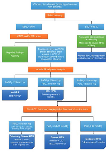
Clinical algorithm for evaluating patients with chronic liver disease for HPS was proposed by Fuhrmann et al. [46] (Figure 1).

FIGURE 1: Clinical algorithm for screening patients with chronic liver disease for hepatopulmonary syndrome (HPS). SaO2: arterial oxygen saturation; CEEC: contrast enhanced echocardiography; 99mTc scan: lung perfusion scintigraphy with technetium 99mTc labeled macro aggregated albumin; AaPO2: alveolar-arterial oxygen tension difference; PaO2: partial pressure of oxygen; IPVD: intrapulmonary vasodilatation; CT: computed tomography; LTOT: long term oxygen therapy; MELD: Model for End-Stage Liver Disease; LT: liver transplantation.

HPS TREATMENT

The pathogenesis of HPS is not completely understood, and no effective pharmacological treatment has been developed yet. Currently, the treatment of choice for HPS is OLT. Without liver transplatation, the prognosis of HPS is poor. In the case of the development of HPS, the probability of death in the next year is 41% [9]. Although HPS can be an indication for liver transplantation [9,47-49], severe hypoxia in the case of HPS is associated with a high risk of complications of liver transplantation (a 30% chance during the first 90 days) and thus increases the gap between transplantation and improving arterial oxygenation [9,50]. Patients can develop severe posttransplant hypoxemia which means that administration of 100% inspired oxygen is required to maintain an oxygen saturation of ≥85%. This complication is observed in 6-21% of patients, with a mortality rate of 45% [51].

In another study, the strongest predictors of mortality after liver transplatation were preoperative arterial oxygen tension (PaO2) of ≤50 mmHg alone or in combination with a MAA shunt fraction ≥20% [52,53]. In addition, survival rates between 68% and 80% within one year after liver transplatation were suggested in patients with HPS [53].

A complete regression of HPS symptoms after liver transplantation is observed only in 80% of patients, and may take 6 months or more [9]. Factors that contribute to the regression of HPS after liver transplantation are a young age, minimal hypoxemia, and good response to the therapy with 100% oxygen (PaO2 > 200 mmHg). After liver transplantation, 76% of patients had a 5 year life span [54].

Swanson et al. [55] investigated long-term survival in 61 HPS patients diagnosed at Mayo Clinic between 1985 and 2002 compared to case controls, and they assessed the impact of OLT on survival in HPS and control group. Overall, HPS patients who did not undergo transplantation had worse 5-year survival compared to their matched controls (p = 0.0003). The authors indicated that the effect of comorbidity on the survival difference cannot be underestimated, and that baseline PaO2 at the time of diagnosis was also associated with worse survival in HPS patients [55]. Gupta et al. [56] evaluated mortality, complications, and gas exchange in 21 HPS patients who underwent liver transplantation between 2002 and 2008 (11/21 patients were with severe HPS and 5/21 had living donor liver transplantation). Oxygenation improved in 19 patients for which PaO2 or SaO2 were recorded. Survival of liver transplant HPS and severe HPS patients was higher in their study compared to previous reports and the authors indicated that severity of HPS should not be the reason to deny a transplant [56].

DIFFERENTIAL DIAGNOSIS OF HPS

General criteria [6,27] for differential diagnosis of pulmonary vascular disorders associated with liver disease are presented in Table 2.

TABLE 2: Differential diagnosis of pulmonary vascular disorders associated with liver disease

Portopulmonary hypertension (PPH)

PPH is a pulmonary complication of liver disease, which should be differentially diagnosed from HPS. According to the classification adopted in 1998 at the World Conference on the Problems of Primary Pulmonary Hypertension in France, PPH is defined as pulmonary hypertension associated with chronic liver disease or portal hypertension. It is characterized by increased pressure in the pulmonary artery, i.e., more than 25 mmHg at rest and more than 30 mmHg during exercise, as well as increased pulmonary vascular resistance (PVR), i.e., more than 240 dynes/sec/sm-5 [57-59].

The incidence of PPH is relatively low; according to different studies [60-64] it is from 2-8.5% in patients with portal hypertension and 16-20% in patients with cirrhosis. However, diagnostics of PPH needs to be improved due to extremely poor prognosis of the disease (without proper treatment a 5-year survival rate is observed in 14% cases) and high mortality rate (up to 35%) even in the case of successful liver transplantation [65,66]. Risk factors previously determined for PPH include: female sex, autoimmune hepatitis, and genetic variation in estrogen signaling and cell growth regulators [67,68], while hepatitis C infection was associated with a decreased risk of PPH [67].

It appears that the development of PPH is not associated with the causes of portal hypertension. Although a majority of PPH patients have liver cirrhosis as the primary disorder, PPH has been found in patients with portal hypertension due to reasons other than liver, such as portal vein thrombosis without chronic liver disease [61]. Therefore, portal hypertension could likely be an etiology of pulmonary hypertension [69,70].

The mechanism by which portal hypertension causes pulmonary hypertension remains unclear. In the early stages of the disease, almost all patients with PPH have hyperdynamic circulation. However, high cardiac output and hyperdynamic circulation are also the main clinical features in almost all patients with progressive liver disease who develop portal hypertension [70].

Predisposing factors of pulmonary hypertension include: blood volume overload in the splanchnic circulation and congestion in the intestine that leads to the release of endotoxins and cytokines into the splanchnic circulation. The increased cardiac output generates shear stress on the pulmonary circulation. This can result either in maintaining adequate PVR which prevents development of related syndromes or in abnormal pulmonary vascular dilatation that causes an abnormal decrease in vascular resistance and leads to the development of HPS. In contrast, increased vascular resistance due to vasoconstriction and remodeling of pulmonary vessels leads to pulmonary hypertension. In the cases of PPH and HPS, altered vascular resistance is probably more due to remodeling of pulmonary vasculature and less due to vascular tone changes [58,70].

A prominent histological feature of PPH is proliferative pulmonary arteriopathy with obliteration of the vascular lumen by endothelial cells and smooth muscle cells and formation of plexiform lesions identical to those observed in primary pulmonary hypertension [57,70,71,72].

Factors affecting the development of proliferative vasculopathy are still not clear. Among these factors may be mutations in the bone morphogenetic protein receptor type 2 (BMPR2), which belongs to the transforming growth factor β (TGF-β) superfamily. These mutations have been associated with proliferative pulmonary vasculopathy and identified in patients with primary pulmonary hypertension, but no PPH case has been described with BMPR2 mutations. Mutations of activin receptor-like kinase 1 (ALK-1), another member of the TGF-β family, have been associated with pulmonary hypertension in patients with hereditary hemorrhagic telangiectasia (HHT). Nevertheless, uninvestigated genetic mechanisms may also contribute to the development of proliferative pulmonary vasculopathy, that are specific only to patients with pulmonary hypertension [70,73,74].

Increased cardiac output may lead to mild pulmonary hypertension if PVR is normal or close to normal, resulting in an overestimation of the incidence rate of PPH [70].

In most patients diagnosed with PPH of mild and moderate severity, vasoconstriction and moderate hypertrophy of pulmonary arteries are predominant clinical features [72]. However, in some people, mild or moderate PPH progresses to severe disease and becomes a complication in chronic liver disease or portal hypertension. Patients with progressive PPH tend to deteriorate rapidly and have right heart failure [70].

PPH symptoms

A typical clinical symptom of PPH is breathlessness during exercise. Other symptoms such as fatigue, palpitations, loss of consciousness due to a fall in blood pressure, or chest pain occur less frequently. Clinical findings indicating pulmonary hypertension may even be completely absent. The emphasis of the second tone on the pulmonary artery and systolic heart murmur, suggesting failure of tricuspid valve, are more common. Enlarged jugular veins, edema, and ascites are clinical features associated with decompensated liver cirrhosis and right ventricular failure [70].

Hypoxemia and increased alveolar-arterial (A-a) gradient are observed in the arterial blood of patients with PPH. Signs of right ventricular hypertrophy, right atrial enlargement and displacement of the heart to the right can be observed in electrocardiogram (ECG). X-ray of the lungs is usually normal or with signs of enlarged pulmonary arteries and cardiomegaly [67]. Pulmonary function tests may be normal or indicate lower lung diffusion capacity [60].

Clinical features that support the diagnosis of PPH in patients with liver cirrhosis or portal hypertension include increased pulmonary artery pressure and PVR (Table 3), when other possible causes are excluded such as left ventricular failure, pathology of heart valves, and interstitial and obstructive lung diseases. Although electrocardiography, vectorcardiography, phonocardiography, and pulmonary function tests can be useful for diagnosing PPH, the most accurate method for PPH diagnosis is echocardiography [70].

TABLE 3: Classification of portopulmonary hypertension (PPH)

According to the recommendations of the American Association for the Study of Liver Diseases [60,75] the primary diagnostic method for screening patients suspected of having PPH is transthoracic echocardiography, i.e. estimation of the right ventricular systolic pressure (>50 mmHg indicates PPH in about 65% of patients). Diagnosis confirmation is based on pulmonary artery (right heart) catheterization which includes the measurement of pulmonary arterial pressure, cardiac output, and PVR [11,64].

In addition, vasoreactivity testing with nitric oxide or epoprostenol (prostacyclin) is performed to determine if a patient may benefit from an oral calcium channel blocker. The mean pulmonary arterial pressure >35 mmHg is a risk factor of increased mortality after liver transplantation and requires adequate therapy in the perioperative period [76,77].

There are only a few reports on the co occurrence of HPS and PPH [78,79]. HPS usually develops before PPH [76], although Ioachimescu et al. [80] reported a patient in whom HPS developed following PPH [80].

Hereditary hemorrhagic telangiectasia (HHT)

Another condition which should be differentially diagnosed from HPS is HHT, also known as Osler–Weber–Rendu syndrome. HHT is a relatively common, under-recognized autosomal dominant genetic disorder that results from multisystemic vascular dysplasia and is characterized by mucocutaneous telangiectases and arteriovenous malformations (AVMs) [81].

HHT was first described by Sutton in 1864; the author reported a case of a man with vascular malformations and recurrent hemorrhage. In 1896, Rendu reported the complex of hereditary epistaxis and telangiectases in a 52-year-old patient, who suffered anemia and epistaxis since the age of 12. In 1901, Osler presented three cases of the disease and highlighted its hereditary nature using the phrase «family recurrent nasal bleeding, associated with multiple telangiectasia» [82,83]. Weber (1907) recognized Osler-Weber-Rendu Syndrome (OWRS) as a clinical entity distinct from hereditary hemophilia. The term “hereditary hemorrhagic telangiectasia” was coined by Hanes [84] in 1909.

The first morphological change of HHT is a focal dilatation of postcapillary venules. As the venules increase in size they become folded and connect to dilating arterioles through capillary segments, forming eventually arteriovenous communication [85,86]. The disease occurs at different geographical areas and the frequency varies between 1-2 cases per 10000-100000 people. Different symptoms are associated with HHT such as hematological, neurological, pulmonary, skin and gastrointestinal tract symptoms. In many cases, clinical symptoms are limited only to nasal bleeding.

The diagnosis of HHT is considered if at least three of the four diagnostic criteria are met: spontaneous, recurrent epistaxis, multiple telangiectases at different sites (lips, oral cavity, fingers, and nose), visceral lesions including gastrointestinal telangiectasia, pulmonary PAVMs, hepatic, cerebral and spinal AVMs, as well as family history of HHT [87].

HHT is particularly associated with mutations in two genes, and classified into two types accordingly: HHT1 and HHT2 [88,89]. In HHT1, the related gene ENG is located on the chromosome 9 and encodes protein endoglin, found on the surface of the cells that line the interior of the blood vessels. The gene ACVRL1 (chromosome 12) is associated with HHT2 and produces the enzyme activin receptor-like kinase 1 (ACVRL1 or ALK1). This protein is also found on the surface of cells, especially on the cells lining developing arteries.

Clinical manifestations of disease vary depending on the genotype [90]. PAVMs are associated with mutations in the ENG gene, while altered ACVRL1 gene is related to the occurrence of liver disease [91]. In some cases HHT phenotype was not associated with these two mutations, suggesting that other genes may be involved in the pathogenesis of HHT.

HHT symptoms

Spontaneous, recurrent epistaxes are due to telangiectasias in the nasal mucosa, and occur in more than 90% of patients with HHT. The severity of the disease can range from mild, requiring no treatment, to severe when patients need repeated blood transfusions. Usually, nasal bleeding occurs in early childhood, and becomes more frequent and prolonged later in life.

Vascular malformations in the lungs are found in 15-35% of patients with HHT, and they are common after 30 years of age [86]. Clinically, they may be asymptomatic when shunt fraction is less then 25%. Large AVMs may manifest by acute intolerance to physical activity, respiratory insufficiency, secondary polycythemia, and even pulmonary bleeding. It should be noted that vascular malformations in the lungs may be also complicated by neurological factors. In normal physiological conditions, pulmonary capillaries provide a filter system for blood, i.e., gas exchange occurs, removing carbon dioxide and adding oxygen to the blood. In the case of PAVMs, which are anatomical right-to-left shunts between pulmonary arteries and pulmonary veins, small clots can bypass the pulmonary capillaries and eventually reach the brain which causes transient ischemic attack (TIA) [92]. In some patients, these complications may indicate the beginning of HHT.

Using multislice computed tomography, Memeo et al. [93] showed that liver is affected in 41-78% of patients with HHT. In most of these patients, there were no clinical symptoms of the liver disease [93]. Abnormalities of the blood vessels connected to the liver vary in size greatly, from microscopic to large arteriovenous shunts and portal venous shunts. Portal hypertension in HHT patients is due to shunting of blood from the hepatic artery to the portal vein [86].

Liver transplantation reduce the possibility of sepsis of the biliary tract and prevents the development of cardiopulmonary failure, greatly improving the quality of patient life. The long-term survival rate was 75% in patients undergoing OLT (i.e., 9 patients from 12 that underwent the transplantation). These patients experienced improvements in epistaxis and quality of life and had more physical activity [94,95].

CONCLUSION

HPS is a severe complication of advanced liver disease associated with an extremely poor prognosis. The pathogenesis of HPS is not fully understood, and no effective pharmacological treatment has been developed yet. Currently, the treatment of choice for HPS is OLT. However, additional studies are necessary to identify the optimal criteria for the selection of patients for liver transplantation. Furthermore, the mechanism of pulmonary vasodilatation, associated humoral factors, and the reversal of symptoms after liver transplantation require further investigation. Finally, because HPS is still an under-recognized complication of end-stage liver disease, it should be considered in every patient with advanced liver disease showing symptoms of dyspnea and hypoxemia. Also, modern diagnostic methods should be used for early identification of pulmonary vascular complications and their differential diagnostics.

DECLARATION OF INTERESTS

The authors declare no conflict of interests.

REFERENCES

  1. (). Vorkommen von trommelschagel formigen fingerendphalangen ohne chronische veranderungen an der lungen oder am herzen. Wien Med Wochenschr.
  2. (). The effects of chronic disease of the liver on the composition and physiochemical properties of blood: Changes in the serum proteins; reduction in the oxygen saturation of the arterial blood. Ann Intern Med. https://doi.org/10.7326/0003-4819-9-6-690
  3. , (). Multiple pulmonary arteriovenous fistulas in juvenile cirrhosis. Am J Med. https://doi.org/10.1016/0002-9343(56)90043-2
  4. , (). Exercise-aggravated hypoxemia and orthodeoxia in cirrhosis. Chest. https://doi.org/10.1378/chest.72.3.305
  5. , (). Hepatopulmonary syndrome: Update on recent advances in pathophysiology, investigation, and treatment. J Gastroenterol Hepatol. https://doi.org/10.1111/jgh.12061
  6. , (). Hepatopulmonary syndrome: A liver induced oxygenation defect. Eur Respir Mon. https://doi.org/10.1183/1025448x.10008510
  7. , (). Hepatopulmonary syndrome – A liver-induced lung vascular disorder. N Engl J Med. https://doi.org/10.1056/NEJMra0707185
  8. , (). Hepatopulmonary syndrome: Update on pathogenesis and clinical features. Nat Rev Gastroenterol Hepatol. https://doi.org/10.1038/nrgastro.2012.123
  9. , , (). Gepatopulmonalnyy sindrom: Sostoyaniye problemy [Hepatopulmonary syndrome: State of problem]. Sovremennyye tekhnologii v meditsine.
  10. , (). The hepatopulmonary syndrome: No way out?. Eur Respir J. https://doi.org/10.1183/09031936.04.00028204
  11. , , , (). ERS Task Force Pulmonary-Hepatic Vascular Disorders (PHD) Scientific Committee. Pulmonary-hepatic vascular disorders (PHD). Eur Respir J. https://doi.org/10.1183/09031936.04.00010904
  12. , , (). Hepatopulmonary syndrome: Pathogenesis, diagnosis and treatment [Article in Portuguese]. Arq Gastroenterol. https://doi.org/10.1590/S0004-28032004000400010
  13. , , (). Gepatopulmonalnyy sindrom [Hepatopulmonary syndrome]. Transplantologiya.
  14. , , , , , (). Frequency of hepatopulmonary syndrome in cirrhotic patients. J Coll Physicians Surg Pak. DOI: 05.2008/JCPSP.278281
  15. , (). Hepatopulmonary syndrome: An update. Sao Paulo Med J. https://doi.org/10.1590/S1516-31802009000400008
  16. , , , , , (). Genetic risk factors for hepatopulmonary syndrome in patients with advanced liver disease. Gastroenterology. https://doi.org/10.1053/j.gastro.2010.03.044
  17. , (). Hepatopulmonary syndrome: Current concepts in diagnostic and therapeutic considerations. Chest. https://doi.org/10.1378/chest.105.5.1528
  18. , (). The hepatopulmonary syndrome. Ann Intern Med. https://doi.org/10.7326/0003-4819-122-7-199504010-00008
  19. , , , , , (). Prognostic significance of the hepatopulmonary syndrome in patients with cirrhosis. Gastroenterology. https://doi.org/10.1016/S0016-5085(03)01207-1
  20. , , , , , (). Pulmonary dysfunction in non-cirrhotic patients with chronic viral hepatitis. Eur J Intern Med. https://doi.org/10.1016/S0953-6205(02)00066-3
  21. , (). Gepatopulmonalnyy sindrom: Patologicheskaya fiziologiya, klinicheskoye techeniye, diagnostika i lecheniye [Hepatopulmonary syndrome: Pathological physiology, clinical course, diagnostics and treatment]. Klinicheskiye Perspektivy Gastroenterologii, Gepatologii.
  22. , , , , , (). Occurrence of hepatopulmonary syndrome in Budd-Chiari syndrome and the role of venous decompression. Gastroenterology. https://doi.org/10.1053/gast.2002.32419
  23. , , , , , (). Transient hepatopulmonary syndrome in a patient with acute hepatitis A. J Viral Hep.
  24. , (). Hepatopulmonary syndrome and portopulmonary hypertension. Hepatol Res. https://doi.org/10.1111/j.1872-034X.2009.00552.x
  25. , , (). Non-cirrhotic portal hypertension with hypoxaemia. Gut. https://doi.org/10.1136/gut.29.1.129
  26. , , , , (). Hepatopulmonary syndrome in children with cirrhotic and non-cirrhotic portal hypertension: A single-center experience. Dig Dis Sci. https://doi.org/10.1007/s10620-011-1832-6
  27. , , , , , (). Pulmonary vascular disorders in portal hypertension. Europ Respir J. https://doi.org/10.1183/09031936.98.11051153
  28. , , (). Hepatopulmonary syndrome: An unusual presentation of chronic hypervitaminosis A. J Paediatr.
  29. , , , , , (). Hepatopulmonary syndrome occurring after orthotopic liver transplantation. Liver Transpl. https://doi.org/10.1053/jlts.2001.29416
  30. , (). Hepatopulmonary syndrome, severe cyanosis and marfanoid habitus. J Assoc Physicians India.
  31. , (). The hepatopulmonary syndrome. Ann Intern Med. https://doi.org/10.7326/0003-4819-122-7-199504010-00008
  32. , , , , , (). Clinical features of hepatopulmonary syndrome in cirrhotic patients. World J Gastroenterol. https://doi.org/10.3748/wjg.v12.i12.1954
  33. , , , , (). The normal alveolar-arterial oxygen-tension gradient in man. Clin Sci Mol Med. https://doi.org/10.1042/cs0460089
  34. , (). Progress in investigating the pathogenesis of hepatopulmonary syndrome. Hepatobiliary Pancreat Dis Int.
  35. , , (). Left atrial volume: A novel predictor of hepatopulmonary syndrome. Am J Gastroenterol. https://doi.org/10.1111/j.1572-0241.2007.01228.x
  36. , , , , , (). Left ventricle enlargement and increased systolic velocity in the mitral valve are indirect markers of the hepatopulmonary syndrome. Liver Int. https://doi.org/10.1111/j.1478-3231.2011.02591.x
  37. , (). The hepatopulmonary syndrome - known symptoms and new name. Case Rep Clin Pract Rev.
  38. , , , (). Utility of pulse oximetry screening for hepatopulmonary syndrome. Clin Gastroenterol Hepatol. https://doi.org/10.1016/j.cgh.2006.12.003
  39. , , , , , (). Hepatopulmonary syndrome in patients with chronic liver disease: Role of pulse oximetry. BMC Gastroenterol. https://doi.org/10.1186/1471-230X-6-15
  40. , , , , , (). Von Willebrand factor antigen for detection of hepatopulmonary syndrome in patients with cirrhosis. J Hepatol. https://doi.org/10.1016/j.jhep.2014.04.025
  41. , , , , , (). Serum bile acids in patients with hepatopulmonary syndrome. Z Gastroenterol. DOI: 10.1055/s-0042-121268
  42. (). Current concepts in the management of hepatopulmonary syndrome. Vasc Health Risk Manag. https://doi.org/10.2147/VHRM.S3608
  43. (). Recommended standardized procedures for pulmonary testing. Amer Rev Respir Dis.
  44. (). Gepatopulmonalniy sindrom: osoblivostí patogenezu, díagnostiki, klíníchnogo perebígu ta líkuvannya [Hepatopulmonalnyy syndrome: features of patogenesis, diagnostics, clinical course and treatment]. Ukrayinskyy medychnyy almanakh.
  45. (). Portopulmonary hypertension and hepatopulmonary syndrome. J Gastroenterol Hepatol. https://doi.org/10.1046/j.1440-1746.17.s3.9.x
  46. , , , (). HPS: Diagnosis, clinical features, and medical therapy. Clin Liver Dis. https://doi.org/10.1002/cld.402
  47. , , , , , (). Hepatopulmonary syndrome among cirrhotic candidates for liver transplantation. Transplant Proc. https://doi.org/10.1016/j.transproceed.2012.06.001
  48. (). Management of pulmonary complications in pretransplant patients. Clin Liver Dis. https://doi.org/10.1016/j.cld.2011.08.012
  49. , (). Hepatopulmonary syndrome. Semin Respir Crit Care Med. https://doi.org/10.1055/s-0032-1301730
  50. , , , , , (). Liver transplantation for hepatopulmonary syndrome: A ten-year experience in Paris, France. Transplantation. https://doi.org/10.1097/01.TP.0000061612.78954.6C
  51. , , , , (). Proposed management algorithm for severe hypoxemia after liver transplantation in the hepatopulmonary syndrome. Am J Transplant. https://doi.org/10.1111/ajt.13177
  52. , , , , , (). Two cases of hepatopulmonary syndrome with improved liver function following long-term oxygen therapy. J Gastroenterol. https://doi.org/10.1007/s00535-006-1965-0
  53. , , , (). Prospective evaluation of outcomes and predictors of mortality in patients with hepatopulmonary syndrome undergoing liver transplantation. Hepatology. https://doi.org/10.1053/jhep.2003.50023
  54. , , , (). Gepatopulmonalniy sindrom [Hepatopulmonalnyy syndrome]. Meditsina Neotlozhnykh Sostoyaniy.
  55. , , (). Natural history of hepatopulmonary syndrome: Impact of liver transplantation. Hepatology. https://doi.org/10.1002/hep.20658
  56. , , , , , (). Improved survival after liver transplantation in patients with hepatopulmonary syndrome. Am J Transplant. https://doi.org/10.1111/j.1600-6143.2009.02822.x
  57. (). Portopulmonalnaya gipertenziya i gepatopulmonalnyy sindrom u bolnykh tsirrozom pecheni [Portopulmonary hypertension and hepatopulmonary syndrome in patients with cirrhosis]. Pulmonologiya.
  58. (). Hepatopulmonary syndrome and portopulmonary hypertension. Swiss Med Wkly.
  59. , , , (). Hepatopulmonary syndrome and portopulmonary hypertension: What’s new?. Acta Gastroenterol Belg.
  60. , (). Porto Pul’monal’na gípertenzíya: osoblivostí patogenezu, díagnostiki, klíníchnogo perebígu ta líkuvannya [Portopulmonary hypertension: features of patogenesis, diagnostics, clinical course and treatment]. Ukrayinskyy Medychnyy Almanakh.
  61. , (). Portopulmonary hypertension: A tale of two circulations. Chest. https://doi.org/10.1378/chest.123.2.562
  62. , , , , , (). Frequency and clinical implications of increased pulmonary artery pressures in liver transplant patients. Mayo Clinic Proc. https://doi.org/10.4065/71.6.543
  63. , (). Pulmonary hypertension in chronic liver disease. Clin Chest Med. https://doi.org/10.1016/S0272-5231(05)70296-3
  64. (). Portopulmonary hypertension and right heart failure in patients with cirrhosis. Curr Opin Anaesthesiol. https://doi.org/10.1097/ACO.0b013e32833725c4
  65. , , , , (). Survival in portopulmonary hypertension: Mayo Clinic experience categorized by treatment subgroups. Am J Transplant. https://doi.org/10.1111/j.1600-6143.2008.02384.x
  66. , , , , , (). Parenchymal alterations in cirrhotic livers in patients with hepatopulmonary syndrome or portopulmonary hypertension. Liver Transpl. https://doi.org/10.1002/lt.23632
  67. , , , , , (). Clinical risk factors for portopulmonary hypertension. Hepatology. https://doi.org/10.1002/hep.22275
  68. , , , , , (). Genetic risk factors for portopulmonary hypertension in patients with advanced liver disease. Amer J Respir Crit Care Med. https://doi.org/10.1164/rccm.200809-1472OC
  69. , , , , (). Pulmonary dysfunction and hepatopulmonary syndrome in cirrhosis and portal hypertension. Liver Int. https://doi.org/10.1111/j.1478-3231.2009.02103.x
  70. , , (). Portopulmonary hypertension and hepatopulmonary syndrome. Lancet. https://doi.org/10.1016/S0140-6736(04)16107-2
  71. , (). Different distribution of plexiform lesions in primary and secondary pulmonary hypertension. Hum Pathol. https://doi.org/10.1016/0046-8177(95)90088-8
  72. , , , , , (). Coexistent pulmonary and portal hypertension: Morphologic and clinical features. J Amer Coll Cardiol. https://doi.org/10.1016/S0735-1097(87)80123-7
  73. , , , , , (). Familial primary pulmonary hypertension (Gene PPH-1) is caused by mutations in the bone morphogenetic protein receptor-II gene. Am J Hum Genet. https://doi.org/10.1086/303059
  74. , , , , , (). Clinical and molecular genetic features of pulmonary hypertension in patients with hereditary hemorrhagic teleangiectasia. N Engl J Med. https://doi.org/10.1056/NEJM200108023450503
  75. , (). AASLD practice guidelines: Evaluation of the patient for liver transplantation. Hepatology. https://doi.org/10.1002/hep.20704
  76. , , , , , (). Hepatopulmonary syndrome and portopulmonary hypertension: A report of the multicenter liver transplant database. Liver Transpl. https://doi.org/10.1002/lt.20016
  77. , , , , , (). Pulmonary hemodynamics and perioperative cardiopulmonary-related mortality in patients with portopulmonary hypertension undergoing liver transplantation. Liver Transpl. https://doi.org/10.1053/jlts.2000.6356
  78. , , , , , (). The coexistence of portopulmonary hypertension and hepatopulmonary syndrome. Anesthesiology. https://doi.org/10.1097/00000542-199902000-00041
  79. , , , (). Development of hepatopulmonary syndrome and portopulmonary hypertension in a paediatric liver transplantat patient. Pediatr Transplant. https://doi.org/10.1111/j.1399-3046.2004.00221.x
  80. , , (). Hepatopulmonary syndrome following portopulmonary hypertension. Eur Respir J. https://doi.org/10.1183/09031936.00140306
  81. , (). Hereditary haemorrhagic telangiectasia. Haemophilia. https://doi.org/10.1111/j.1365-2516.2008.01774.x
  82. , (). Nasledstvennaya hemorrahycheskaya teleanhyéktazyya (bolezn-Randyu-Oslera) [Hereditary hemorrhagic telangiectasia (Rendu-Osler disease)]. Bolezni Serdtsa i Sosudov.
  83. , , (). Osler-Weber-Rendu syndrome – Dental implications. J Can Dent Assoc.
  84. (). Multiple hereditary telangiectases causing hemorrhage (hereditary hemorrhagic telangiectasia). Bull Johns Hopkins Hosp.
  85. , (). Osobennosti klinicheskogo techeniya nasledstvennoy gemorragicheskoy teleangiektazii [Clinical features of hereditary hemorrhagic telangiectasia]. Trudnyy Patsiyent.
  86. , , (). Sosudistyye malformatsii legkikh i pecheni u bolnogo s nasledstvennoy gemorragicheskoy teleangiektaziyey [Vascular malformation of the lungs and liver in a patient with hereditary hemorrhagic telangiectasia]. Ros Zh Gastroenterol Gepatol Koloproktol.
  87. , , , , , (). Diagnostic criteria for hereditary hemorrhagic telangiectasia (Rendu-Osler-Weber syndrome). Am J Med Genet. https://doi.org/10.1002/(SICI)1096-8628(20000306)91:1<66 : AID-AJMG12> 3.0.CO;2-P
  88. , , , , , (). Hereditary haemorrhagic telangiectasia: Mutation detection, test sensitivity and novel mutations. J Med Genet. https://doi.org/10.1136/jmg.2006.042606
  89. , , , , , (). Hereditary hemorrhagic telangiectasia: Clinical features in ENG and ALK1 mutation carriers. J Thromb Haemost. https://doi.org/10.1111/j.1538-7836.2007.02531.x
  90. , , , , , (). Genotype-phenotype relationship for localization and age distribution of telangiectases in hereditary hemorrhagic telangiectasia. Am J Med Genet. https://doi.org/10.1002/ajmg.a.32243
  91. (). Liver involvement in hereditary hemorrhagic teleangiectasia (HHT). J Hepatol. https://doi.org/10.1016/j.jhep.2006.12.008
  92. , , (). Hereditary hemorrhagic telangiectasia: Transient ischemic attacks. CMAJ. https://doi.org/10.1503/cmaj.081550
  93. , , , , , (). Hepatic involvement in hereditary hemorrhagic telangiectasia: CT findings. Abdom Imaging. https://doi.org/10.1007/s00261-003-0101-3
  94. , (). Sovremennyye aspekty terapii nasledstvennoy gemorragicheskoy teleangiektazii (bolezni Randyu-Oslera) [Modern aspects of hereditary hemorrhagic telangiectasia (Rendu-Osler disease) treatment]. Vrach Skoroy Pomoshchi.
  95. , , , , , (). Long-term outcome of patients with hereditary hemorrhagic telangiectasia and severe hepatic involvement after orthotopic liver transplantation: A single-center study. Liver Transpl. DOI: 10.1002/lt.21990