Vascular endothelial growth factor (VEGF)-related single nucleotide polymorphisms rs10738760 and rs6921438 are not associated with diabetic retinopathy (DR) in Slovenian patients with type 2 diabetes mellitus (T2DM)

Authors

  • Rifet Terzić Department of Biology, Faculty of Science, University of Tuzla, Tuzla, Bosnia and Herzegovina
  • Ines Cilenšek Institute of Histology and Embryology, Faculty of Medicine, University of Ljubljana, Ljubljana, Slovenia
  • Ruda Zorc Pleskovič Institute of Histology and Embryology, Faculty of Medicine, University of Ljubljana, Ljubljana, Slovenia
  • Sara Mankoč Institute of Histology and Embryology, Faculty of Medicine, University of Ljubljana, Ljubljana, Slovenia
  • Aleksandra Milutinović Institute of Histology and Embryology, Faculty of Medicine, University of Ljubljana, Ljubljana, Slovenia

DOI:

https://doi.org/10.17305/bjbms.2017.2068

Keywords:

Vascular endothelial growth factor, VEGF, VEGFA, diabetic retinopathy, DR, type 2 diabetes mellitus, T2DM, single nucleotide polymorphism, SNP

Abstract

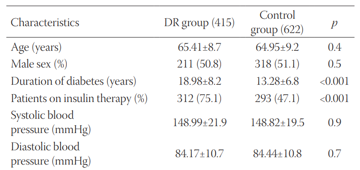
Diabetic retinopathy (DR) is a complication of diabetes characterized by vascular permeability, increased tissue ischemia, and angiogenesis. One of the most important proteins involved in angiogenesis is vascular endothelial growth factor (VEGF, also known as VEGFA). A previous study demonstrated that two single nucleotide polymorphisms (SNPs), rs6921438 and rs10738760, account for nearly half the variation in circulating VEGF levels. The aim of our study was to assess the association between rs6921438 and rs10738760 and DR in Slovenian patients with type 2 diabetes mellitus (T2DM). This case-control study enrolled 1037 unrelated Slovenian individuals (Caucasians) with T2DM. DR group included 415 T2DM patients with DR, while control group included 622 T2DM patients with no clinical signs of DR. The clinical and laboratory data were obtained from the medical records of the patients. The genotyping of rs6921438 and rs10738760 SNPs was carried out with real-time PCR assays. Significant differences were observed between patients with DR and controls in the duration of diabetes (p < 0.001), insulin therapy (p < 0.001), glycated hemoglobin (p = 0.001), body mass index (p = 0.002), total cholesterol (p = 0.002), and low-density lipoprotein cholesterol (p < 0.001). However, we did not observe significant differences in the genotype and allele distribution of the two SNPs, between DR and control group (p < 0.05). Logistic regression analysis showed that rs6921438 and rs10738760 were not independent genetic risk factors for DR in the co-dominant model adjusted for the above-mentioned clinical and laboratory data. In conclusion, VEGF-related SNPs rs10738760 and rs6921438 are not associated with DR in our group of Slovenian patients (Caucasians) with T2DM.

Downloads

Download data is not yet available.

Author Biography

Rifet Terzić, Department of Biology, Faculty of Science, University of Tuzla, Tuzla, Bosnia and Herzegovina

Department of Biology

References

Pavljasević S, Pranjić N, Sarajlić D. Argon laser photo-coagulation complications in diabetic retinopathy. Bosn J Basic Med Sci 2004;4(2):41-4.

Kulenovic I, Rasic S, Karcic S. Development of microvascular complications in type 1 diabetic patients 10 years follow-up. Bosn J Basic Med Sci 2006;6(2):47-50.

Cekić S, Cvetković T, Jovanović I, Jovanović P, Pesić M, Stanković Babić G, et al. C-reactive protein and chitinase 3-like protein 1 as biomarkers of spatial redistribution of retinal blood vessels on digital retinal photography in patients with diabetic retinopathy. Bosn J Basic Med Sci 2014;14(3):177-84. https://doi.org/10.17305/bjbms.2014.3.21.

Petrovic MG, Kunej T, Peterlin B, Dovc P, Petrovic D. Gly482Ser polymorphism of the peroxisome proliferator-activated receptor-gamma coactivator-1 gene might be a risk factor for diabetic retinopathy in Slovene population (Caucasians) with type 2 diabetes and the Pro12Ala polymorphism of the PPARgamma gene is not. Diabetes Metab Res Rev 2005;21(5):470-4. https://doi.org/10.1002/dmrr.546.

Petrovic MG, Osredkar J, Saraga-Babić M, Petrovic D. K469E polymorphism of the intracellular adhesion molecule 1 gene is associated with proliferative diabetic retinopathy in Caucasians with type 2 diabetes. Clin Exp Ophthalmol 2008;36(5):468-72. https://doi.org/10.1111/j.1442-9071.2008.01785.x.

Nikolajević-Starčević J, Globočnik Petrovič M, Petrovič D. A1/A2 polymorphism of the glycoprotein IIIa gene and diabetic retinopathy in Caucasians with type 2 diabetes. Clin Exp Ophthalmol 2011;39(7):665-72. https://doi.org/10.1111/j.1442-9071.2011.02520.x.

Urbančič M, Kloboves, Prevodnik V, Petrovič D, Globočnik Petrovič M. A flow cytometric analysis of vitreous inflammatory cells in patients with proliferative diabetic retinopathy. Biomed Res Int 2013;2013:251528. https://doi.org/10.1155/2013/251528.

Petrovic MG, Kruzliak P, Petrovic D. The rs6060566 of the reactive oxygen species modulator 1 (Romo-1) gene affects Romo-1 expression and the development of diabetic retinopathy in Caucasians with type 2 diabetes. Acta Ophthalmol 2015;93(8):654-7. https://doi.org/10.1111/aos.12723.

Awata T, Inoue K, Kurihara S, Ohkubo T, Watanabe M, Inukai K, et al. A common polymorphism in the 5'-untranslated region of the VEGF gene is associated with diabetic retinopathy in type 2 diabetes. Diabetes 2002;51(5):1635-9. https://doi.org/10.2337/diabetes.51.5.1635.

Mankoč Ramuš S, Kumše T, Globočnik Petrovič M, Petrovič D, Cilenšek I. SNP rs2073618 of the osteoprotegerin gene is associated with diabetic retinopathy in Slovenian patients with type 2 diabetes. Biomed Res Int 2013;2013:364073. https://doi.org/10.1155/2013/364073.

Bates DO, Harper SJ. Regulation of vascular permeability by vascular endothelial growth factors. Vascul Pharmacol 2002;39(4-5):225-37.

https://doi.org/10.1016/S1537-1891(03)00011-9.

Jiang Y, Sun L, Xuan X, Wang J. Impacts of N-Butylphthalide on expression of growth factors in rats with focal cerebral ischemia. Bosn J Basic Med Sci 2016;16(2):102-7. https://doi.org/10.17305/bjbms.2016.560.

Wirostko B, Wong TY, Simó R. Vascular endothelial growth factor and diabetic complications. Prog Retin Eye Res 2008;27(6):608-21. https://doi.org/10.1016/j.preteyeres.2008.09.002.

Bonnefond A, Saulnier PJ, Stathopoulou MG, Grarup N, Ndiaye NC, Roussel R, et al. What is the contribution of two genetic variants regulating VEGF levels to type 2 diabetes risk and to microvascular complications? PLoS One 2013;8(2):e55921. https://doi.org/10.1371/journal.pone.0055921.

Berrahmoune H, Herbeth B, Lamont JV, Masson C, Fitzgerald PS, Visvikis-Siest S. Heritability for plasma VEGF concentration in the Stanislas family study. Ann Hum Genet 2007;71(Pt 1):54-63. https://doi.org/10.1111/j.1469-1809.2006.00298.x.

Globočnik Petrovič M, Korošec P, Košnik M, Osredkar J, Hawlina M, Peterlin B, et al. Local and genetic determinants of vascular endothelial growth factor expression in advanced proliferative diabetic retinopathy. Mol Vis 2008;14:1382-7.

Debette S, Visvikis-Siest S, Chen MH, Ndiaye NC, Song C, Destefano A, et al. Identification of cis- and trans-acting genetic variants explaining up to half the variation in circulating vascular endothelial growth factor levels. Circ Res 2011;109(5):554-63. https://doi.org/10.1161/CIRCRESAHA.111.243790.

Genuth S, Alberti KG, Bennett P, Buse J, Defronzo R, Kahn R, et al. Follow-up report on the diagnosis of diabetes mellitus. Diabetes Care 2003;26(11):3160-7. https://doi.org/10.2337/diacare.26.11.3160.

[No authors listed]. Grading diabetic retinopathy from stereoscopic color fundus photographs - an extension of the modified Airlie House classification. ETDRS report number 10. Early Treatment Diabetic Retinopathy Study Research Group. Ophthalmology 1991;98(5 Suppl):786-806. https://doi.org/10.1016/S0161-6420(13)38012-9.

Petrovic MG, Peterlin B, Hawlina M, Petrovic D. Aldose reductase (AC)n gene polymorphism and susceptibility to diabetic retinopathy in Type 2 diabetes in Caucasians. J Diabetes Complications 2005;19(2):70-3. https://doi.org/10.1016/j.jdiacomp.2004.08.004.

Petrovič D. Candidate genes for proliferative diabetic retinopathy. Biomed Res Int 2013;2013:540416. https://doi.org/10.1155/2013/540416.

Hampton BM, Schwartz SG, Brantley MA Jr, Flynn HW Jr. Update on genetics and diabetic retinopathy. Clin Ophthalmol 2015;9:2175-93. DOI: 10.2147/OPTH.S94508.

Turnpenny P, Ellard S. Emery's Elements of Medical Genetics. 15th ed. Exeter, UK: Elsevier; 2017.

Nakayama M, Yasue H, Yoshimura M, Shimasaki Y, Kugiyama K, Ogawa H, et al. T-786C mutation in the 5'-flanking region of the endothelial nitric oxide synthase gene is associated with coronary spasm. Circulation 1999;99(22):2864-70. https://doi.org/10.1161/01.CIR.99.22.2864.

Vascular endothelial growth factor (VEGF)-related single nucleotide polymorphisms rs10738760 and rs6921438 are not associated with diabetic retinopathy (DR) in Slovenian patients with type 2 diabetes mellitus (T2DM)

Downloads

Additional Files

Published

2017-11-20

How to Cite

1.
Terzić R, Cilenšek I, Zorc Pleskovič R, Mankoč S, Milutinović A. Vascular endothelial growth factor (VEGF)-related single nucleotide polymorphisms rs10738760 and rs6921438 are not associated with diabetic retinopathy (DR) in Slovenian patients with type 2 diabetes mellitus (T2DM). Biomol Biomed [Internet]. 2017Nov.20 [cited 2023Sep.23];17(4):328-32. Available from: https://www.bjbms.org/ojs/index.php/bjbms/article/view/2068

Issue

Section

Molecular Biology

INTRODUCTION

Diabetic retinopathy (DR)is a microvascular complication of diabetes characterized by progressive damage to the eyes. DR is the leading cause of new cases of blindness in adults [1,2,3], and the development of DR is influenced by both genetic and environmental factors [4-8]. The clinical characteristics of DR include vascular permeability, increased tissue ischemia, and angiogenesis [9,10]. Vascular endothelial growth factor A (VEGFA, also known as VEGF)is a chemokine involved in the process of angiogenesis, acting as a key regulator of vascular permeability [11,12]. Higher serum VEGF levels have been linked to DR [10,13,14].

The heritability of circulating VEGF levels is high [15] and several common single nucleotide polymorphisms (SNPs)were shown to be significantly related to serum VEGF levels [16,17]. A genome-wide association (GWA), community-based study [17], enrolling 3,527 individuals of European descent, analyzed the association between SNPs and circulating VEGF levels. A total of 140 SNPs reached genome-wide significance and four of these SNPs were independently associated with VEGF levels: rs6921438, rs4416670, rs6993770, and rs10738760 [17]. rs6921438 SNP is located on the short arm of chromosome 6 at 6p21.1, 171 kb downstream of the VEGFA gene, and close to the mitochondrial ribosomal protein L14 gene (MRPL14)[16,17]. rs10738760 SNP is located on chromosome 9p24.2, close to the very low-density lipoprotein receptor (VLDLR)and potassium voltage-gated channel subfamily V, member 2 (KCNV2)genes [16,17]. In the GWA study, rs6921438 explained 41.2% and rs10738760 5% of the variability in circulating VEGF levels [17].

To date, only one study assessed the effect of the VEGF-related SNPs, rs6921438 and rs10738760, on the risk of type 2 diabetes mellitus (T2DM)and associated microvascular complications [14]. They found that the G allele of rs6921438 was associated with increased glycated haemoglobin (HbA1c)levels and increased risk for T2DM in a French population [14].

Considering the potential contribution of VEGF-related SNPs to the risk of T2DM as well as the lack of relevant studies, here, we assessed the association between rs6921438 and rs10738760 and DR in Slovenian patients with T2DM.

MATERIALS AND METHODS

Patients

This case-control study enrolled 1037 unrelated Caucasians with T2DM and defined ocular status. The patients were not evaluated with regard to the history of glycemic control. T2DM was diagnosed according to the current American Diabetes Association criteria [18]. Dilated fundus examination was performed by a senior ophthalmologist (M.P.)after pupil dilation with 2.5% tropicamide and phenylephrine, using a slit lamp biomicroscope with non-contact lens. The result was electronically documented by a fundus camera with a 50-degree angle (Topcon-TRC 40-IX, Tokyo, Japan). The clinical and laboratory data were obtained from the medical records of the patients. DR staging was performed according to the Early Treatment Diabetic Retinopathy Study (ETDRS)diabetic retinopathy severity scale [19]. The study group (DR)included 415/1037 patients with T2DM and DR, while the control group involved 622 individuals with T2DM who had no clinical signs of DR. To avoid the confounding effect of impaired kidney function, we did not enroll patients with overt nephropathy (i.e., end-stage renal failure). The study was approved by the National Medical Ethics Committee (number 118/12/2011). After obtaining informed consent from the patients, a detailed interview was undertaken.

Genotyping

Genomic DNA was extracted from 200 µL of the whole blood using a FlexiGene DNA kit, according to the recommended protocol (Qiagen, Germany). We selected VEGF-related rs6921438 and rs10738760 SNPs for analysis. rs6921438 is located on the short arm of chromosome 6 at 6p21.1, 171 kb downstream of the VEGFA, and close to the C6orf223 gene, which encodes an uncharacterized protein. rs10738760 is located on the short arm of chromosome 9 (i.e., 9p24.2), between the VLDLR and KCNV2 genes [14]. Two SNPs were genotyped using predesigned TaqMan SNP Genotyping Assays (Applied Biosystems, Foster City, CA, USA), C 1971047 1 and C 27464534 20. We used the StepOne Real-Time PCR system (Applied Biosystems, Foster City, CA, USA)for SNP analysis, according to the manufacturer’s instructions. The real-time PCR reactions were set up in a final volume of 5 µL, containing 2.5 µL of 2 × TaqMan Genotyping Master Mix (Applied Biosystems, Foster City, CA, USA), 0.12 µL of 40 × SNP Genotyping Assay (Applied Biosystems, Foster City, CA, USA), 1.88 µL of nuclease-free water, and 25 ng of genomic DNA. The PCR was carried out under the following conditions: enzyme activation for 10 minutes at 95°C, followed by 55 cycles of amplification at 95°C for 15 seconds and at 60°C for 1 minute.

Statistical analysis

Statistical analysis was conducted using SPSS Statistics for Windows, Version 20.0. (IBM Corp., Armonk, NY). Two researchers (D.P. and I.C.), blinded to the DR status of the patients, performed SNP genotyping. We used a Chi-square test to compare discrete variables. Continuous variables were compared by the unpaired Student’s t-test. In addition, all variables that showed significant differences in the univariate analysis (i.e., Chi-square test and unpaired Student’s t-test)were included in a logistic regression analysis. A value of p < 0.05 was considered statistically significant. The deviation from Hardy–Weinberg equilibrium (HWE)was assessed by the Fisher’s exact test (http://ihg.gsf.de/).

RESULTS

This study enrolled 1037 individuals with T2DM of at least 10 years’ duration. The control group included 622 patients with T2DM but with no evidence of DR, while 415 patients with T2DM and DR were included in DR group. The clinical and laboratory data, obtained from the medical records of the patients, are shown in Table 1. There were no significant differences in the age, sex, systolic and diastolic blood pressure, history of hypertension, smoking status, high-density lipoprotein (HDL)cholesterol, and triglycerides between the groups (Table 1). On the contrary, a statistically significant difference was observed between the groups in the following parameters: duration of diabetes (p < 0.001), insulin therapy (p < 0.001), HbA1c (p = 0.001), body mass index [BMI] (p = 0.002), total cholesterol (p = 0.002), and low-density lipoprotein (LDL)cholesterol [p < 0.001](Table 1). The duration of diabetes was more than 5 years longer in DR compared to control group [p < 0.001](Table 1). A significantly higher proportion of the patients with DR required insulin therapy compared to control group (Table 1). The patients with DR had higher HbA1c, total cholesterol, and LDL cholesterol levels, while BMI was significantly lower in this group compared to the controls (Table 1).

TABLE 1: Clinical and laboratory characteristics of T2DM patients with DR and those without DR (control group)

As shown in Table 2, the frequencies of the GG, GA and AA genotypes of rs10738760 were 23.1%, 54.9%, and 21.9% in DR group, and 22.0%, 51.3%, and 26.7% in control group, respectively. The average frequencies of rs10738760 alleles were 50.6% for G allele and 49.4% for A allele in DR group, and 47.7% for G allele and 52.3% for A allele in control group (Table 2).

TABLE 2: Distribution of rs10738760 and rs6921438 genotypes and alleles in T2DM patients with DR and those without DR (controls)

The distributions of the GG, GA and AA genotypes of rs6921438 were 24.8%, 50.1%, and 25.1% in DR group and 28.8%, 49.7%, and 21.5% in control group, respectively (Table 2). The average frequencies of rs6921438 alleles in DR group were 49.9% for G allele and 50.1% for A allele, and in control group 53.6% for G allele and 46.4% for A allele (Table 2).

The genotype distribution of rs6921438 conformed to HWE in both groups [p = 0.9 for DR and p = 0.9 for control group] (Table 2). The genotype distribution of rs10738760 conformed to HWE in control group (p = 0.5), but not in DR group [p = 0.04] (Table 2).

We used a logistic regression analysis to evaluate whether the two SNPs were independently associated with DR, after adjusting for duration of diabetes, insulin therapy, BMI, HbA1c, total cholesterol, and LDL cholesterol (Table 3). We found no significant association between rs10738760 or rs6921438 and DR [co-dominant model] (Table 3).

TABLE 3: Association between rs10738760 and rs6921438 polymorphisms and the risk for DR

DISCUSSION

Previously, a GWA study on 3,527 individuals of European descent reported four SNPs that were independently associated with VEGF levels: rs6921438, rs4416670, rs6993770, and rs10738760 [17]. In this cross-sectional study, we analyzed the association between rs6921438 and rs10738760 SNPs and DR in a group of Slovenian patients with T2DM. Our results showed no association between the two SNPs and DR in the patients with T2DM. These results are in agreement with the French case-control study [14]on 1336 T2DM patients with DR and 1231 T2DM controls, where they also reported no significant association between rs6921438 or rs10738760 and DR. In addition, no significant association between the two SNPs and macular oedema was found in their study [14].

A previous study on a subset of Slovenian patients with T2DM reported the effect of another VEGF polymorphism, rs2010963, on the serum and vitreous levels of VEGF; however, this SNP did not contribute to the genetic susceptibility to proliferative diabetic retinopathy (PDR) [16].

Four proteins are proposed to have important roles in the pathogenesis of diabetes, DR, PDR, or diabetic macular oedema (DMO), including: VEGF, the receptor for advanced glycation end products (RAGE), endothelial nitric oxide synthase (eNOS), and aldose reductase (AR)[13,16,20,21]. These proteins are encoded by four well-studied genes, and in the context of DR, the VEGFA has been the most commonly investigated gene [22]. The best-studied VEGF-related polymorphisms as potential risk factors of DR include rs833061, rs699947, rs2010963, and rs3025039. Many, but not all studies, confirmed the association between candidate SNPs and DR [13, 16, 20-22]. These studies included different populations or races (i.e., Caucasians, Asians, or both)[22]. Similar to our study, another study including Caucasians (i.e., French patients)assessed the effect of rs6921438 and rs10738760 on DR in T2DM patients, and showed no association between these SNPs and the risk of DR [14].

In our study, rs10738760 SNP did not conformed to HWE in DR group, meaning that the allele and genotype frequencies for this SNP are not constant from generation to generation [23]. This could be the result of evolutionary forces, such as mate choice, mutation, selection, genetic drift, gene flow, and meiotic drive. The deviation from HWE present only in DR and not in the control group, indicates a possible association of the SNP locus with DR [24].

The strengths of our prospective study are the community-based sample of Caucasians with T2DM and exact assessment of DR. A limitation of the study may be the small number of participants. Nevertheless, the study was appropriately designed to detect the differences in the distribution of genotypes in this cohort of T2DM patients with and without DR.

Further studies should clarify the contribution of VEGF-related SNPs to the development of DR in patients with T2DM.

CONCLUSION

In summary, we showed that VEGF-related SNPs rs10738760 and rs6921438 are not associated with DR in our group of Slovenian patients (Caucasians)with T2DM.

Acknowledgements

ACKNOWLEDGMENTS

The authors thank prof. dr. Mojca Globočnik for the help in determining the stages of DR. The authors also thank Ms. Visam Bajt, BA for revising the English language in the manuscript.

This work was supported by the program grants from the Slovenian research agency, ARRS P3-0019.

DECLARATION OF INTERESTS

The authors declare no conflict of interests.

REFERENCES

  1. , , (). Argon laser photo-coagulation complications in diabetic retinopathy. Bosn J Basic Med Sci.
  2. , , (). Development of microvascular complications in type 1 diabetic patients 10 years follow-up. Bosn J Basic Med Sci.
  3. , , , , , (). C-reactive protein and chitinase 3-like protein 1 as biomarkers of spatial redistribution of retinal blood vessels on digital retinal photography in patients with diabetic retinopathy. Bosn J Basic Med Sci. https://doi.org/10.17305/bjbms.2014.3.21
  4. , , , , (). Gly482Ser polymorphism of the peroxisome proliferator-activated receptor-gamma coactivator-1 gene might be a risk factor for diabetic retinopathy in Slovene population (Caucasians)with type 2 diabetes and the Pro12Ala polymorphism of the PPARgamma gene is not. Diabetes Metab Res Rev. https://doi.org/10.1002/dmrr.546
  5. , , , (). K469E polymorphism of the intracellular adhesion molecule 1 gene is associated with proliferative diabetic retinopathy in Caucasians with type 2 diabetes. Clin Exp Ophthalmol. https://doi.org/10.1111/j.1442-9071.2008.01785.x
  6. , , (). A1/A2 polymorphism of the glycoprotein IIIa gene and diabetic retinopathy in Caucasians with type 2 diabetes. Clin Exp Ophthalmol. https://doi.org/10.1111/j.1442-9071.2011.02520.x
  7. , , , (). A flow cytometric analysis of vitreous inflammatory cells in patients with proliferative diabetic retinopathy. Biomed Res Int. https://doi.org/10.1155/2013/251528
  8. , , (). The rs6060566 of the reactive oxygen species modulator 1 (Romo-1)gene affects Romo-1 expression and the development of diabetic retinopathy in Caucasians with type 2 diabetes. Acta Ophthalmol. https://doi.org/10.1111/aos.12723
  9. , , , , , (). A common polymorphism in the 5’-untranslated region of the VEGF gene is associated with diabetic retinopathy in type 2 diabetes. Diabetes. https://doi.org/10.2337/diabetes.51.5.1635
  10. , , , , (). SNP rs2073618 of the osteoprotegerin gene is associated with diabetic retinopathy in Slovenian patients with type 2 diabetes. Biomed Res Int. https://doi.org/10.1155/2013/364073
  11. , (). Regulation of vascular permeability by vascular endothelial growth factors. Vascul Pharmacol. https://doi.org/10.1016/S1537-1891(03)00011-9
  12. , , , (). Impacts of N-Butylphthalide on expression of growth factors in rats with focal cerebral ischemia. Bosn J Basic Med Sci. https://doi.org/10.17305/bjbms.2016.560
  13. , , (). Vascular endothelial growth factor and diabetic complications. Prog Retin Eye Res. https://doi.org/10.1016/j.preteyeres.2008.09.002
  14. , , , , , (). What is the contribution of two genetic variants regulating VEGF levels to type 2 diabetes risk and to microvascular complications?. PLoS One. https://doi.org/10.1371/journal.pone.0055921
  15. , , , , , (). Heritability for plasma VEGF concentration in the Stanislas family study. Ann Hum Genet. https://doi.org/10.1111/j.1469-1809.2006.00298.x
  16. , , , , , (). Local and genetic determinants of vascular endothelial growth factor expression in advanced proliferative diabetic retinopathy. Mol Vis.
  17. , , , , , (). Identification of cis- and trans-acting genetic variants explaining up to half the variation in circulating vascular endothelial growth factor levels. Circ Res. https://doi.org/10.1161/CIRCRESAHA.111.243790
  18. , , , , , (). Follow-up report on the diagnosis of diabetes mellitus. Diabetes Care. https://doi.org/10.2337/diacare.26.11.3160
  19. (). Grading diabetic retinopathy from stereoscopic color fundus photographs - an extension of the modified Airlie House classification. ETDRS report number 10. Early Treatment Diabetic Retinopathy Study Research Group. Ophthalmology. https://doi.org/10.1016/S0161-6420(13)38012-9
  20. , , , (). Aldose reductase (AC)n gene polymorphism and susceptibility to diabetic retinopathy in Type 2 diabetes in Caucasians. J Diabetes Complications. https://doi.org/10.1016/j.jdiacomp.2004.08.004
  21. (). Candidate genes for proliferative diabetic retinopathy. Biomed Res Int. https://doi.org/10.1155/2013/540416
  22. , , , (). Update on genetics and diabetic retinopathy. Clin Ophthalmol. DOI: 10.2147/OPTH.S94508
  23. , (). . Emery’s Elements of Medical Genetics.
  24. , , , , , (). T-786®C mutation in the 5’-flanking region of the endothelial nitric oxide synthase gene is associated with coronary spasm. Circulation. https://doi.org/10.1161/01.CIR.99.22.2864