The value of 18F-FDG PET/CT imaging in breast cancer staging

Authors

  • Ulkem Yararbas Department of Nuclear Medicine, Ege University Medical Faculty, Izmir, Turkey
  • Neslihan Cetin Avci Department of Nuclear Medicine, Umraniye Training and Research Hospital, Istanbul, Turkey
  • Levent Yeniay Department of General Surgery, Ege University Medical Faculty, Izmir, Turkey
  • Aziz Murat Argon Department of Nuclear Medicine, Ege University Medical Faculty, Izmir, Turkey

DOI:

https://doi.org/10.17305/bjbms.2017.2179

Keywords:

Breast cancer, Breast cancer staging, 18F-FDG, 18F-FDG PET/CT

Abstract

The National Comprehensive Cancer Network (NCCN) guidelines recommend assessment with positron emission tomography with 2-deoxy-2-[fluorine-18]fluoro-D-glucose integrated with computed tomography (18F-FDG PET/CT) in staging of breast cancer, starting from the stage IIIA. Previously, PET/CT contributed to the accurate staging from the stage IIB. Our aim is to evaluate the contribution of 18F-FDG PET/CT in staging of breast cancer patients. A total of 234 patients were retrospectively evaluated. PET/CT was performed preoperatively in 114/234 and postoperatively in 120/234 patients. Initial staging was performed based on histopathological results in 125/234 and clinical results in 109/234 patients, according to the American Joint Committee on Cancer (AJCC) classification. All patients had a normal abdominal ultrasound and chest x-ray. Following PET/CT imaging, modification in the staging was performed in patients with the metastatic findings. In 42/234 (17.9%) patients hypermetabolic extra-axillary regional lymph nodes and in 65/234 patients (27.7%) distant metastatic involvement were detected with PET/CT. Modification in the staging was applied in 82/234 (35%) patients. Patient management was changed in 69/234 (29.4%) cases. The percentage of patients with upstaging, according to each stage, was as follows: IIA: 18.6%, IIB: 30%, IIIA: 46.3%, IIIB: 68.8%, and IIIC: 20.8%. In 43/43 patients, 99mTc-methylene diphosphonate (MDP) bone scan did not show additional bone metastasis. In 5/32 patients, metastatic involvement was detected with sentinel lymph node biopsy (SLNB), but preoperative PET/CT scan did not reveal hypermetabolic lymph nodes. Although our study was limited by the referral bias and lack of homogeneity in the referral group, PET/CT still significantly contributed to the accurate staging and management of our breast cancer patients, starting from the stage IIA.

Downloads

Download data is not yet available.

Author Biographies

Ulkem Yararbas, Department of Nuclear Medicine, Ege University Medical Faculty, Izmir, Turkey

Department of Nuclear Medicine

Neslihan Cetin Avci, Department of Nuclear Medicine, Umraniye Training and Research Hospital, Istanbul, Turkey

Department of Nuclear Medicine

Levent Yeniay, Department of General Surgery, Ege University Medical Faculty, Izmir, Turkey

Department of General Surgery

Aziz Murat Argon, Department of Nuclear Medicine, Ege University Medical Faculty, Izmir, Turkey

Department of Nuclear Medicine

References

Jemal A, Siegel R, Ward E, Hao Y, Xu J, Thun MJ. Cancer statistics, 2009. CA Cancer J Clin 2009;59(4):225-49.

https://doi.org/10.3322/caac.20006.

Groheux D, Espié M, Giacchetti S, Hindié E. Performance of FDG PET/CT in the clinical management of breast cancer. Radiology 2013;266(2):388-405.

https://doi.org/10.1148/radiol.12110853.

Veronesi U, De Cicco C, Galimberti VE, Fernandez JR, Rotmensz N, Viale G, et al. A comparative study on the value of FDG-PET and sentinel node biopsy to identify occult axillary metastases. Ann Oncol 2007;18(3):473-8.

https://doi.org/10.1093/annonc/mdl425.

Evangelista L, Baretta Z, Vinante L, Cervino AR, Gregianin M, Ghiotto C, et al. Tumour markers and FDG PET/CT for prediction of disease relapse in patients with breast cancer. Eur J Nucl Med Mol Imaging 2011;38(2):293-301.

https://doi.org/10.1007/s00259-010-1626-7.

Aukema TS, Rutgers EJ, Vogel WV, Teertstra HJ, Oldenburg HS, Vrancken Peeters MT, et al. The role of FDG PET/CT in patients with locoregional breast cancer recurrence: A comparison to conventional imaging techniques. Eur J Surg Oncol 2010;36(4):387-92.

https://doi.org/10.1016/j.ejso.2009.11.009.

NCCN. Clinical Practice Guidelines in Oncology. Breast Cancer. Version 3; 2017. [cited 2017 Aug 10]. Available from: https://www.nccn.org/professionals/physician_gls/pdf/breast.pdf.

Groheux D, Giacchetti S, Espié M, Vercellino L, Hamy AS, Delord M, et al. The yield of 18F-FDG PET/CT in patients with clinical stage IIA, IIB, or IIIA breast cancer: A prospective study. J Nucl Med 2011;52(10):1526-34.

https://doi.org/10.2967/jnumed.111.093864.

Segaert I, Mottaghy F, Ceyssens S, De Wever W, Stroobants S, Van Ongeval C, et al. Additional value of PET-CT in staging of clinical stage IIB and III breast cancer. Breast J 2010;16(6):617-24.

https://doi.org/10.1111/j.1524-4741.2010.00987.x.

Yararbas U, Argon AM, Yeniay L, Kapkac M. Problematic aspects of sentinel lymph node biopsy and its relation to previous excisional biopsy in breast cancer. Clin Nucl Med 2009;34(12):854-8.

https://doi.org/10.1097/RLU.0b013e3181becec2.

AJCC. Breast Cancer Staging. 8th ed. [cited 2017 Aug 15]. Available from: https://cancerstaging.org/references-tools/deskreferences/Documents/AJCC%20Breast%20Cancer%20Staging%20System.pdf.

Caldarella C, Treglia G, Giordano A. Diagnostic performance of dedicated positron emission mammography using fluorine-18-fluorodeoxyglucose in women with suspicious breast lesions: A meta-analysis. Clin Breast Cancer 2014;14(4):241-8.

https://doi.org/10.1016/j.clbc.2013.12.004.

Schilling K, Narayanan D, Kalinyak JE, The J, Velasquez MV, Kahn S, et al. Positron emission mammography in breast cancer presurgical planning: Comparisons with magnetic resonance imaging. Eur J Nucl Med Mol Imaging 2011;38(1):23-36.

https://doi.org/10.1007/s00259-010-1588-9.

Gil-Rendo A, Martínez-Regueira F, Zornoza G, García-Velloso MJ, Beorlegui C, Rodriguez-Spiteri N. Association between [18F] fluorodeoxyglucose uptake and prognostic parameters in breast cancer. Br J Surg 2009;96(2):166-70.

https://doi.org/10.1002/bjs.6459.

Fujii T, Yajima R, Kurozumi S, Higuchi T, Obayashi S, Tokiniwa H, et al. Clinical significance of 18F-FDG-PET in invasive lobular carcinoma. Anticancer Res 2016;36(10):5481-5.

https://doi.org/10.21873/anticanres.11129.

Jung NY, Kim SH, Choi BB, Kim SH, Sung MS. Associations between the standardized uptake value of (18)F-FDG PET/CT and the prognostic factors of invasive lobular carcinoma: In comparison with invasive ductal carcinoma. World J Surg Oncol 2015;13:113.

https://doi.org/10.1186/s12957-015-0522-9.

Soerjomataram I, Louwman MW, Ribot JG, Roukema JA, Coebergh JW. An overview of prognostic factors for long-term survivors of breast cancer. Breast Cancer Res Treat 2008;107(3):309-30.

https://doi.org/10.1007/s10549-007-9556-1.

Galimberti V, Cole BF, Zurrida S, Viale G, Luini A, Veronesi P, et al. Axillary dissection versus no axillary dissection in patients with sentinel-node micrometastases (IBCSG 23-01): A phase 3 randomised controlled trial. Lancet Oncol 2013;14(4):297-305.

https://doi.org/10.1016/S1470-2045(13)70035-4.

Reimer T, Hartmann S, Stachs A, Gerber B. Local treatment of the axilla in early breast cancer: Concepts from the national surgical adjuvant breast and bowel project B-04 to the planned intergroup sentinel mamma trial. Breast Care (Basel) 2014;9(2):87-95.

https://doi.org/10.1159/000360411.

Manca G, Volterrani D, Mazzarri S, Duce V, Svirydenka A, Giuliano A, et al. Sentinel lymph node mapping in breast cancer: A critical reappraisal of the internal mammary chain issue. Q J Nucl Med Mol Imaging 2014;58(2):114-26.

Jatoi I, Hilsenbeck SG, Clark GM, Osborne CK. Significance of axillary lymph node metastasis in primary breast cancer. J Clin Oncol 1999;17(8):2334-40.

https://doi.org/10.1200/JCO.1999.17.8.2334.

Galasko C. The anatomy and pathways of bone metastases. In: Weiss L, Gilbert A, editors. Bone Metastases. Boston: G.K. Hall; 1981. p. 49-63.

Plunkett TA, Smith P, Rubens RD. Risk of complications from bone metastases in breast cancer. Implications for management. Eur J Cancer 2000;36(4):476-82.

https://doi.org/10.1016/S0959-8049(99)00331-7.

Shie P, Cardarelli R, Brandon D, Erdman W, Abdulrahim N. Meta-analysis: Comparison of F-18 fluorodeoxyglucose-positron emission tomography and bone scintigraphy in the detection of bone metastases in patients with breast cancer. Clin Nucl Med 2008;33(2):97-101.

https://doi.org/10.1097/RLU.0b013e31815f23b7.

Groheux D, Cochet A, Humbert O, Alberini JL, Hindié E, Mankoff D. 18F-FDG PET/CT for staging and restaging of breast cancer. J Nucl Med 2016;57(Suppl 1):17S-26S. DOI: 10.2967/jnumed.115.157859.

Riedl CC, Slobod E, Jochelson M, Morrow M, Goldman DA, Gonen M, et al. Retrospective analysis of 18F-FDG PET/CT for staging asymptomatic breast cancer patients younger than 40 years. J Nucl Med 2014;55(10):1578-83.

https://doi.org/10.2967/jnumed.114.143297.

Koolen BB, Vrancken Peeters MJ, Aukema TS, Vogel WV, Oldenburg HS, van der Hage JA, et al. 18F-FDG PET/CT as a staging procedure in primary stage II and III breast cancer: Comparison with conventional imaging techniques. Breast Cancer Res Treat 2012;131(1):117-26.

https://doi.org/10.1007/s10549-011-1767-9.

Cochet A, Dygai-Cochet I, Riedinger JM, Humbert O, Berriolo-Riedinger A, Toubeau M, et al. 18F-FDG PET/CT provides powerful prognostic stratification in the primary staging of large breast cancer when compared with conventional explorations. Eur J Nucl Med Mol Imaging 2014;41(3):428-37.

https://doi.org/10.1007/s00259-013-2595-4.

Groheux D, Hindié E, Delord M, Giacchetti S, Hamy AS, de Bazelaire C, et al. Prognostic impact of (18) FDG-PET-CT findings in clinical stage III and IIB breast cancer. J Natl Cancer Inst 2012;104(24):1879-87.

https://doi.org/10.1093/jnci/djs451.

Jain S, Fisher C, Smith P, Millis RR, Rubens RD. Patterns of metastatic breast cancer in relation to histological type. Eur J Cancer 1993;29A(15):2155-7.

https://doi.org/10.1016/0959-8049(93)90053-I.

He H, Gonzalez A, Robinson E, Yang WT. Distant metastatic disease manifestations in infiltrating lobular carcinoma of the breast. AJR Am J Roentgenol 2014;202(5):1140-8.

https://doi.org/10.2214/AJR.13.11156.

Ferlicot S, Vincent-Salomon A, Médioni J, Genin P, Rosty C, Sigal-Zafrani B, et al. Wide metastatic spreading in infiltrating lobular carcinoma of the breast. Eur J Cancer 2004;40(3):336-41.
https://doi.org/10.1016/j.ejca.2003.08.007.

The value of 18F-FDG PET/CT imaging in breast cancer staging

Downloads

Additional Files

Published

2018-02-20

How to Cite

1.
Yararbas U, Avci NC, Yeniay L, Argon AM. The value of 18F-FDG PET/CT imaging in breast cancer staging. Biomol Biomed [Internet]. 2018Feb.20 [cited 2023Mar.29];18(1):72-9. Available from: https://www.bjbms.org/ojs/index.php/bjbms/article/view/2179

Issue

Section

Translational and Clinical Research

INTRODUCTION

Molecular imaging modalities visualize physiological processes at the cellular and molecular levels. Current nuclear medicine and molecular imaging techniques, including positron emission tomography with 2-deoxy-2-[fluorine-18]fluoro- D-glucose integrated with computed tomography (18F-FDG PET/CT or PET/CT), bone scintigraphy, lymphoscintigraphy, and gamma probe guided sentinel lymph node biopsy (SLNB) have an important role in the management of breast cancer. Although mortality rates have been decreasing over the past few decades, breast cancer remains the most commonly diagnosed cancer in women [1].

Accurate staging is important for the management of the disease. In breast cancer staging, the tumor, node, and metastasis (TNM) classification is used. T (stage) refers to the size of the primary tumor. In the evaluation of tumor size, conventional techniques including mammography and ultrasound (US) give sufficient information, while magnetic resonance imaging (MRI) is the preferred method for the assessment of multifocal tumors [2]. For axillary nodal staging (N stage), in patients with clinically negative axilla, SLNB has become a standard approach, where histopathological analysis of the sentinel nodes is conducted intraoperatively [3].

Studies evaluating the role of the whole-body 18F-FDG PET/CT imaging in defining primary tumor characteristics and occult axillary lymph node metastases show no benefit over the standard methods. However, the detection of extra-axillary lymph node involvement and distant metastases in the initial staging has been indicated as the major contribution of 18F-FDG PET imaging to breast cancer management [4,5]. According to the National Comprehensive Cancer Network (NCCN) guidelines, the utilization of 18F-FDG PET/CT in breast cancer staging is recommended (category 2B: Based on lower-level evidence, there is NCCN consensus that the intervention is appropriate), starting from the stage IIIA (T3, N1, M0) [6]. Other studies indicated that the contribution of 18F-FDG PET/CT increases with the stage and that the use 18F-FDG PET/CT from the stage IIB and operable IIIA would be beneficial [7,8].

The aim of this retrospective study is to evaluate the contribution of 18F-FDG PET/CT in staging of breast cancer patients, at different stages of the disease.

MATERIALS AND METHODS

Patients

We retrospectively evaluated 234 female breast cancer patients for which cancer staging was performed between January 2012 and December 2015. The age range of the patients was 23-87 (mean: 52.5). The grouping of patients according to major histological subtypes of breast cancer is given in Table 1.

TABLE 1: Classification of patients according to major histological subtypes of breast cancer

PET/CT was performed preoperatively in 114/234 patients (48.7%) diagnosed with Tru-Cut biopsy or needle biopsy, and postoperatively in 120/234 patients (51.7%) after excision biopsy, partial or total mastectomy, or axillary surgery including SLNB with or without axillary clearance.

Staging of the patients

Initial staging was performed based on histopathological results in 125 patients. In the remaining 109 patients, the staging was performed clinically based on the physical examination, mammography, breast and axillary US, and MRI in a few cases.

SLNB was performed according to our previously published studies [9]. Technetium-99m (99mTc) tin colloid was injected intradermally (ID) into 4 quadrants around the areola in all patients. Each injection contained 0.25 mCi (9 MBq) of Tc99m-nanocolloid with 0.25 ml of sterile saline. A handheld gamma probe (Navigator GPS, Tyco Healthcare, Princeton, New Jersey, US) was used to detect radioactive lymph nodes during the operation. Axillary clearance followed SLNB when positive nodes were detected during SLNB procedure. All patients had a normal abdominal US and chest X-ray. The TNM staging was done according the American Joint Committee on Cancer (AJCC) classification [6]. Following PET/CT imaging, modification in the staging was performed in patients with metastatic findings. The grouping of patients according to the initial cancer staging is shown in Table 2.

TABLE 2: Classification of patients according to the initial staging (prior to 18F-FDG PET/CT)

At our institution, a bone scan was not routinely performed in the patients who were assessed with PET/CT. In 43/234 patients with suspicious symptoms and no bone lesions on PET/CT, 99mTc-methylene diphosphonate (MDP) bone scan with or without single photon emission computed tomography (SPECT)/CT was performed.

Imaging protocol

18F-FDG PET/CT imaging was performed before chemotherapy or radiotherapy in all cases. The patients fasted for at least 6 hours before 18F-FDG injection. The dose varied in relation to the body weight and was between 6 and 13 mCi. The plasma glucose level was <200 mg/dL in all patients. Following 18F-FDG injection, patients rested for 60 minutes, during the period of radioisotope uptake. The PET/CT scans were obtained in the supine position, from the base of the skull to the mid-thigh area, using a Siemens Biograph 16 PET/CT scanner. The CT data were acquired without contrast enhancement. Informed consent was obtained from all patients.

Interpretation of the images

18F-FDG PET/CT scans were read by two experienced nuclear medicine physicians. In addition to the visual interpretation of the images, maximum standardized uptake value (SUVmax), reflecting the degree of FDG uptake within a lesion, was also calculated. The interpretation criteria for local and distant metastases were in accordance with previously published studies [3]. Lymph nodes with 18F-FDG uptake exceeding that of the surrounding soft tissue were reported as positive. Apart from typical benign lesions including degenerative or traumatic bone disease, hypermetabolic findings observed in the bones were considered malignant. For other solid organs (i.e., pleura, adrenal glands, and liver), hot spots corresponding to a morphological abnormality on CT, suggestive of metastasis, were also considered metastatic. For solitary lesions or hot spots without density change on CT, histopathological confirmation was performed. If the lesion was not easily accessible, MRI of bone lesions and US and/or MRI of liver lesions were performed. Suspicious lung nodules were determined according to the final clinical status. The nodules were assumed metastatic if the regression upon therapy or progression in the follow-up period occurred.

Impact of PET/CT data on patient management

Modification in the staging was performed in the presence of extra-axillary lymph node involvement (infraclavicular area, supraclavicular area, and internal mammary basin) and/or distant metastases [10]. Although the results of positron emission mammography (PEM) in the evaluation of breast masses is promising, whole-body PET/CT scanners that are used in routine oncology practice are not optimal for the evaluation of primary lesion characteristics, and therefore, the T stage in this study was not re-evaluated according to the PET/CT results [11,12].

Statistical analysis

Data were analyzed using IBM SPSS Statistics for Windows, Version 21.0. (IBM Corp, Armonk, NY). The Mann–Whitney U test was used to test the difference in the SUVmax values between the histological subtypes. A p ≤ 0.05 was considered statistically significant.

RESULTS

18F-FDG PET/CT in the evaluation of primary breast lesion

Among the 234 patients, 120 had breast removal (partial/total mastectomy or excisional biopsy) before PET/CT imaging; therefore, 18F-FDG uptake of the primary lesion could be evaluated in 114 cases.

Apart from 3 cases of invasive ductal carcinoma (IDC), all lesions showed different degrees of 18F-FDG avidity. The SUVmax values according to the major histological subtypes are given in Table 3. Although the patient number was not homogenous in each group, the statistical analysis showed that IDC and inflammatory breast cancer (IBC) lesions had a significantly higher 18F-FDG uptake (p < 0.05).

TABLE 3: SUVmax values of four major histopathological subtypes of breast cancer

18F-FDG PET/CT in the evaluation of the axilla

Among 114/234 patients treated without axillary surgery, 101 patients demonstrated hypermetabolic lymph nodes in the axillary basin on the PET/CT.

Thirty-two of 114 patients had axillary surgery after PET/CT; therefore, preoperative data and corresponding histopathological results were available in these patients. In 27 of 32 patients, metastatic lymph nodes were detected histopathologically after the axillary surgery. The PET/CT results and histopathological diagnosis were in agreement, as the scans demonstrated hypermetabolic axillary lymph nodes in each patient. In the remaining 5/32 patients, pre-operative PET/CT scans did not reveal hypermetabolic lymph nodes, while metastatic involvement was detected by SLNB. Among these 5 patients, 3 had millimetric metastatic foci (3-6 mm). In the remaining 2 patients, metastatic foci were 1.5 and 1.7 cm in size. The histological types in these 2 patients were IDC + invasive lobular carcinoma (ILC) and IDC, respectively.

Among 120/234 patients evaluated by PET/CT postoperatively, 8 (6.6%) had postoperative residual, hypermetabolic Rotter’s lymph nodes. Sixty-nine patients out of 120 underwent neoadjuvant chemotherapy. In this group of patients, the following lymph node biopsies were applied in 17 patients: Fine-needle aspiration biopsy (FNA) with US guidance in 9 patients, SLNB in 6, excision biopsy in 1, and tru-cut biopsy in 1 patient. FNA biopsies revealed metastases of lymph nodes in all 9 patients. PET/CT showed hypermetabolic nodes in 8 of these 9 cases. In 6 SLNB cases, 4 underwent the procedure before PET/CT. Therefore, comparable data were available only for 2 patients in whom PET/CT was false negative for nodal metastasis. In these 2 patients, histopathological analysis showed metastatic deposits of 3 mm in diameter. In the remaining two cases in whom excision and tru-cut biopsy had been performed, histopathology and PET/CT results were in concordance, showing nodal metastasis.

18F-FDG PET/CT in the evaluation of extra-axillary regional lymph nodes

In 42/234 (17.9%) patients, hypermetabolic extra-axillary regional lymph nodes were detected. In 28 of 42 patients, hypermetabolic lymph nodes were present in more than one location. The numbers of patients with hypermetabolic lymph nodes and in relation to their location were as follows: Supraclavicular: 20; infraclavicular: 7; and internal mammarian: 20.

18F-FDG PET/CT in the evaluation of distant metastases

Distant metastases were detected in 65/234 (27.7%) cases, which led to upstaging to the stage IV in these patients. Metastatic spread was found in non-regional lymph nodes in 35/234 and in solid organs in 56/234 patients. In 26/234 patients, both non-regional lymph node and solid organ metastases were present. Table 4 shows the distribution of distant metastatic lesions. When solid organ metastases were considered, 28/56 cases had one metastatic focus (in the liver: 2 patients, in the lungs: 4, and in the bones: 22). The remaining patients had multiple metastatic involvement.

TABLE 4: Classification of distant metastatic lesions detected by 18F-FDG PET/CT in 65 patients with breast cancer

18F-FDG PET/CT findings that led to stage modification and change in patient management

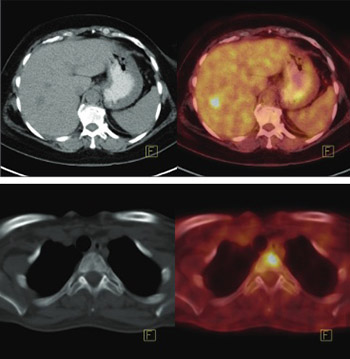
Among 234 patients, modification of the staging system following 18F-FDG PET/CT was applied in 82 (35%) patients. The percentage of patients with upstaging, according to each stage, was as follows: IIA: 18.6% (Figure 1 and 2), IIB: 30%, IIIA: 46.3%, IIIB: 68.8%, and IIIC: 20.8%. The total number and percentage of patients with upstaging in each stage group is given in Table 5. Detailed information on extra-axillary regional lymph nodes and distant metastatic involvements that led to upstaging is shown in Table 6. The percentage of stage modification according to the main histological subtypes was as follows: 32.5% in IDC, 40% in ILC, 33.3% in IDC + ILC, 53.3% in IBC, and 39.1% in other rare histological types (Table 7).

FIGURE 1: A 61-year-old female patient with invasive ductal carcinoma. She had T2N0 (IIA) disease. A 1.3 cm hypermetabolic (maximum standardized uptake value (SUVmax): 8.1), hypodense lesion at the right lobe, segment 5 of the liver, was detected. The patient stage was modified as “4” following positron emission tomography with 2-deoxy-2-[fluorine-18]fluoro- D-glucose integrated with computed tomography (18F-FDG PET/CT).
FIGURE 2: A 51-year-old female patient with infiltrating lobular carcinoma. She had T1N1 (IIA) disease. Positron emission tomography with 2-deoxy-2-[fluorine-18]fluoro- D-glucose integrated with computed tomography (18F-FDG PET/CT) revealed multiple bone metastases. Lytic and hypermetabolic lesion (maximum standardized uptake value [SUVmax]: 7.9) located at the T3 vertebra is shown in the figure.
TABLE 5: Number and percentage of cases that were upstaged following 18F-FDG PET/CT
TABLE 6: A comparison of initial breast cancer staging and that following 18F-FDG PET/CT
TABLE 7: Number and percentages of patients with stage modification following 18F-FDG PET/CT

Breast cancer is classified into three groups in relation to therapy selection: a) clinical stage 1-2 and operable locally advanced (T3N1M0) breast cancer; b) inoperable (at presentation) locally advanced breast cancer: Clinical stage 3A (except for T3N1M0), 3B, and 3C; and c) clinical stage 4 (metastatic disease). According to this classification, the PET/CT changed therapy in 67 of 82 upstaged cases (81.7%). In addition, in two upstaged patients, the radiotherapy field was widened, since hypermetabolic metastatic internal mammary lymph nodes were detected by PET/CT. Overall, patient management was changed in 69/234 cases (29.4%).

The 8 patients with hypermetabolic, residual interpectoral lymph nodes detected on postoperative PET/CT already had widespread metastatic involvement in the level I and II, therefore the radiotherapy field was not changed in this group of patients.

Contribution of additional bone scan to the patient management

In 43/234 patients with no bone lesion on 18F-FDG PET/CT, the Tc99m-MDP bone scan with or without SPECT/CT was performed. In 38 patients, the results were normal. In 5 patients, suspicious hyperactive processes were observed (1 diffuse bone uptake and 4 hypermetabolic foci). According to MRI, all lesions were further diagnosed as benign (1 metabolic bone disease, 1 bone island, and 3 severe degenerative changes).

DISCUSSION

Previously, it was indicated that FDG avidity of a breast cancer lesion depends on multiple factors [13]. One of these factors is the histological type of the tumor, where ILCs are known to have lower FDG avidity [14,15]. Although the number of patients in each of our histological subgroup was not identical, the SUVmax was significantly higher in IDC and IBC groups compared to ILC and IDC + ILC tumors. However, the important note is that “variable FDG affinity” has little effect on the clinical utility of 18F-FDG PET/CT in various histological subtypes.

Axillary lymph node status is one of the main prognostic factors in breast cancer [16]. If there are no palpable lymph nodes on clinical examination, the currently accepted approach is SLNB for axillary staging. This technique has the advantage of detecting even micrometastases (<2 mm) or isolated tumor cells. Our results are in agreement with previous studies and showed that the size of the metastatic deposit in a lymph node is a limiting factor for PET/CT [7]. Among 5 patients with false-negative axillary lymph nodes on PET/CT, 3 had millimetric metastatic foci. Furthermore, the clinical significance of millimetric metastatic involvement and even the necessity of axillary node dissection have been debated in the recent years [17,18]. Another point is that the detection/visualization of extra-axillary lymph nodes with SLNB in lymphatic mapping studies differs in relation to the technique used. Lymphatic mapping done with superficial injection techniques (subareolar or periareolar) is preferred in many centers, with the advantages of being easier to perform and having higher detection rates of sentinel lymph nodes. On the other hand, with peritumoral deep injections, the internal mammary (IM) chain drainage could be visualized more efficiently compared with superficial sub/periareolar or subdermal injections [19]. The inability to detect regional extra-axillary lymph nodes with SLNB, especially when performed with superficial injections, can be overcome using 18F-FDG PET/CT. In our study group, the SLNB with periareolar subdermal injection showed no SLN in the IM chain or infra/supraclavicular region. However, the PET/CT revealed metastatic regional extra-axillary lymph nodes in 42 (17.9%) patients.

In addition to the detection of axillary lymph nodes, the number and location of metastatic nodes are also important in cancer staging and therapy planning. This has a prognostic significance and may widen the radiotherapy field [6,20]. In our study group, among the patients with axillary clearance, 8 (6.6%) had residual Rotter’s lymph nodes. This region is not routinely reached during the surgery. Although in 8 patients with widespread metastatic axillary lymph nodes, the detection of postoperative residual lymph nodes did not change the radiotherapy field, PET/CT may affect the radiotherapy field in patients with limited disease of the axilla.

Bone metastases are frequently observed in breast cancer, especially in its advanced stages. Post-mortem studies reveal bone metastases in 70% of cases with advanced breast cancer [21]. On the other hand, in patients in which bone metastasis is the only metastatic site, the survival time is significantly longer compared to patients with visceral metastases [22]. The prevention of morbidity from metastatic bone disease is a major issue and needs early and accurate diagnosis. The visualization of bone metastasis with Tc99m-MDP bone scintigraphy is based on the osteoblastic response to the bone destruction by cancer cells. Tc99m-MDP may have lower specificity due to benign conditions with osteoblastic response, including the flare response of metastatic bone disease to the treatment. In their meta-analysis, Shie et al. [23] indicated that the main advantages of 18F-FDG PET/CT in the evaluation of bone metastasis are higher rates of specificity and the potential for monitoring the therapy response [23]. In our study group, a bone scan was performed only in symptomatic patients with negative 18F-FDG PET/CT results. In these 43 patients, there was no additional benefit of the bone scan; furthermore, a number of false-positive findings related to benign lesions were observed.

The presence of either metastatic extra-axillary regional lymph nodes or distant metastases changes the stage, and therefore, the management of the disease. In our study group, comprised mainly of patients with the stage IIA to IIIC disease, the overall upstaging rate was 35%. The percentage of patients with upstaging in each stage group indicated a significant contribution of PET/CT to the upstaging, starting from the stage IIA. Currently, the NCCN guidelines recommend the utilization of 18F-FDG PET/CT in staging breast cancer, starting from the stage IIIA (T3, N1, and M0) [6]. Moreover, other studies showed the benefit of using 18F-FDG PET/CT in staging from the IIB stage [7,8,24]. The 30.3% rate of upstaging in our IIB group supports these findings. In the literature, the contribution of 18F-FDG PET/CT to stage IIA disease varies significantly (4.5%-17%) [7,25-27]. In our study, 9 of 44 (20.5%) patients with stage IIA disease were upstaged following 18F-FDG PET/CT. Both T2N0 and T1N1 disease belong to the stage IIA, and one of the factors contributing to the variable results obtained with 18F-FDG PET/CT for stage IIA disease may be a different number of T2N0 and T1N1 patients. Nevertheless, other factors have also been indicated; for example, while Koolen et al. [26] reported a higher rate of upstaging in T2N0 patients (11%), in the study of Groheux et al. [28], stage IIA group consisted predominantly of T2N0 patients (T2N0: 34, T1N1:2), and the rate of upstaging was only 5.5%. Another factor to consider in the upstaged patients in our IIA group are the histological types of tumor. While the overall incidence was 6.4% for ILC and 5.1% for ILC + IDC mixed tumors among the 8 upstaged patients from IIA group, 3 patients had ILC (37%) and 1 patient had IDC + ILC (12.5%). Three of these 4 patients with ILC/IDC+ILC had bone and 1 had lymph node metastasis. Although not confirmed in all studies, a significant number of studies reported a higher prevalence of bone and bone marrow metastasis in early stage ILC tumors [29-31]. A relatively higher number of ILC patients in our stage IIA group may explain the higher rate of upstaged patients when compared to other studies. The tendency of a tumor to metastasize depends on multiple factors, including histological subtypes, size of tumor, and pathophysiological status. Studies evaluating the imaging modalities lack homogeneity in terms of tumor and patient characteristics. Therefore, large meta-analytic studies are needed to reach the consensus.

Our comparison of different histological subtypes in relation to the rate of upstaging showed that IBC had the highest rate of stage modification. This finding is in accordance with a previous report [2].

The following are the limitations of our study. There was a lack of homogeneity in the referral group in terms of the PET/CT imaging timing, since it was performed during the postoperative period in 51.7% of the cases. Although this limitation decreased the number of patients available for comparative axillary evaluation, it did not affect the evaluation of extra-axillary lymph nodes and distant metastases. The referral bias can be reduced with well-structured, prospective studies including detailed information on histological subtypes, size of tumor, and patient demographics.

CONCLUSION

Our results showed that 18F-FDG PET/CT significantly contributed the accurate staging and management of breast cancer, starting from the stage IIA. For axillary staging, although comparable histopathological and PET/CT data were limited, PET/CT was not favored over SLNB. In the patients evaluated postoperatively, the detection of residual metastatic lymph nodes did not change the radiotherapy field, since all patients in this group already had widespread metastatic disease in axillary level I and II. However, residual lymph nodes detected on postoperative PET/CT may affect the subsequent management in patients with isolated metastatic axillary lymph nodes. In the selected group of patients with no signs of bone metastasis on the PET/CT scan, the Tc99m-MDP bone scan revealed only false-positive lesions.

DECLARATION OF INTERESTS

The authors declare no conflict of interests.

REFERENCES

  1. , , , , , (). Cancer statistics, 2009. CA Cancer J Clin. https://doi.org/10.3322/caac.20006
  2. , , , (). Performance of FDG PET/CT in the clinical management of breast cancer. Radiology. https://doi.org/10.1148/radiol.12110853
  3. , , , , , (). A comparative study on the value of FDG-PET and sentinel node biopsy to identify occult axillary metastases. Ann Oncol. https://doi.org/10.1093/annonc/mdl425
  4. , , , , , (). Tumour markers and FDG PET/CT for prediction of disease relapse in patients with breast cancer. Eur J Nucl Med Mol Imaging. https://doi.org/10.1007/s00259-010-1626-7
  5. , , , , , (). The role of FDG PET/CT in patients with locoregional breast cancer recurrence: A comparison to conventional imaging techniques. Eur J Surg Oncol. https://doi.org/10.1016/j.ejso.2009.11.009
  6. (). Clinical Practice Guidelines in Oncology. Breast Cancer. Version 3. Available from: https://www.nccn.org/professionals/physician_gls/pdf/breast.pdf
  7. , , , , , (). The yield of18F-FDG PET/CT in patients with clinical stage IIA, IIB, or IIIA breast cancer: A prospective study. J Nucl Med. https://doi.org/10.2967/jnumed.111.093864
  8. , , , , , (). Additional value of PET-CT in staging of clinical stage IIB and III breast cancer. Breast J. https://doi.org/10.1111/j.1524-4741.2010.00987.x
  9. , , , (). Problematic aspects of sentinel lymph node biopsy and its relation to previous excisional biopsy in breast cancer. Clin Nucl Med. https://doi.org/10.1097/RLU.0b013e3181becec2
  10. , , (). Diagnostic performance of dedicated positron emission mammography using fluorine-18-fluorodeoxyglucose in women with suspicious breast lesions: A meta-analysis. Clin Breast Cancer. https://doi.org/10.1016/j.clbc.2013.12.004
  11. , , , , , (). Positron emission mammography in breast cancer presurgical planning: Comparisons with magnetic resonance imaging. Eur J Nucl Med Mol Imaging. https://doi.org/10.1007/s00259-010-1588-9
  12. , , , , , (). Association between [18F] fluorodeoxyglucose uptake and prognostic parameters in breast cancer. Br J Surg. https://doi.org/10.1002/bjs.6459
  13. , , , , , (). Clinical significance of18F-FDG-PET in invasive lobular carcinoma. Anticancer Res. https://doi.org/10.21873/anticanres.11129
  14. , , , , (). Associations between the standardized uptake value of (18)F-FDG PET/CT and the prognostic factors of invasive lobular carcinoma: In comparison with invasive ductal carcinoma. World J Surg Oncol. https://doi.org/10.1186/s12957-015-0522-9
  15. , , , , (). An overview of prognostic factors for long-term survivors of breast cancer. Breast Cancer Res Treat. https://doi.org/10.1007/s10549-007-9556-1
  16. , , , , , (). Axillary dissection versus no axillary dissection in patients with sentinel-node micrometastases (IBCSG 23-01): A phase 3 randomised controlled trial. Lancet Oncol. https://doi.org/10.1016/S1470-2045(13)70035-4
  17. , , , (). Local treatment of the axilla in early breast cancer: Concepts from the national surgical adjuvant breast and bowel project B-04 to the planned intergroup sentinel mamma trial. Breast Care (Basel). https://doi.org/10.1159/000360411
  18. , , , , , (). Sentinel lymph node mapping in breast cancer: A critical reappraisal of the internal mammary chain issue. Q J Nucl Med Mol Imaging.
  19. , , , (). Significance of axillary lymph node metastasis in primary breast cancer. J Clin Oncol. https://doi.org/10.1200/JCO.1999.17.8.2334
  20. (). The anatomy and pathways of bone metastases. Bone Metastases.
  21. , , (). Risk of complications from bone metastases in breast cancer. Implications for management. Eur J Cancer. https://doi.org/10.1016/S0959-8049(99)00331-7
  22. , , , , (). Meta-analysis: Comparison of F-18 fluorodeoxyglucose-positron emission tomography and bone scintigraphy in the detection of bone metastases in patients with breast cancer. Clin Nucl Med. https://doi.org/10.1097/RLU.0b013e31815f23b7
  23. , , , , , (). 18F FDG PET/CT for staging and restaging of breast cancer. J Nucl Med. DOI: 10.2967/jnumed.115.157859
  24. , , , , , (). Retrospective analysis of18F-FDG PET/CT for staging asymptomatic breast cancer patients younger than 40 years. J Nucl Med. https://doi.org/10.2967/jnumed.114.143297
  25. , , , , , (). 18F-FDG PET/CT as a staging procedure in primary stage II and III breast cancer: Comparison with conventional imaging techniques. Breast Cancer Res Treat. https://doi.org/10.1007/s10549-011-1767-9
  26. , , , , , (). 18F-FDG PET/CT provides powerful prognostic stratification in the primary staging of large breast cancer when compared with conventional explorations. Eur J Nucl Med Mol Imaging. https://doi.org/10.1007/s00259-013-2595-4
  27. , , , , , (). Prognostic impact of (18) FDG-PET-CT findings in clinical stage III and IIB breast cancer. J Natl Cancer Inst. https://doi.org/10.1093/jnci/djs451
  28. , , , , (). Patterns of metastatic breast cancer in relation to histological type. Eur J Cancer. https://doi.org/10.1016/0959-8049(93)90053-I
  29. , , , (). Distant metastatic disease manifestations in infiltrating lobular carcinoma of the breast. AJR Am J Roentgenol. https://doi.org/10.2214/AJR.13.11156
  30. , , , , , (). Wide metastatic spreading in infiltrating lobular carcinoma of the breast. Eur J Cancer. https://doi.org/10.1016/j.ejca.2003.08.007