Oligoasthenoteratozoospermic (OAT) men display altered phospholipase C ζ (PLCζ) localization and a lower percentage of sperm cells expressing PLCζ and post-acrosomal sheath WW domain-binding protein (PAWP)

Authors

  • Nahid Azad Department of Biology and Anatomical Sciences, School of Medicine, Shahid Beheshti University of Medical Sciences, Tehran, Iran
  • Hamid Nazarian Department of Biology and Anatomical Sciences, School of Medicine, Shahid Beheshti University of Medical Sciences, Tehran, Iran
  • Marefat Ghaffari Novin Infertility and Reproductive Health Research Center, Shahid Beheshti University of Medical Sciences, Tehran, Iran
  • Reza Masteri Farahani Department of Biology and Anatomical Sciences, School of Medicine, Shahid Beheshti University of Medical Sciences, Tehran, Iran
  • Abbas Piryaei Department of Biology and Anatomical Sciences, School of Medicine, Shahid Beheshti University of Medical Sciences, Tehran, Iran; Urogenital Stem Cell Research Center, Shahid Beheshti University of Medical Sciences, Tehran, Iran
  • Mohammad Hassan Heidari Department of Biology and Anatomical Sciences, School of Medicine, Shahid Beheshti University of Medical Sciences, Tehran, Iran
  • Meghdad Abdollahpour Alitappeh Basic and Molecular Epidemiology of Gastrointestinal Disorders Research Center, Research Institute for Gastroenterology and Liver Diseases, Shahid Beheshti University of Medical Sciences, Tehran, Iran

DOI:

https://doi.org/10.17305/bjbms.2017.2208

Keywords:

Oligoasthenoteratozoospermia, OAT, phospholipase C ζ, PLCζ, post-acrosomal sheath WW domain-binding protein, PAWP, semen quality, sperm cells

Abstract

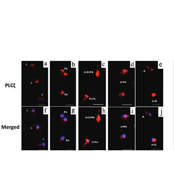
Oligoasthenoteratozoospermia (OAT) is demonstrated to be one of the most common causes of male subfertility. Phospholipase C ζ (PLCζ), a sperm-specific protein, is considered to be one of the sperm-borne oocyte activating factors (SOAFs), which play a vital role in fertilization. The post-acrosomal sheath WW domain-binding protein (PAWP) is another candidate for SOAF. The aim of this study was to compare the PLCζ localization patterns and percentage of PLCζ- and PAWP-positive sperm cells in patients with OAT and fertile men with normozoospermia. A total of 40 men included in this study were classified into two groups: OAT (n = 25) and control group (n = 15). Semen samples were collected and analyzed using conventional semen analysis according to the World Health Organization guidelines. The percentage of PLCζ- and PAWP-positive sperm cells and localization patterns of PLCζ were evaluated using immunofluorescence staining. The mean percentage of sperm cells expressing PAWP and PLCζ was significantly lower in OAT compared to control group (52.8 ± 4.2 vs. 76.8 ± 5 and 63.4 ± 3.5 vs. 86.7 ± 2.1, respectively). In addition, statistically significant differences were found with regard to the PLCζ localization patterns, including equatorial, acrosomal + equatorial, and equatorial + post-acrosomal pattern, between the two groups (p < 0.01). The present study showed a lower percentage of sperm cells expressing PLCζ and PAWP, as well as altered localization patterns of PLCζ in men with OAT. Given the role of PLCζ and PAWP in fertilization, as two major candidates for SOAFs, our findings indicate that PLCζ and PAWP impairments may be one of the possible etiologies of decreased fertility in OAT.

Downloads

Download data is not yet available.

Author Biographies

Nahid Azad, Department of Biology and Anatomical Sciences, School of Medicine, Shahid Beheshti University of Medical Sciences, Tehran, Iran

Department of Biology and Anatomical Sciences

Hamid Nazarian, Department of Biology and Anatomical Sciences, School of Medicine, Shahid Beheshti University of Medical Sciences, Tehran, Iran

Department of Biology and Anatomical Sciences

Reza Masteri Farahani, Department of Biology and Anatomical Sciences, School of Medicine, Shahid Beheshti University of Medical Sciences, Tehran, Iran

Department of Biology and Anatomical Sciences

Abbas Piryaei, Department of Biology and Anatomical Sciences, School of Medicine, Shahid Beheshti University of Medical Sciences, Tehran, Iran; Urogenital Stem Cell Research Center, Shahid Beheshti University of Medical Sciences, Tehran, Iran

Department of Biology and Anatomical Sciences

Mohammad Hassan Heidari, Department of Biology and Anatomical Sciences, School of Medicine, Shahid Beheshti University of Medical Sciences, Tehran, Iran

Department of Biology and Anatomical Sciences

References

Tournaye H, Krausz C, Oates RD. Novel concepts in the aetiology of male reproductive impairment. Lancet Diabetes Endocrinol 2017;5(7):544-53.

https://doi.org/10.1016/S2213-8587(16)30040-7.

Amdani SN, Jones C, Coward K. Phospholipase C zeta (PLCζ): Oocyte activation and clinical links to male factor infertility. Adv Biol Regul 2013;53(3):292-308. https://doi.org/10.1016/j.jbior.2013.07.005.

Jungwirth A, Giwercman A, Tournaye H, Diemer T, Kopa Z, Dohle G, et al. European association of urology guidelines on male infertility: The 2012 update. Eur Urol 2012;62(2):324-32. https://doi.org/10.1016/j.eururo.2012.04.048.

World Health Organization (WHO). WHO Laboratory Manual for the Examination and Processing of Human Semen. 5th ed. Geneva: World Health Organization Press; 2010.

Loutradi KE, Tarlatzis BC, Goulis DG, Zepiridis L, Pagou T, Chatziioannou E, et al. The effects of sperm quality on embryo development after intracytoplasmic sperm injection. J Assist Reprod Genet 2006;23(2):69-74. https://doi.org/10.1007/s10815-006-9022-8.

Zheng J, Lu Y, Qu X, Wang P, Zhao L, Gao M, et al. Correction: Decreased sperm motility retarded ICSI fertilization rate in severe oligozoospermia but good-quality embryo transfer had achieved the prospective clinical outcomes. PLoS One 2016:11(10):e0165684. https://doi.org/10.1371/journal.pone.0165684.

Lu YH, Gao HJ, Li BJ, Zheng YM, Ye YH, Qian YL, et al. Different sperm sources and parameters can influence intracytoplasmic sperm injection outcomes before embryo implantation. J Zhejiang Univ Sci B 2012;13(1):1-10. https://doi.org/10.1631/jzus.B1100216.

Kashir J, Heindryckx B, Jones C, De Sutter P, Parrington J, Coward K. Oocyte activation, phospholipase C zeta and human infertility. Hum Reprod Update 2010;16(6):690-703. https://doi.org/10.1093/humupd/dmq018.

Park JH, Kim SK, Kim J, Kim JH, Chang JH, Jee BC, et al. Relationship between phospholipase C zeta immunoreactivity and DNA fragmentation and oxidation in human sperm. Obstet Gynecol Sci 2015;58(3):232-8. https://doi.org/10.5468/ogs.2015.58.3.232.

Yelumalai S, Yeste M, Jones C, Amdani SN, Kashir J, Mounce G, et al. Total levels, localization patterns, and proportions of sperm exhibiting phospholipase C zeta are significantly correlated with fertilization rates after intracytoplasmic sperm injection. Fertil Steril 2015;104(3):561-8.e4. https://doi.org/10.1016/j.fertnstert.2015.05.018.

Amdani SN, Yeste M, Jones C, Coward K. Phospholipase C zeta (PLCζ) and male infertility: Clinical update and topical developments. Adv Biol Regul 2016;61:58-67. https://doi.org/10.1016/j.jbior.2015.11.009.

Yu Y, Nomikos M, Theodoridou M, Nounesis G, Lai FA, Swann K. PLCζ causes Ca2+ oscillations in mouse eggs by targeting intracellular and not plasma membrane PI(4,5)P2. Mol Biol Cell 2012;23(2):371-80. https://doi.org/10.1091/mbc.E11-08-0687.

Swann K, Lai FA. PLCζ and the initiation of Ca2+ oscillations in fertilizing mammalian eggs. Cell Calcium 2013;53(1):55-62. https://doi.org/10.1016/j.ceca.2012.11.001.

Swann K, Lai FA. The sperm phospholipase C-ζ and Ca2+ signalling at fertilization in mammals. Biochem Soc Trans 2016;44(1):267-72. https://doi.org/10.1042/BST20150221.

Kashir J, Jones C, Mounce G, Ramadan WM, Lemmon B, Heindryckx B, et al. Variance in total levels of phospholipase C zeta (PLC-ζ) in human sperm may limit the applicability of quantitative immunofluorescent analysis as a diagnostic indicator of oocyte activation capability. Fertil Steril 2013;99(1):107-17. https://doi.org/10.1016/j.fertnstert.2012.09.001.

Yoon SY, Jellerette T, Salicioni AM, Lee HC, Yoo MS, Coward K, et al. Human sperm devoid of PLC, zeta 1 fail to induce Ca2+ release and are unable to initiate the first step of embryo development. J Clin Invest 2008;118(11):3671-81. https://doi.org/10.1172/JCI36942.

Aarabi M, Balakier H, Bashar S, Moskovtsev SI, Sutovsky P, Librach CL, et al. Sperm content of postacrosomal WW binding protein is related to fertilization outcomes in patients undergoing assisted reproductive technology. Fertil Steril 2014;102(2):440-7. https://doi.org/10.1016/j.fertnstert.2014.05.003.

Aarabi M, Balakier H, Bashar S, Moskovtsev SI, Sutovsky P, Librach CL, et al. Sperm-derived WW domain-binding protein, PAWP, elicits calcium oscillations and oocyte activation in humans and mice. FASEB J 2014;28(10):4434-40. https://doi.org/10.1096/fj.14-256495.

Aarabi M, Qin Z, Xu W, Mewburn J, Oko R. Sperm-borne protein, PAWP, initiates zygotic development in Xenopus laevis by eliciting intracellular calcium release. Mol Reprod Dev 2010;77(3):249-56. DOI: 10.1002/mrd.21140.

Grasa P, Coward K, Young C, Parrington J. The pattern of localization of the putative oocyte activation factor, phospholipase C zeta, in uncapacitated, capacitated, and ionophore-treated human spermatozoa. Hum Reprod 2008;23(11):2513-22. https://doi.org/10.1093/humrep/den280.

Nomikos M, Sanders JR, Kashir J, Sanusi R, Buntwal L, Love D, et al. Functional disparity between human PAWP and PLCζ in the generation of Ca2+ oscillations for oocyte activation. Mol Hum Reprod 2015;21(9):702-10. https://doi.org/10.1093/molehr/gav034.

Sutovsky P, Aarabi M, Miranda-Vizuete A, Oko R. Negative biomarker based male fertility evaluation: Sperm phenotypes associated with molecular-level anomalies. Asian J Androl 2015;17(4):554-60. https://doi.org/10.4103/1008-682X.153847.

Ramadan WM, Kashir J, Jones C, Coward K. Oocyte activation and phospholipase C zeta (PLCζ): Diagnostic and therapeutic implications for assisted reproductive technology. Cell Commun Signal 2012;10(1):12. https://doi.org/10.1186/1478-811X-10-12.

Tavalaee M, Parivar K, Nasr-Esfahani MH, Shahverdi A, Ghaedi K. A comparison of chromatin structure and PLCζ in sperms of subfertile oligoasthenoteratozoospermic and fertile men. J Shahrekord Univ Med Sci 2016;18(4):9-19.

Taylor SL, Yoon SY, Morshedi MS, Lacey DR, Jellerette T, Fissore RA, et al. Complete globozoospermia associated with PLCζ deficiency treated with calcium ionophore and ICSI results in pregnancy. Reprod Biomed Online 2010;20(4):559-64. https://doi.org/10.1016/j.rbmo.2009.12.024.

Kashir J, Sermondade N, Sifer C, Oo SL, Jones C, Mounce G, et al. Motile sperm organelle morphology evaluation-selected globozoospermic human sperm with an acrosomal bud exhibits novel patterns and higher levels of phospholipase C zeta. Hum Reprod 2012;27(11):3150-60. https://doi.org/10.1093/humrep/des312.

Heytens E, Parrington J, Coward K, Young C, Lambrecht S, Yoon SY, et al. Reduced amounts and abnormal forms of phospholipase C zeta (PLCzeta) in spermatozoa from infertile men. Hum Reprod 2009;24(10):2417-28. https://doi.org/10.1093/humrep/dep207.

Kamali-Dolat Abadi M, Tavalaee M, Shahverdi A, Nasr-Esfahani MH. Evaluation of PLCζ and PAWP expression in globozoospermic individuals. Cell J 2016;18(3):438-45. DOI: 10.22074/cellj.2016.4572.

Janghorban-Laricheh E, Ghazavi-Khorasgani N, Tavalaee M, Zohrabi D, Abbasi H, Nasr-Esfahani MH, et al. An association between sperm PLCζ levels and varicocele? J Assist Reprod Genet 2016;33(12):1649-55. https://doi.org/10.1007/s10815-016-0802-5.

Lee HC, Arny M, Grow D, Dumesic D, Fissore RA, Jellerette-Nolan T. Protein phospholipase C Zeta1 expression in patients with failed ICSI but with normal sperm parameters. J Assist Reprod Genet 2014;31(6):749-56. https://doi.org/10.1007/s10815-014-0229-9.

Chithiwala ZH, Lee HC, Hill DL, Jellerette-Nolan T, Fissore R, Grow D, et al. Phospholipase C-zeta deficiency as a cause for repetitive oocyte fertilization failure during ovarian stimulation for in vitro fertilization with ICSI: A case report. J Assist Reprod Genet 2015;32(9):1415-9. https://doi.org/10.1007/s10815-015-0531-1.

Durban M, Barragán M, Colodron M, Ferrer-Buitrago M, De Sutter P, Heindryckx B, et al. Erratum to: PLCζ disruption with complete fertilization failure in normozoospermia. J Assist Reprod Genet 2015;32(8):1295. https://doi.org/10.1007/s10815-015-0522-2.

Tavalaee M, Kiani-Esfahani A, Nasr-Esfahani MH. Relationship between potential sperm factors involved in oocyte activation and sperm DNA fragmentation with intra-cytoplasmic sperm injection clinical outcomes. Cell J 2017;18(4):588-96. DOI: 10.22074/cellj.2016.4725.

Ferrer-Vaquer A, Barragan M, Freour T, Vernaeve V, Vassena R. PLCζ sequence, protein levels, and distribution in human sperm do not correlate with semen characteristics and fertilization rates after ICSI. J Assist Reprod Genet 2016;33(6):747-56. https://doi.org/10.1007/s10815-016-0718-0.

Freour T, Barragan M, Ferrer-Vaquer A, Rodríguez A, Vassena R. WBP2NL/PAWP mRNA and protein expression in sperm cells are not related to semen parameters, fertilization rate, or reproductive outcome. J Assist Reprod Genet 2017;34(6):803-10.

https://doi.org/10.1007/s10815-017-0902-x.

Oligoasthenoteratozoospermic (OAT) men display altered phospholipase C ζ (PLCζ) localization and a lower percentage of sperm cells expressing PLCζ and post-acrosomal sheath WW domain-binding protein (PAWP)

Downloads

Additional Files

Published

2018-05-20

How to Cite

1.
Azad N, Nazarian H, Ghaffari Novin M, Masteri Farahani R, Piryaei A, Heidari MH, Abdollahpour Alitappeh M. Oligoasthenoteratozoospermic (OAT) men display altered phospholipase C ζ (PLCζ) localization and a lower percentage of sperm cells expressing PLCζ and post-acrosomal sheath WW domain-binding protein (PAWP). Biomol Biomed [Internet]. 2018May20 [cited 2023Jun.4];18(2):178-84. Available from: https://www.bjbms.org/ojs/index.php/bjbms/article/view/2208

Issue

Section

Translational and Clinical Research

INTRODUCTION

Male factor infertility affects 20-70% of couples suffering from infertility [1]. Although the main cause is still unexplained, alterations in semen parameters seem to play a role in male infertility [2]. Oligoasthenoteratozoospermia (OAT), with abnormalities in sperm count, motility and morphology, is considered as one of the most common causes of infertility [3,4]. Some studies indicated that poor sperm quality can negatively affect fertilization outcome following the assisted reproductive technique (ART) [5-7].

Phospholipase C ζ (PLCζ), a sperm-specific protein, is considered to be one of the sperm-borne oocyte activating factors (SOAFs) and a potential biomarker for the prediction of male fertility [8-11]. In addition, significant evidence shows that PLCζ released into the oocyte after the fusion of gametes triggers a series of molecular events named “oocyte activation”, including meiotic resumption, pronuclear formation, and embryo cleavage. Indeed, oocyte activation occurs when PLCζ induces calcium oscillations in the cytoplasm [11-14]. The absence or decreased amount of PLCζ as well as its abnormal localization patterns were reported in infertile patients with oocyte activation deficiency (OAD) and failed intracytoplasmic sperm injection (ICSI) [15,16].

The post-acrosomal sheath WW domain-binding protein (PAWP) is another major candidate for SOAF, and is associated with fertilization outcomes following ICSI [17]. Microinjection of a recombinant PAWP protein or PAWP complementary RNA (cRNA) into humans, mice, and Xenopus oocytes induces calcium oscillations similar to those observed following fertilization [18,19].

Nonetheless, no previous studies have simultaneously investigated PLCζ and PAWP proteins in patients with OAT and poor sperm quality.

The aim of our study was to compare the PLCζ localization patterns and percentage of PLCζ- and PAWP-positive sperm cells between patients with OAT and fertile men with normozoospermia.

MATERIALS AND METHODS

Chemicals

A PLCζ rabbit polyclonal antibody was purchased from LifeSpan BioSciences, Inc. (USA). A PAWP rabbit polyclonal antibody was obtained from Proteintech Company (UK). Goat anti-rabbit IgG (H + L) [Alexa Fluor®555] was obtained from Abcam Company (UK). Normal goat serum, Ham’s F-10 nutrient medium, human serum albumin (HSA), Tween 20, Dulbecco’s phosphate-buffered saline (DPBS), and paraformaldehyde (PFA) were purchased from Sigma-Aldrich (Germany). Diff-Quick rapid stain for sperm morphology was obtained from Avicenna (I.R.I).

Patients and ethics

The study was carried out between January 2015 and March 2017. We obtained semen samples from 15 fertile men with normozoospermia (control) and 25 patients with OAT, from couples who had been referred to the infertility center for the examination and treatment. Individuals with any history of testicular trauma, surgical intervention, inflammation of the testis, cryptorchidism, and globozoospermia were excluded from the study. The ethics committee of the Shahid Beheshti University of Medical Sciences and appropriate regulatory bodies of the Infertility and Reproductive Health Research Center approved the study. We obtained written informed consents from all participating men. Patients with sperm concentration <15×106/mL, total motility <40%, and normal morphology <4%, as confirmed by at least two tests, were considered as having OAT and were included in the study. Fertile men with normozoospermia were sperm donors with proven fertility.

Semen analysis and processing

All semen samples were collected by masturbation after 2-5 days of sexual abstinence. After the liquefaction (30-45 minutes), semen parameters including sperm concentration (×106/mL), motility (%), and morphology (%) were analyzed according to the World Health Organization (WHO) guidelines [4]. Sperm concentration was manually evaluated using the Makler counting chamber with an inverted microscope (Nikon, Japan) and sperm morphology was assessed by a Diff-Quick staining kit.

Semen processing for the separation of motile sperm cells from dead sperm and other cells was performed using mild centrifugation and several washings. Then, the pellet was gently covered by 0.3 ml Ham’s F10 supplemented with 10% HSA and incubated at 37°C for 30-60 minutes for a swim-up. After that, the supernatant was removed and used for immunofluorescence staining.

Diff-Quick staining

The smears were prepared with a 10-µl drop of semen placed on a microscope slide and, after the fixation, the sperm cells were stained with a Diff-Quick staining kit, to evaluate the morphology. Following the slide examination under a bright-field microscope (Nikon, Japan) at ×40 and ×100 magnifications, photographs were captured using a Nikon camera [Japan] (Figure 1). The morphology of 100 sperm cells was analyzed according to the WHO guidelines, in duplicate. Normal size, shape, and acrosome of the sperm head, without midpiece or tail defects, were considered as normal sperm morphology [4], and the percentage of normal and abnormal sperm cells was determined.

FIGURE 1: Evaluation of sperm cell morphology with Diff-Quick staining. Normal size, shape, and acrosome of the sperm head, without midpiece or tail defects, are considered as normal sperm morphology. Sperm cells that do not show these properties are considered as abnormal. Five sperm cells with abnormal morphology are shown in the figure. Bar = 10 µm.

PLCζ immunofluorescence detection

Immunofluorescent staining of PLCζ was performed as previously described [16,20], with some alterations. Briefly, the washed sperm cells were fixed with a freshly prepared PFA in phosphate-buffered saline (PBS, 4%) for 10 minutes. After permeabilization with 0.2% Triton X-100/PBS on ice for 10 minutes, non-specific binding sites were blocked with 5% normal goat serum/PBS for 1 hour, and incubated with PLCζ rabbit anti-human polyclonal antibody (1:50) in the blocking medium, overnight at 4°C. Then, the sperm cells were incubated with goat anti-rabbit IgG (H + L) antibody (1:200) in the blocking medium at room temperature for 1 hour (light protected). Subsequently, the sperm cells were counterstained with 4’, 6-diamidino-2-phenylindole [DAPI] (10 µg/ml) for 5 minutes, and mounted with glycerol. The washing of sperm cells between the described steps was performed using PBST (0.1% Tween 20/PBS). Figure 2A shows different localization patterns of PLCζ in sperm cells stained with DAPI and PLCζ immunofluorescence.

FIGURE 2: Phospholipase C ζ (PLCζ) immunostaining. (A) Different localization patterns of PLCζ in the sperm head. The sperm cells were stained with anti-PLCζ (red, a-e) and 4’,6-diamidino-2-phenylindole [DAPI] (blue). Parts f-j represent merged images of anti-PLCζ and DAPI staining [bar = 10 µm]. (B) Comparison of the mean percentages of sperm cells expressing PLCζ between OAT and control groups. (C) Comparison of different localization patterns of PLCζ between the two groups. Significant differences: ****p < 0.0001 and **p < 0.01. All data are presented as mean ± SEM. OAT: Oligoasthenoteratozoospermia. PLCζ localization patterns: A = Acrosomal, E = Equatorial, PA = Post-acrosomal, A+E = Acrosomal and equatorial, A+PA = Acrosomal and post-acrosomal, E+PA = Equatorial and post-acrosomal, and A+ E+PA = Acrosomal, equatorial, and post-acrosomal.

PAWP immunofluorescence detection

Immunofluorescent staining of PAWP was carried out as previously described by Nomikos et al. [21], with some modifications. Briefly, the washed sperm cells were fixed with 4% PFA/PBS for 10 minutes. Following the permeabilization of sperm cells with 0.2% Triton X-100/PBS on ice for 10 minutes, the non-specific antigens were blocked with 5% normal goat serum/PBS for 1 hour at 37°C, and incubated with PAWP rabbit polyclonal antibody (1:200) in the blocking medium, overnight at 4°C. Then, the sperm cells were incubated with goat anti-rabbit IgG (H + L), diluted 1:100, in the blocking medium at room tepmerature for 1 hour (in the dark). Subsequently, the sperm cells were counterstained with DAPI (10 µg/ml) for 5 minutes, and mounted with a mounting medium. The washing of sperm cells between the described steps was performed using PBST. Figure 3A shows sperm cells stained with DAPI and PAWP immunofluorescence.

FIGURE 3: Post-acrosomal sheath WW domain-binding protein (PAWP) immunostaining. (A) PAWP immunofluorescence staining (red) in sperm cells and counterstaining with 4’, 6-diamidino-2-phenylindole [DAPI] (blue). (B) Comparison of the mean percentages of sperm cells expressing PAWP between OAT and control group. Significant difference: **p < 0.01. (C) A correlation between phospholipase C ζ- and PAWP-positive sperm cells in all individuals; r = 0.6, p < 0.0001. All data are presented as mean ± SEM. OAT: Oligoasthenoteratozoospermia.

PAWP and PLCζ immunofluorescence analyses

The microscope slides were visualized under a Nikon fluorescence microscope (TS100, Japan) at ×20 magnification, and photographs were captured using a Nikon camera (DS-Fi1c, Japan). The photos were merged using the ImageJ software (National Institutes of Health).

Different localization patterns of PLCζ in sperm cells from each patient were classified as previously described by Grasa et al. [20] and Kashir et al. [15]. The localization patterns, including acrosomal (A), equatorial (E), post-acrosomal (PA), acrosomal and equatorial (A+E), acrosomal and post-acrosomal (A+PA), equatorial and post-acrosomal (E+PA), and acrosomal, equatorial and post-acrosomal (A+E+PA) were observed in sperm cells and recorded (n = 200).

The percentages of PAWP and PLCζ-positive sperm cells (the cells with PAWP and PLCζ immunofluorescence in the sperm head) were detected in each individual and recorded (n = 200).

Statistical analysis

All data were analyzed with GraphPad Prism 6 software (GraphPad Software, San Diego, CA, USA). The results were first analyzed using the D’Agostino-Pearson omnibus normality test. According to the normality test, the percentage of PAWP- and PLCζ-positive sperm cells and different localization patterns of PLCζ were compared between control and OAT group using the t-test or Mann–Whitney U-test. The correlation between PLCζ- and PAWP-positive sperm cells was calculated using the Pearson’s correlation coefficient. Data was shown as mean ± standard error of the mean (SEM). The level of significance was considered at p < 0.05.

RESULTS

Semen analysis

A total of 40 men (25 patients with OAT and 15 controls) were included in this study and their semen parameters were assessed according to the WHO criteria [4]. Table 1 shows the mean age and semen parameters in OAT and control groups. The mean of sperm concentration (×106/mL), total motility (progressive + non-progressive, %), and normal morphology (%) in OAT group were significantly lower compared to control group (p < 0.0001).

TABLE 1: Age and sperm parameters in two groups

Comparative analysis of PLCζ immunofluorescence between the groups

The proportion of sperm cells positive for PLCζ, and different localization patterns of PLCζ (i.e., A, E, PA, A+E, A+PA, E+PA, and A+E+PA) were analyzed using the ImageJ software, and compared between the two groups. As shown in Figure 2B, OAT group displayed a significantly lower proportion of sperm cells expressing PLCζ compared with control group (63.4 ± 3.5 vs. 86.7 ± 2.1, p < 0.0001). As depicted in Figure 2C, significant differences were found in the percentage of E, A+E, and E+PA localization patterns between OAT and control groups (p < 0.01). However, no significant differences were observed in the other localizations of PLCζ between the two groups.

Comparative analysis of PAWP immunofluorescence between the groups

The mean percentage of sperm cells positive for PAWP was analyzed and compared between the two groups. As shown in Figure 3B, OAT group showed a lower percentage of PAWP-positive sperm cells compared to control group (52.8 ± 4.2 vs 76.8 ± 5, p < 0.01).

Correlation between PLCζ- and PAWP-positive sperm cells

A significant positive correlation was observed between PLCζ-positive and PAWP-positive sperm cells in all participants, using the Pearson analysis [r = 0.6, p < 0.0001] (Figure 3C).

DISCUSSION

To the best of our knowledge, this is the first report simultaneously assessing PLCζ and PAWP in patients with OAT. In this study, men with OAT had PLCζ deficiency and altered localization patterns of PLCζ. In addition, a lower percentage of sperm cells expressing PAWP was found in those patients. A decreased ability of sperm cells for oocyte activation due to alterations in SOAFs may be one of the possible etiologies of decreased fertility in men with OAT.

Biomarker analysis, accompanied by the conventional semen analysis, can improve the treatment of human male infertility [22]. PLCζ, as one of the candidates for SOAFs, was suggested as a diagnostic, prognostic, and therapeutic biomarker in male infertility [8-10,23]. Since the discovery of PLCζ in 2002, a number of studies have demonstrated that PLCζ causes calcium oscillations in oocytes after gamete fusion [23]. PAWP, another major candidate for SOAFs, may also be used as a diagnostic and therapeutic biomarker of infertility [17,22]. In this study, we evaluated PLCζ and PAWP proteins in men with OAT and showed their alterations in the OAT group compared to fertile men with normozoospermia. We observed a significant decrease in the percentage of sperm cells expressing PLCζ and PAWP in patients with OAT. Furthermore, significantly lower proportions of E pattern and its combinations (A+E and E+PA) were found in OAT compared with control group. Finally, a significant positive correlation was observed between the sperm cells expressing PLCζ and PAWP in all individuals.

Grasa et al. [20], demonstrated for the first time that PLCζ is predominantly localized in the E region of the human sperm head. In addition, Yoon et al. [16] showed that PLCζ, while absent in the sperm cells from patients who had experienced fertilization failure, is localized in the E region of the sperm head in fertile men. However, in another study, a variation in PLCζ localization was documented in both control men and infertile patients with OAD; the E localization pattern alone or in combination with other patterns was predominant in controls, while the PA localization pattern was frequent in OAD patients [15]. Similarly, in our study, a significantly decreased proportion of E pattern and its combinations in patients with OAT indicate altered localization patterns of PLCζ in these patients compared to controls (Figure 2C).

Tavalaee et al. [24] reported a significantly lower percentage of PLCζ-positive sperm cells in subfertile patients with OAT compared to fertile men [24]. Consistent with that study, the percentage of sperm cells expressing PLCζ was significantly lower in our patients with OAT, indicating PLCζ impairment in these patients (Figure 2B). Other studies have also demonstrated decreased expression and/or atypical localization of PLCζ in globozoospermic patients [25-28], patients with grade II and III varicocele [29], and patients with previous fertilization failure [16,25,30-32].

Until now, conflicting results have been reported on the correlation between semen parameters and PLCζ. Yelumalai et al. [10] revealed that the total levels of PLCζ are correlated with sperm count and percentage of progressive motile sperm. Recently, Tavalaee et al. [33] have shown a positive correlation between sperm concentration and PLCζ. Moreover, a significant negative correlation between sperm morphology and PLCζ protein has been demonstrated in another study [28]. On the contrary, Park et al. [9] and Ferrer-Vaquer et al. [34] showed no correlation between PLCζ and semen parameters. Collectively, our results are in agreement with the studies reporting a correlation between PLCζ and sperm parameters.

In the present study, we found a lower percentage of PAWP-positive sperm cells in patients with OAT and poor sperm quality compared to control group (Figure 3B), which is in agreement with other two studies showing a significant correlation between PAWP and sperm concentration, motility, and morphology [28,33]. Recently, Freour et al. [35] indicated a significant negative correlation between sperm morphology and PAWP protein expression and positive correlation between sperm motility and proportion of PAWP-positive sperm cells; however, no correlation was found between PAWP mRNA and semen parameters.

Moreover, similar to our results (Figure 3C), two studies reported a positive correlation between two major candidates for SOAFs [28,33].

Our results suggested that patients with poor sperm quality may be candidates for assisted oocyte activation (a highly specialized method) in infertility clinics, which should be evaluated in the future studies. Due to low sperm count in our samples, only immunofluorescence staining was carried out, with no further analyses.

CONCLUSION

Our study showed lower percentages of sperm cells expressing PLCζ and PAWP, as well as altered localization of PLCζ in men with OAT, as compared with fertile men. Given the role of PLCζ and PAWP, as two major candidates for SOAFs, in male fertility, our findings indicated that SOAF(s) impairments may be considered as one of the possible etiologies of decreased fertility in patients with OAT.

Acknowledgements

ACKNOWLEDGMENTS

We would like to express our appreciation to Mrs. Seyedeh Susan Sadjadpour for her technical help in this study. This article has been extracted from the Ph.D thesis written by Mrs Nahid Azad in School of Medicine, Shahid Beheshti University of Medical Sciences, Tehran, Iran (Registration No: 218).

DECLARATION OF INTERESTS

The authors declare no conflict of interests.

REFERENCES

  1. , , (). Novel concepts in the aetiology of male reproductive impairment. Lancet Diabetes Endocrinol. https://doi.org/10.1016/S2213-8587(16)30040-7
  2. , , (). Phospholipase C zeta (PLCζ): Oocyte activation and clinical links to male factor infertility. Adv Biol Regul. https://doi.org/10.1016/j.jbior.2013.07.005
  3. , , , , , (). European association of urology guidelines on male infertility: the 2012 update. Eur Urol. https://doi.org/10.1016/j.eururo.2012.04.048
  4. (). . WHO Laboratory Manual for the Examination and Processing of Human Semen.
  5. , , , , , (). The effects of sperm quality on embryo development after intracytoplasmic sperm injection. J Assist Reprod Genet. https://doi.org/10.1007/s10815-006-9022-8
  6. , , , , , (). Correction: Decreased sperm motility retarded ICSI fertilization rate in severe oligozoospermia but good-quality embryo transfer had achieved the prospective clinical outcomes. PLoS One. https://doi.org/10.1371/journal.pone.0165684
  7. , , , , , (). Different sperm sources and parameters can influence intracytoplasmic sperm injection outcomes before embryo implantation. J Zhejiang Univ Sci B. https://doi.org/10.1631/jzus.B1100216
  8. , , , , , (). Oocyte activation, phospholipase C zeta and human infertility. Hum Reprod Update. https://doi.org/10.1093/humupd/dmq018
  9. , , , , , (). Relationship between phospholipase C zeta immunoreactivity and DNA fragmentation and oxidation in human sperm. Obstet Gynecol Sci. https://doi.org/10.5468/ogs.2015.58.3.232
  10. , , , , , (). Total levels, localization patterns, and proportions of sperm exhibiting phospholipase C zeta are significantly correlated with fertilization rates after intracytoplasmic sperm injection. Fertil Steril. https://doi.org/10.1016/j.fertnstert.2015.05.018
  11. , , , (). Phospholipase C zeta (PLCζ) and male infertility: Clinical update and topical developments. Adv Biol Regul. https://doi.org/10.1016/j.jbior.2015.11.009
  12. , , , , , (). PLCζ causes Ca2+oscillations in mouse eggs by targeting intracellular and not plasma membrane PI(4,5)P2 . Mol Biol Cell. https://doi.org/10.1091/mbc.E11-08-0687
  13. , (). PLCζ and the initiation of Ca2+oscillations in fertilizing mammalian eggs. Cell Calcium. https://doi.org/10.1016/j.ceca.2012.11.001
  14. , (). The sperm phospholipase C-ζ and Ca2+signalling at fertilization in mammals. Biochem Soc Trans. https://doi.org/10.1042/BST20150221
  15. , , , , , (). Variance in total levels of phospholipase C zeta (PLC-ζ) in human sperm may limit the applicability of quantitative immunofluorescent analysis as a diagnostic indicator of oocyte activation capability. Fertil Steril. https://doi.org/10.1016/j.fertnstert.2012.09.001
  16. , , , , , (). Human sperm devoid of PLC, zeta 1 fail to induce Ca2+release and are unable to initiate the first step of embryo development. J Clin Invest. https://doi.org/10.1172/JCI36942
  17. , , , , , (). Sperm content of postacrosomal WW binding protein is related to fertilization outcomes in patients undergoing assisted reproductive technology. Fertil Steril. https://doi.org/10.1016/j.fertnstert.2014.05.003
  18. , , , , , (). Sperm-derived WW domain-binding protein, PAWP, elicits calcium oscillations and oocyte activation in humans and mice. FASEB J. https://doi.org/10.1096/fj.14-256495
  19. , , , , (). Sperm-borne protein, PAWP, initiates zygotic development in Xenopus laevis by eliciting intracellular calcium release. Mol Reprod Dev. DOI: 10.1002/mrd.21140
  20. , , , (). The pattern of localization of the putative oocyte activation factor, phospholipase C zeta, in uncapacitated, capacitated, and ionophore-treated human spermatozoa. Hum Reprod. https://doi.org/10.1093/humrep/den280
  21. , , , , , (). Functional disparity between human PAWP and PLCζ in the generation of Ca2+oscillations for oocyte activation. Mol Hum Reprod. https://doi.org/10.1093/molehr/gav034
  22. , , , (). Negative biomarker based male fertility evaluation: Sperm phenotypes associated with molecular-level anomalies. Asian J Androl. https://doi.org/10.4103/1008-682X.153847
  23. , , , (). Oocyte activation and phospholipase C zeta (PLCζ): diagnostic and therapeutic implications for assisted reproductive technology. Cell Commun Signal. https://doi.org/10.1186/1478-811X-10-12
  24. , , , , (). A comparison of chromatin structure and PLCζ in sperms of subfertile oligoasthenoteratozoospermic and fertile men. J Shahrekord Univ Med Sci.
  25. , , , , , (). Complete globozoospermia associated with PLCζ deficiency treated with calcium ionophore and ICSI results in pregnancy. Reprod Biomed Online. https://doi.org/10.1016/j.rbmo.2009.12.024
  26. , , , , , (). Motile sperm organelle morphology evaluation-selected globozoospermic human sperm with an acrosomal bud exhibits novel patterns and higher levels of phospholipase C zeta. Hum Reprod. https://doi.org/10.1093/humrep/des312
  27. , , , , , (). Reduced amounts and abnormal forms of phospholipase C zeta (PLCzeta) in spermatozoa from infertile men. Hum Reprod. https://doi.org/10.1093/humrep/dep207
  28. , , , (). Evaluation of PLCζ and PAWP expression in globozoospermic individuals. Cell J. DOI: 10.22074/cellj.2016.4572
  29. , , , , , (). An association between sperm PLCζ levels and varicocelé. J Assist Reprod Genet. https://doi.org/10.1007/s10815-016-0802-5
  30. , , , , , (). Protein phospholipase C Zeta1 expression in patients with failed ICSI but with normal sperm parameters. J Assist Reprod Genet. https://doi.org/10.1007/s10815-014-0229-9
  31. , , , , , (). Phospholipase C-zeta deficiency as a cause for repetitive oocyte fertilization failure during ovarian stimulation for in vitro fertilization with ICSI: A case report. J Assist Reprod Genet. https://doi.org/10.1007/s10815-015-0531-1
  32. , , , , , (). Erratum to: PLCζ disruption with complete fertilization failure in normozoospermia. J Assist Reprod Genet. https://doi.org/10.1007/s10815-015-0522-2
  33. , , (). Relationship between potential sperm factors involved in oocyte activation and sperm DNA fragmentation with intra-cytoplasmic sperm injection clinical outcomes. Cell J. DOI: 10.22074/cellj.2016.4725
  34. , , , , (). PLCζ sequence, protein levels, and distribution in human sperm do not correlate with semen characteristics and fertilization rates after ICSI. J Assist Reprod Genet. https://doi.org/10.1007/s10815-016-0718-0
  35. , , , , (). WBP2NL/PAWP mRNA and protein expression in sperm cells are not related to semen parameters, fertilization rate, or reproductive outcome. J Assist Reprod Genet. https://doi.org/10.1007/s10815-017-0902-x