Vitamin D status, serum lipid concentrations, and vitamin D receptor (VDR) gene polymorphisms in Familial Mediterranean fever

Authors

  • Turan Turhan Department of Biochemistry, Ankara Numune Training and Research Hospital, Ankara, Turkey
  • Halef Okan Doğan Department of Biochemistry, Faculty of Medicine, University of Cumhuriyet, Sivas, Turkey https://orcid.org/0000-0001-8738-0760
  • Nihal Boğdaycioğlu Department of Biochemistry, Samsun Training and Research Hospital, Samsun, Turkey
  • Nilnur Eyerci Department of Medical Biology, University of Kafkas, Kars, Turkey
  • Ahmet Omma Department of Rheumatology, Ankara Numune Training and Research Hospital, Ankara, Turkey
  • İsmail Sari Department of Biochemistry, Faculty of Medicine, University of Niğde Ömer Halis Demir, Niğde, Turkey https://orcid.org/0000-0003-3732-2102
  • Ahmet Yeşilyurt Department of Genetics, Dıskapı Yıldırım Beyatız Education and Training Hospital, Ankara, Turkey
  • Yaşar Karaaslan Department of Rheumatology, Ankara Numune Training and Research Hospital, Ankara, Turkey

DOI:

https://doi.org/10.17305/bjbms.2017.2259

Keywords:

25(OH)D3, FMF, Familial Mediterranean fever, serum lipids, VDR polymorphisms

Abstract

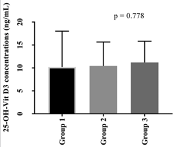
Vitamin D (VitD) is critical for the regulation of inflammatory processes, and VitD deficiency has been linked to several chronic inflammatory disorders. We aimed to investigate the concentrations of serum 25(OH)D3, lipid parameters, and three known VDR polymorphisms (BsmI, FokI, and TaqI) in patients with Familial Mediterranean fever (FMF), an autosomal recessive autoinflammatory disease. The study included 123 FMF patients and 105 controls. Seventy patients had no attack (group 1), 30 had 1-2 attacks (group 2), and 23 had 3 or more attacks (group 3) within last three months. Serum 25(OH)D3 concentrations were determined using liquid chromatography–tandem mass spectrometry. BsmI, FokI, and TaqI polymorphisms were analyzed by a competitive allele specific polymerase chain reaction assay (KASPar). Serum lipid parameters were measured with enzymatic colorimetric methods. 25(OH)D3 concentrations were lower in FMF patients compared to controls (p < 0.001). No difference was observed in 25(OH)D3 concentration between groups 1, 2, and 3. The distributions of FokI and TaqI genotypes were not significantly different between FMF patients and controls. There was a significant difference in the distribution of AA BsmI genotype between male FMF patients and male controls. Increased concentrations of triglycerides (p = 0.012) and decreased concentrations of high-density lipoprotein cholesterol [HDL-C] (p = 0.006) were found in FMF patients compared to controls. Although lower 25(OH)D3 concentrations were observed in FMF patients versus controls, no association was determined between FMF attack frequency and 25(OH)D3 concentrations. We showed that the AA genotype of BsmI polymorphism is associated with FMF in males but not in females. The effects of decreased HDL-C and increased triglyceride concentrations on cardiovascular events in FMF patients should be further investigated.

Downloads

Download data is not yet available.

Author Biographies

Turan Turhan, Department of Biochemistry, Ankara Numune Training and Research Hospital, Ankara, Turkey

Department of Biochemistry

Halef Okan Doğan, Department of Biochemistry, Faculty of Medicine, University of Cumhuriyet, Sivas, Turkey

Department of Biochemistry

Nihal Boğdaycioğlu, Department of Biochemistry, Samsun Training and Research Hospital, Samsun, Turkey

Department of Biochemistry

Nilnur Eyerci, Department of Medical Biology, University of Kafkas, Kars, Turkey

Department of Medical Biology

Ahmet Omma, Department of Rheumatology, Ankara Numune Training and Research Hospital, Ankara, Turkey

Department of Rheumatology

İsmail Sari, Department of Biochemistry, Faculty of Medicine, University of Niğde Ömer Halis Demir, Niğde, Turkey

Department of Biochemistry

Ahmet Yeşilyurt, Department of Genetics, Dıskapı Yıldırım Beyatız Education and Training Hospital, Ankara, Turkey

Department of Genetics

Yaşar Karaaslan, Department of Rheumatology, Ankara Numune Training and Research Hospital, Ankara, Turkey

Department of Rheumatology

References

Shohat M, Halpern GJ. Familial Mediterranean fever - A review. Genet Med 2011;13(6):487-98. https://doi.org/10.1097/GIM.0b013e3182060456.

Zadeh N, Getzug T, Grody WW. Diagnosis and management of familial Mediterranean fever: Integrating medical genetics in a dedicated interdisciplinary clinic. Genet Med 2011;13(3):263-9. https://doi.org/10.1097/GIM.0b013e31820e27b1.

Dogan HO, Koca Y, Erden G, Karaaslan Y, Bozat H. Evaluating MEFV mutation frequency in Turkish familial Mediterranean fever suspected patients and gender correlation: A retrospective study. Mol Biol Rep 2012;39(5):6193-6.

https://doi.org/10.1007/s11033-011-1437-3.

Sugiura T, Kawaguchi Y, Fujikawa S, Hirano Y, Igarashi T, Kawamoto M, et al. Familial Mediterranean fever in three Japanese patients, and a comparison of the frequency of MEFV gene mutations in Japanese and Mediterranean populations. Mod Rheumatol 2008;18(1):57-9. https://doi.org/10.3109/s10165-007-0003-2.

Ben-Chetrit E, Touitou I. Familial Mediterranean fever in the world. Arthritis Rheum 2009;61(10):1447-53. https://doi.org/10.1002/art.24458.

Martinon F, Tschopp J. Inflammatory caspases: Linking an intracellular innate immune system to autoinflammatory diseases. Cell 2004;117(5):561-74. https://doi.org/10.1016/j.cell.2004.05.004.

Özen S, Batu ED, Demir S. Familial Mediterranean fever: Recent developments in pathogenesis and new recommendations for management. Front Immunol 2017;8:253. https://doi.org/10.3389/fimmu.2017.00253.

Henderson C, Goldbach-Mansky R. Monogenic autoinflammatory diseases: New insights into clinical aspects and pathogenesis. Curr Opin Rheumatol 2010;22(5):567-78. https://doi.org/10.1097/BOR.0b013e32833ceff4.

Netea MG, Simon A, van de Veerdonk F, Kullberg BJ, Van der Meer JW, Joosten LA. IL-1beta processing in host defense: Beyond the inflammasomes. PLoS Pathog 2010;6(2):e1000661. https://doi.org/10.1371/journal.ppat.1000661.

Wöbke TK, Sorg BL, Steinhilber D. Vitamin D in inflammatory diseases. Front Physiol 2014;5:244. https://doi.org/10.3389/fphys.2014.00244.

Baker AR, McDonnell DP, Hughes M, Crisp TM, Mangelsdorf DJ, Haussler MR, et al. Cloning and expression of full-length cDNA encoding human Vitamin D receptor. Proc Natl Acad Sci U S A 1988;85(10):3294-8. https://doi.org/10.1073/pnas.85.10.3294.

Uitterlinden AG, Fang Y, van Meurs JB, van Leeuwen H, Pols HA. Vitamin D receptor gene polymorphisms in relation to Vitamin D related disease states. J Steroid Biochem Mol Biol 2004;89-90(1-5):187-93. https://doi.org/10.1016/j.jsbmb.2004.03.083.

Keles N, Aksu F, Aciksari G, Yılmaz Y, Demircioğlu K, Köstek O, et al. Is triglyceride/HDL ratio a reliable screening test for assessment of atherosclerotic risk in patients with chronic inflammatory disease? North Clin Istanbul 2016;3(1):39-45. DOI: 10.14744/nci.2016.52824.

Acay A, Ulu MS, Ahsen A, Ozkececi G, Demir K, Ozuguz U, et al. Atherogenic index as a predictor of atherosclerosis in subjects with familial Mediterranean fever. Medicina (Kaunas) 2014;50(3):329-33. https://doi.org/10.1016/j.medici.2014.11.009.

Candan Z, Akdogan A, Karadag Ö, Kalyoncu U, Sahin A, Bilgen S, et al. Serum lipid changes and insulin resistance in familial Mediterranean fever. Eur J Rheumatol 2014;1(4):140-3. https://doi.org/10.5152/eurjrheumatol.2014.140045.

Onur H, Aral H, Arica V, Bercem GA, Kasapcopur O. Vitamin D levels in children with familial Mediterranean fever. Pediatr Rheumatol Online J 2016;14(1):28. https://doi.org/10.1186/s12969-016-0089-1.

Erten S, Altunoglu A, Ceylan GG, Maras Y, Koca C, Yüksel A. Low plasma Vitamin D levels in patients with familial Mediterranean fever. Rheumatol Int 2012;32(12):3845-9. https://doi.org/10.1007/s00296-011-2281-4.

Anik A, Catli G, Makay B, Abaci A, Küme T, Unsal E, et al. Decreased Vitamin D levels in children with familial Mediterranean fever. Int J Rheum Dis 2014;17(3):321-6. https://doi.org/10.1111/1756-185X.12253.

Kisacik B, Kaya SU, Pehlivan Y, Tasliyurt T, Sayarlioglu M, Onat AM. Decreased Vitamin D levels in patients with familial mediterranean fever. Rheumatol Int 2013;33(5):1355-7. https://doi.org/10.1007/s00296-011-2278-z.

Livneh A, Langevitz P, Zemer D, Zaks N, Kees S, Lidar T, et al. Criteria for the diagnosis of familial Mediterranean fever. Arthritis Rheum 1997;40(10):1879-85. https://doi.org/10.1002/art.1780401023.

Hollis BW, Wagner CL. Normal serum Vitamin D levels. N Engl J Med 2005;352(5):515-6. https://doi.org/10.1056/NEJM200502033520521.

Fradkin A, Yahav J, Zemer D, Jonas A. Colchicine-induced lactose malabsorption in patients with familial Mediterranean fever. Isr J Med Sci 1995;31(10):616-20.

Webb DI, Chodos RB, Mahar CQ, Faloon WW. Mechanism of vitamin B12 malabsorption in patients receiving colchicine. N Engl J Med 1968;279(16):845-50. https://doi.org/10.1056/NEJM196810172791602.

Karatay S, Yildirim K, Karakuzu A, Kiziltunc A, Engin RI, Eren YB, et al. Vitamin D status in patients with Behcet’s disease. Clinics (Sao Paulo) 2011;66(5):721-3.

Song GG, Bae SC, Lee YH. Vitamin D receptor FokI, BsmI, and TaqI polymorphisms and susceptibility to rheumatoid arthritis: A meta-analysis. Z Rheumatol 2016;75(3):322-9. https://doi.org/10.1007/s00393-015-1581-6.

Ateş Ö, Dölek B, Dalyan Y, Sarıkaya AT. Vitamin D receptor gene polymorphisms in rheumatoid arthritis. Arch Rheumatol 2011;26(2):145-9. https://doi.org/10.5152/tjr.2011.021.

Carvalho C, Marinho A, Leal B, Bettencourt A, Boleixa D, Almeida I, et al. Association between Vitamin D receptor (VDR) gene polymorphisms and systemic lupus erythematosus in Portuguese patients. Lupus 2015;24(8):846-53. https://doi.org/10.1177/0961203314566636.

Kolahi S, Khabbazi A, Khodadadi H, Estiar MA, Hajialiloo M, Emrahi L, et al. Vitamin D receptor gene polymorphisms in Iranian Azary patients with Behçet’s disease. Scand J Rheumatol 2015;44(2):163-7. https://doi.org/10.3109/03009742.2014.945477.

Kizildag S, Dedemoglu F, Anik A, Catli G, Kizildag S, Abaci A, et al. Association between Vitamin D receptor polymorphism and familial mediterranean fever disease in Turkish children. Biochem Genet 2016;54(2):169-76. https://doi.org/10.1007/s10528-015-9710-0.

Bodoki L, Chen JQ, Zeher M, Nagy-Vincze M, Griger Z, Zilahi E, et al. Vitamin D receptor gene polymorphisms and haplotypes in Hungarian patients with idiopathic inflammatory myopathy. Biomed Res Int 2015;2015:809895. https://doi.org/10.1155/2015/809895.

Ban Y, Taniyama M, Ban Y. Vitamin D receptor gene polymorphism is associated with Graves’ disease in the Japanese population. J Clin Endocrinol Metab 2000;85(12):4639-43. https://doi.org/10.1210/jcem.85.12.7038.

Hajj A, Chedid R, Chouery E, Megarbané A, Helene M, Yared G, et al. Relationship between Vitamin D receptor gene polymorphisms, cardiovascular risk factors and adiponectin in a healthy young population. Pharmacogenomics 2016;17(15):1675-86. https://doi.org/10.2217/pgs-2016-0045.

Kasifoglu T, Bilge SY, Sari I, Solmaz D, Senel S, Emmungil H, et al. Amyloidosis and its related factors in Turkish patients with familial Mediterranean fever: A multicenter study. Rheumatol (Oxford) 2014;53(4):741-5. ttps://doi.org/10.1093/rheumatology/ket400.

Saatçi U, Ozen S, Ozdemir S, Bakkaloglu A, Besbas N, Topaloglu R, et al. Familial Mediterranean fever in children: Report of a large series and discussion of the risk and prognostic factors of amyloidosis. Eur J Pediatr 1997;156(8):619-23. https://doi.org/10.1007/s004310050677.

Rashid MU, Muzaffar M, Khan FA, Kabisch M, Muhammad N, Faiz S, et al. Association between the BsmI Polymorphism in the Vitamin D receptor gene and breast cancer risk: Results from a Pakistani case-control study. PLoS One 2015;10(10):e0141562. https://doi.org/10.1371/journal.pone.0141562.

Azab SF, Ali YF, Farghaly MA, Hamed ME, Allah MA, Emam AA, et al. Vitamin D receptor gene BsmI polymorphisms in Egyptian children and adolescents with systemic lupus erythematosus: A case-control study. Medicine (Baltimore) 2016;95(46):e5233. https://doi.org/10.1097/MD.0000000000005233.

Kaleta B, Bogaczewicz J, Robak E, Sysa-Jedrzejowska A, Wrzoesk M, Szubierajska W, et al. Vitamin D receptor gene BsmI polymorphism in Polish patients with Systemic lupus erythematosus. ISRN Endocriol 2013;2013:427818. https://doi.org/ 10. 1155/2013/427818.

Akdogan A, Calguneri M, Yavuz B, Arslan EB, Kalyoncu U, Sahiner L, et al. Are familial Mediterranean fever (FMF) patients at increased risk for atherosclerosis? Impaired endothelial function and increased intima media thickness are found in FMF. J Am Coll Cardiol 2006;48(11):2351-3. https://doi.org/10.1016/j.jacc.2006.09.013.

Tanne D, Yaari S, Goldbourt U. High-density lipoprotein cholesterol and risk of ischemic stroke mortality. A 21-year follow-up of 8586 men from the Israeli Ischemic Heart Disease Study. Stroke 1997;28(1):83-7. https://doi.org/10.1161/01.STR.28.1.83.

Assmann G, Schulte H. Relation of high-density lipoprotein cholesterol and triglycerides to incidence of atherosclerotic coronary artery disease (the PROCAM experience). Prospective Cardiovascular Münster study. Am J Cardiol 1992;70(7):733-7.

https://doi.org/10.1016/0002-9149(92)90550-I.

Vitamin D status, serum lipid concentrations, and vitamin D receptor (VDR) gene polymorphisms in Familial Mediterranean fever

Downloads

Additional Files

Published

2018-02-20

How to Cite

1.
Turhan T, Doğan HO, Boğdaycioğlu N, Eyerci N, Omma A, Sari İsmail, Yeşilyurt A, Karaaslan Y. Vitamin D status, serum lipid concentrations, and vitamin D receptor (VDR) gene polymorphisms in Familial Mediterranean fever. Biomol Biomed [Internet]. 2018Feb.20 [cited 2023Dec.5];18(1):21-8. Available from: https://www.bjbms.org/ojs/index.php/bjbms/article/view/2259

Issue

Section

Biochemistry

INTRODUCTION

Familial Mediterranean fever (FMF), an autosomal recessive autoinflammatory disease, is characterized by recurrent fever, abdominal attacks, prodromes, and pericarditis [1]. Mutations in the Mediterranean fever (MEFV) gene, encoding the protein pyrin, are found in many FMF cases, including mutations in the exons 1, 2, 3, 5, 9, and 10. The five most frequent mutations of the MEFV gene are E148Q, M680I, M694V, M694I, and V726A [1-3]. FMF is common in Mediterranean and Middle Eastern populations, but sporadic cases have also been reported in other populations [4,5]. Abnormal activation of the innate immune system is associated with the pathogenesis of autoinflammatory diseases [6,7]. The proposed molecular mechanism in the pathogenesis of FMF is increased inflammasome activation due to decreased expression of pyrin [8]. Cytokines activated by inflammasomes stimulate neutrophils and macrophages and induce an inflammatory response [9]. The regulation of pro-inflammatory transcription factor and cytokine gene expressions in the inflammatory process leads to the inhibition of lymphocyte proliferation and secretion of cytokines [10,11].

Vitamin D (VitD) receptor is a member of the nuclear receptor family of transcription factors and the human VDR gene is located on chromosome 12q12-14 [11,12]. Single nucleotide polymorphisms (SNPs) in the VitD receptor gene (VDR), BsmI, FokI and TaqI, have been associated with inflammatory pathways in different diseases [12].

Experimental and clinical studies also demonstrated the role of inflammation in the development of cardiovascular events. Moreover, different factors, such as serum lipid changes and endothelial dysfunction, were associated with cardiovascular events [13]. Recent studies showed that FMF patients have an increased risk for cardiovascular events compared with healthy controls, due to increased inflammation and altered serum lipid profile, but discordant results have also been reported on the serum lipid levels in FMF [14,15].

The aim of this study was to investigate 25-hydroxy Vit D3 [25(OH)D3] and serum lipid concentrations, as well as the three VDR polymorphisms (BsmI, FokI, and TaqI) in patients with FMF. We analyzed the association between the attack frequency and 25(OH)D3 concentration and the relationship between serum lipid concentration and VDR polymorphisms. To the best of our knowledge, no other study has simultaneously investigated VDR polymorphisms, 25(OH)D3 and serum lipid concentrations in patients with FMF. An additional advantage of our study is a larger sample size compared to previous similar studies [16-19]. This study provides important clues on the role of VitD, BsmI, FokI, and TaqI polymorphisms and serum lipids in FMF.

MATERIALS AND METHODS

Patients and controls

The study population comprised 123 FMF patients (57 males and 66 females; aged 18-62 years [mean age: 37.01 ± 10.46 years]) and 105 healthy controls (52 males and 53 females; aged 19-57 years [mean age: 37.71 ± 8.06 years]). The patients were randomly included in the study. No effort has been made to ensure that the number of women and men is equal. FMF diagnosis was made according to the Tel-Hashomer criteria [20]. Thirty-eight patients had acute attacks at the time of investigation. We also grouped 123 FMF patients according to the frequency of attacks within last three months. Seventy patients had no attack (group 1), 30 patients had 1-2 attacks (group 2), and 23 patients had 3 or more attacks (group 3) within last three months. The diagnosis of FMF attacks was confirmed by the presence of fever, clinical findings of serositis/arthritis, skin rash, and elevated C-reactive protein (CRP >5 mg/L).

One hundred thirteen patients had been receiving only maintenance doses of colchicine (1.5 mg/day). In addition, two patients had been taking Anakinra® and colchicine. Eight patients were not taking any medications.

FMF patients with impaired renal or thyroid function, diabetes mellitus, intestinal, musculoskeletal or skin diseases, liver disease, malignancy, or pregnancy were excluded from the study.

For the healthy control group, the exclusion criteria included clinical signs of infections (body temperature not in the range of 36-38°C, heart rate >90 bpm, respiratory rate >20/minute, and white blood count (WBC) >12,000/mm3 or <4000/mm3), the presence of liver, kidney or rheumatic disease, malignancy, pregnancy, and smoking. Moreover, individuals taking VitD supplementation were not included in the study population.

The genotype distributions of MEFV polymorphisms and the values of creatinine, CRP, WBC, and hemoglobin were obtained from the Ankara Numune Education and Training Hospital database. None of the patients or controls had any condition that could affect the lipid profile, such as familial dyslipidemia, obesity, metabolic syndrome, and diabetes. The blood samples were obtained from Ankara Numune Education and Research Hospital, Department of Rheumatology.

The protocol was approved by the Ethics Committee of Ankara Numune Education and Training Hospital (E-15-422). Written informed consent was obtained from all participants.

Samples

After overnight fasting, blood samples were collected from each participant into red top tubes and tubes containing ethylenediaminetetraacetic acid [EDTA] (Becton Dickinson, UK). The red top tube was used for the analysis of 25(OH)D3 and serum lipids. The EDTA tube was used for the molecular analysis of BsmI (rs1544410), FokI (rs2228570) (tagging rs10735810), and TaqI (rs731236) polymorphisms. The blood samples were obtained in the same season (03/16-08/16) from all patients and controls to avoid the variation in sun exposure and its effect on the 25(OH)D3 status.

Analysis of VitD and serum lipid concentrations

25(OH)D3 concentrations were measured using liquid chromatography–tandem mass spectrometry (LC-MS/MS). LC-MS/MS was performed using the Shimadzu Prominence HPLC system (Kyoto, Japan) which is coupled to an AB Sciex API 3200 triple quadrupole mass spectrometer (AB SCIEX, Framingham, MA, USA). Total cholesterol, triglyceride, low-density lipoprotein cholesterol (LDL-C), and high-density lipoprotein cholesterol (HDL-C) concentrations were determined by colorimetric enzymatic methods (Beckman Coulter, USA).

Genotyping

Genomic DNA was extracted using the RTA DNA Kit (RTA, Istanbul, Turkey), according to the manufacturer’s instructions. SNPs were selected based on the functional relevance and minor allele frequency (>0.05) using genotype data obtained from Caucasians in the HapMap project (HapMap Data Rel 24/Phase II Nov08, NCBI B36 assembly, dbSNP b126). We analyzed the following VDR SNPs: BsmI (rs1544410), FokI (rs2228570) (tagging rs10735810), and TaqI (rs731236). Genotyping was performed at the Dıskapı Yıldırım Beyazıt Traning and Research Hospital (Ankara, Turkey) using a previously validated competitive allele specific polymerase chain reaction (PCR) assay [KASPar] (KBiosciences, Hoddesdon, UK). The thermocycling was performed according to the manufacturer’s instructions. The results were analyzed in a Rotor-Gene Q 6plex Platform system with V2.0 software (Qiagen, Germany).

Statistical analysis

The analyses were performed using IBM SPSS Statistics for Windows, Version 20.0. (IBM Corp., Armonk, NY, USA). The conformity to a normal distribution was assessed with the Shapiro–Wilk test. The Mann–Whitney U test was used to compare the differences between non-parametric variables. The χ2 test was used to compare the differences between categorical variables. The differences in 25(OH)D3 concentrations between the groups, in relation to attack frequency, were determined by Kruskal–Wallis test. Genotype frequencies were compared between the patients and controls using the χ2 test. As an estimation of the relative risk of the disease, odds ratios (OR) were calculated on the basis of 95% confidence intervals (CIs). The independent samples t-test was used to compare the HDL-C, LDL-C, total cholesterol, triglycerides, and 25(OH)D3 between the wild-type and mutant genotypes of BsmI, TaqI and FokI polymorphisms in the patient and control groups. A value of p < 0.05 was considered statistically significant.

RESULTS

The baseline characteristics of the study population are shown in Table 1. Mutations in the MEFV gene were detected in 96/123 FMF patients. The genotype distributions of MEFV mutations are shown in Table 2.

TABLE 1: Baseline characteristics of study population
TABLE 2: Genotype distributions of MEFV gene mutations in patients with Familial Mediterranean fever

25(OH)D3 concentrations

In a report by Hollis et al. [21] the circulating levels of 25(OH)D3 of <32 ng/mL were considered as VitD deficiency. In our study, 25(OH)D3 concentrations were >32 ng/mL in 2 FMF patients and 12 controls. The median 25(OH)D3 concentrations were 10.70 (7.70-14.40) and 17.40 (10.75-26.50) ng/mL in FMF and control group, respectively (p < 0.001). The median 25(OH)D3 concentrations were 10.70 (7.74-14.33) ng/mL and 12.20 (7.70-25.61) ng/mL in FMF patients in the acute attack (n = 38) and attack-free period (n = 85), respectively (p = 0.193). The mean 25(OH)D3 concentrations were 10.20 (7.47-13.09), 10.45 (7.98-16.16), and 11.20 (7.95-15.17) ng/mL in groups 1, 2, and 3, respectively. No statistically significant difference was determined between the three groups in terms of 25(OH)D3 concentrations (Figure 1).

FIGURE 1: A comparison of 25-hydroxy Vitamin D3 [25(OH)D3] concentrations between patients with Familial Mediterranean fever (FMF), grouped according to the attack frequency. Seventy out of 123 patients did not have attacks (group 1), 30 patients had 1-2 attacks (group 2), and 23 patients had 3 or more attacks (group 3) within last three months. The mean 25(OH)D3 concentrations were 10.20 (7.47-13.09), 10.45 (7.98-16.16), and 11.20 (7.95-15.17) ng/mL in groups 1, 2, and 3, respectively. No statistically significant difference was determined between the three groups in terms of 25(OH)D3 concentrations.

Serum lipid concentrations

A statistically significant difference was found in HDL-C (p = 0.006) and triglyceride (p = 0.012) concentrations between FMF and control group. A comparison of serum lipid parameters between FMF patients and controls is shown in Table 3. We also compared serum lipid parameters between patients in the acute attack and attack-free period. There was a statistically significant difference between the two groups in HDL-C (p < 0.001) and triglyceride (p = 0.043) concentrations (Table 4).

TABLE 3: Median total cholesterol, triglyceride, LDL-C and HDL-C concentrations in patients with Familial Mediterranean fever and controls
TABLE 4: Median total cholesterol, triglyceride, LDL-C and HDL-C concentrations in patients with Familial Mediterranean fever in the acute attack and attack-free period

VDR BsmI, FokI, and TaqI polymorphisms

The genotype and allele frequencies of BsmI, FokI, and TaqI polymorphisms of the VDR gene in FMF patients and controls are shown in Table 5. There were no significant differences in the genotype distributions of FokI (Chi-square = 2.09; p = 0.35) and TaqI (Chi-square = 0.091, p = 0.95) SNPs between the patients and controls. However, a statistically significant difference was determined between the two groups in BsmI genotype distribution (Chi-square = 6.11, p = 0.047). When genotype frequencies of the three SNPs were analyzed in relation to gender, no statistically significant difference was observed for FokI and TaqI polymorphisms between female (p = 0.08 and p = 0.27, respectively) or male (p = 0.09 and p = 0.15, respectively) participants of each group. There was no significant odds ratio for FMF in either the male or female groups (p > 0.05). Statistically significant differences were found between the patient and control groups in the genotype frequencies of BsmI in males (p = 0.02), but not in females (p = 0.58). The males carrying the AA genotype of BsmI had a 2.62-fold higher risk of FMF (OR: 2.63; 95% CI: 1.12-6.01) compared to males carrying the GG or AG genotypes (Table 6).

TABLE 5: Genotype frequencies and OR values of VDR BsmI, FokI, and TaqI polymorphisms in in patients with Familial Mediterranean fever and controls
TABLE 6: Distribution of genotypes and OR values of VDR BsmI, FokI, and TaqI polymorphisms according to gender in patients with Familial Mediterranean fever and controls

When HDL-C, LDL-C, total cholesterol, triglyceride, and VitD concentrations were analyzed in relation to the three VDR SNPs, there were no statistically significant differences in any of these parameters between the wild-type and polymorphic genotypes of BsmI, TaqI, and FokI, in both the patient and control group.

DISCUSSION

Several important findings emerged in the current study: i) 25(OH)D3 concentrations were lower in FMF patients compared to controls, ii) there was no statistically significant difference between FMF patients in the attack and those in attack-free period in terms of 25(OH)D3 concentrations, iii) no statistically significant difference was determined in serum 25(OH)D3 concentrations between FMF patients grouped according to the attack frequency, iv) no significant association was observed between patients and controls in the genotype frequencies of FokI and TaqI polymorphisms, v) the males carrying the AA genotype of BsmI polymorphism had a 2.62-fold higher risk of FMF (OR: 2.63; 95% CI: 1.12-6.01) compared to the males carrying the BsmI GG or AG genotype, vi) increased triglyceride and decreased HDL-C concentrations were found in FMF patients, and vii) no association was found between BsmI, FokI, TaqI polymorphisms, serum lipid levels, and 25(OH)D3 concentrations in FMF or control group.

Some previous studies also reported lower serum VitD concentrations in FMF patients compared to controls [16-19], while other showed discrepant results, which may be related to the presence of VDR polymorphisms and colchicine use. In this study, no relationship was determined between VitD concentrations and BsmI, FokI, and TaqI polymorphisms of the VDR gene. To date, the association between colchicine use and intestinal malabsorption of low VitD concentrations has not been demonstrated, although colchicine has been linked to impaired absorption of different nutrients such as vitamin B12 and lactose [22,23]. On the contrary, Anik et al. [18] and Karatay et al. [24] showed a strong relationship between colchicine use and low serum VitD concentrations in patients with FMF and Behçet’s disease, respectively. In this study, 115 patients received maintenance doses of colchicine (1.5 mg/day), thus, the lower VitD concentration in FMF patients might be related to the colchicine use. Although a correlation was reported between VitD deficiency and FMF attacks [19], we observed no difference between FMF patients in acute attack and attack-free period. In addition, no significant difference was determined in VitD concentrations between FMF patients grouped according to the attack frequency. Accordingly, we speculate that VitD is not an important factor in triggering the attacks in FMF. However, in our FMF group, a higher number of patients had no attack or had 1-2 attacks, compared to those who had 3 or more attacks within last 3 months. Therefore, further studies with larger study population need to confirm the role of VitD in FMF attacks.

The association between VDR polymorphisms and rheumatologic diseases, such as rheumatoid arthritis, systemic lupus erythematosus (SLE), and Behcet’s disease has been investigated in different studies [23-26], although with conflicting results. FokI, but not BsmI and TaqI, was associated with rheumatoid arthritis in a meta-analysis by Song et al. [25]. In a study by Ateş et al. [26], the distributions of BsmI, FokI, and TaqI genotypes were similar in their patients with rheumatoid arthritis and controls. Carvalho et al. [27] showed a correlation of the CT genotype of FokI polymorphism and TT genotype of TaqI with a worse prognosis in patients with SLE. The association of FokI but not of TaqI and BsmI polymorphisms with Behçet’s disease has also been reported [28]. To date, only one other study investigated the relationship between FMF and VDR polymorphisms. In that study [29], no association was found between the four common VDR polymorphisms (FokI, TaqI, BsmI, and ApaI) and FMF. Their findings for FokI and TaqI polymorphisms are in accordance with our results. This indicates that FokI and TaqI SNPs are not associated with the susceptibility to FMF in Turkish population. Furthermore, we showed a statistically significant difference in the genotype distribution of BsmI between the patient and control group in males (p = 0.02), but not in females (p = 0.58). The males carrying the AA genotype of BsmI had a 2.63-fold higher risk of FMF (OR: 2.63; 95% CI: 1.12-6.01) compared to the males carrying the GG or AG genotypes. This finding is not in agreement with the results of Kizildag et al. [29], but several other studies reported sex-related differences in the distribution of BsmI genotypes [30-32]. For example, Bodoki et al. [30] showed significantly different distributions of BsmI genotypes between male and female patients with idiopathic inflammatory myopathy [30]. Different genotype frequencies of BsmI have also been observed between male and female patients with Graves’ disease [31]. Finally, the AA genotype of BsmI polymorphism has been associated with higher body mass index, higher waist circumference, and lower adiponectin levels in randomly selected healthy men [32]. In the study of Dogan et al. [3], no significant difference was observed between male and female patients with FMF in the rate of heterozygous and homozygous mutations of MEFV gene [3]. A correlation between amyloidosis and male gender in FMF was also reported [33,34].

BsmI polymorphism was also associated with several other conditions. A positive association was found between the b allele and increased risk of breast cancer in Pakistani population [35]. The BB genotype and B allele were overrepresented among SLE patients [36], and the BB genotype was a risk factor for the development of nephropathy in those patients [36]. Moreover, a correlation between the AA genotype and higher levels of antinuclear antibodies in patients with SLE was found [37]. Based on this result, it was suggested that the AA genotype of BsmI might be related to the clinical findings of FMF in men. However, further studies with a larger sample size are required to confirm this hypothesis.

There are conflicting data about serum lipid concentrations in FMF patients. In a study by Acay et al. [14], lower HDL-C and higher triglyceride concentrations were reported in patients with FMF. Candan et al. [15] found the difference only in HDL-C concentrations between FMF and control group. Another study showed that FMF patients had lower concentrations of total cholesterol and HDL-C compared to controls [38]. In addition, higher triglyceride to HDL-C ratio was found in patients with chronic inflammatory diseases, including FMF [13]. In this study, higher triglyceride and lower HDL-C concentrations were observed in FMF patients compared to control group. We also observed these differences between FMF patients in the acute attack and those in attack-free period. Our findings are consistent with the results reported by Acay et al. [14] and Keles et al. [13]. However, we found no association of BsmI, FokI, and TaqI polymorphisms with serum lipids. The inconsistent results for the serum lipid levels may be related to the differences in patient selection criteria, sample size, exposure to disease, sampling of patients at different stages of the disease, and analyzed VDR mutations, between different studies. Because decreased concentrations of HDL-C and increased concentrations of triglycerides are associated with an increased risk of cardiovascular disease [39,40], we assume that the changes in HDL-C and triglyceride concentrations may also be related to the inflammatory process and increased atherosclerotic risk in FMF patients. Changes in HDL-C and triglyceride concentrations should be carefully monitored in patients with FMF to reduce the risk of cardiovascular events.

CONCLUSION

The following conclusions can be drawn from our study: i) although 25(OH)D3 concentrations were lower in patients with FMF compared to control group, no association was found between VitD concentrations and attacks; ii) there was no association between FMF and VDR FokI and TaqI polymorphisms, but the AA genotype of BsmI polymorphism was associated with FMF in males; iii) because of the changes in the serum concentrations of HDL-C and triglycerides further studies are required to investigate the effects of decreased HDL-C and increased triglyceride concentrations on cardiovascular events in FMF patients.

DECLARATION OF INTERESTS

The authors declare no conflict of interests.

REFERENCES

  1. , (). Familial Mediterranean fever - A review. Genet Med. https://doi.org/10.1097/GIM.0b013e3182060456
  2. , , (). Diagnosis and management of familial Mediterranean fever: integrating medical genetics in a dedicated interdisciplinary clinic. Genet Med. https://doi.org/10.1097/GIM.0b013e31820e27b1
  3. , , , , (). Evaluating MEFV mutation frequency in Turkish familial Mediterranean fever suspected patients and gender correlation: a retrospective study. Mol Biol Rep. https://doi.org/10.1007/s11033-011-1437-3
  4. , , , , , (). Familial Mediterranean fever in three Japanese patients, and a comparison of the frequency of MEFV gene mutations in Japanese and Mediterranean populations. Mod Rheumatol. https://doi.org/10.3109/s10165-007-0003-2
  5. , (). Familial Mediterranean fever in the world. Arthritis Rheum. https://doi.org/10.1002/art.24458
  6. , (). Inflammatory caspases: linking an intracellular innate immune system to autoinflammatory diseases. Cell. https://doi.org/10.1016/j.cell.2004.05.004
  7. , , (). Familial Mediterranean fever: Recent developments in pathogenesis and new recommendations for management. Front Immunol. https://doi.org/10.3389/fimmu.2017.00253
  8. , (). Monogenic autoinflammatory diseases: new insights into clinical aspects and pathogenesis. Curr Opin Rheumatol. https://doi.org/10.1097/BOR.0b013e32833ceff4
  9. , , , , , (). IL-1beta processing in host defense: beyond the inflammasomes. PLoS Pathog. https://doi.org/10.1371/journal.ppat.1000661
  10. , , (). Vitamin D in inflammatory diseases. Front Physiol. https://doi.org/10.3389/fphys.2014.00244
  11. , , , , , (). Cloning and expression of full-length cDNA encoding human Vitamin D receptor. Proc Natl Acad Sci U S A. https://doi.org/10.1073/pnas.85.10.3294
  12. , , , , (). Vitamin D receptor gene polymorphisms in relation to Vitamin D related disease states. J Steroid Biochem Mol Biol. https://doi.org/10.1016/j.jsbmb.2004.03.083
  13. , , , , , (). Is triglyceride/HDL ratio a reliable screening test for assessment of atherosclerotic risk in patients with chronic inflammatory disease?. North Clin Istanbul. DOI: 10.14744/nci.2016.52824
  14. , , , , , (). Atherogenic index as a predictor of atherosclerosis in subjects with familial Mediterranean fever. Medicina (Kaunas). https://doi.org/10.1016/j.medici.2014.11.009
  15. , , , , , (). Serum lipid changes and insulin resistance in familial Mediterranean fever. Eur J Rheumatol. https://doi.org/10.5152/eurjrheumatol.2014.140045
  16. , , , , (). Vitamin D levels in children with familial Mediterranean fever. Pediatr Rheumatol Online J. https://doi.org/10.1186/s12969-016-0089-1
  17. , , , , , (). Low plasma Vitamin D levels in patients with familial Mediterranean fever. Rheumatol Int. https://doi.org/10.1007/s00296-011-2281-4
  18. , , , , , (). Decreased Vitamin D levels in children with familial Mediterranean fever. Int J Rheum Dis. https://doi.org/10.1111/1756-185X.12253
  19. , , , , , (). Decreased Vitamin D levels in patients with familial mediterranean fever. Rheumatol Int. https://doi.org/10.1007/s00296-011-2278-z
  20. , , , , , (). Criteria for the diagnosis of familial Mediterranean fever. Arthritis Rheum. https://doi.org/10.1002/art.1780401023
  21. , (). Normal serum Vitamin D levels. N Engl J Med. https://doi.org/10.1056/NEJM200502033520521
  22. , , , (). Colchicine-induced lactose malabsorption in patients with familial Mediterranean fever. Isr J Med Sci.
  23. , , , (). Mechanism of vitamin B12 malabsorption in patients receiving colchicine. N Engl J Med. https://doi.org/10.1056/NEJM196810172791602
  24. , , , , , (). Vitamin D status in patients with Behcet’s disease. Clinics (Sao Paulo).
  25. , , (). Vitamin D receptor FokI, BsmI, and TaqI polymorphisms and susceptibility to rheumatoid arthritis: A meta-analysis. Z Rheumatol. https://doi.org/10.1007/s00393-015-1581-6
  26. , , , (). Vitamin D receptor gene polymorphisms in rheumatoid arthritis. Arch Rheumatol. https://doi.org/10.5152/tjr.2011.021
  27. , , , , , (). Association between Vitamin D receptor (VDR) gene polymorphisms and systemic lupus erythematosus in Portuguese patients. Lupus. https://doi.org/10.1177/0961203314566636
  28. , , , , , (). Vitamin D receptor gene polymorphisms in Iranian Azary patients with Behçet’s disease. Scand J Rheumatol. https://doi.org/10.3109/03009742.2014.945477
  29. , , , , , (). Association between Vitamin D receptor polymorphism and familial mediterranean fever disease in Turkish children. Biochem Genet. https://doi.org/10.1007/s10528-015-9710-0
  30. , , , , , (). Vitamin D receptor gene polymorphisms and haplotypes in Hungarian patients with idiopathic inflammatory myopathy. Biomed Res Int. https://doi.org/10.1155/2015/809895
  31. , , (). Vitamin D receptor gene polymorphism is associated with Graves’ disease in the Japanese population. J Clin Endocrinol Metab. https://doi.org/10.1210/jcem.85.12.7038
  32. , , , , , (). Relationship between Vitamin D receptor gene polymorphisms, cardiovascular risk factors and adiponectin in a healthy young population. Pharmacogenomics. https://doi.org/10.2217/pgs-2016-0045
  33. , , , , , (). Amyloidosis and its related factors in Turkish patients with familial Mediterranean fever: A multicenter study. Rheumatol (Oxford). ttps://doi.org/10.1093/rheumatology/ket400
  34. , , , , , (). Familial Mediterranean fever in children: report of a large series and discussion of the risk and prognostic factors of amyloidosis. Eur J Pediatr. https://doi.org/10.1007/s004310050677
  35. , , , , , (). Association between the BsmI Polymorphism in the Vitamin D receptor gene and breast cancer risk: Results from a Pakistani case-control study. PLoS One. https://doi.org/10.1371/journal.pone.0141562
  36. , , , , , (). Vitamin D receptor gene BsmI polymorphisms in Egyptian children and adolescents with systemic lupus erythematosus: A case-control study. Medicine (Baltimore). https://doi.org/10.1097/MD.0000000000005233
  37. , , , , , (). Vitamin D receptor gene BsmI polymorphism in Polish patients with Systemic lupus erythematosus. ISRN Endocriol. https://doi.org/10. 1155/2013/427818
  38. , , , , , (). Are familial Mediterranean fever (FMF) patients at increased risk for atherosclerosis? Impaired endothelial function and increased intima media thickness are found in FMF. J Am Coll Cardiol. https://doi.org/10.1016/j.jacc.2006.09.013
  39. , , (). High-density lipoprotein cholesterol and risk of ischemic stroke mortality. A 21-year follow-up of 8586 men from the Israeli Ischemic Heart Disease Study. Stroke. https://doi.org/10.1161/01.STR.28.1.83
  40. , (). Relation of high-density lipoprotein cholesterol and triglycerides to incidence of atherosclerotic coronary artery disease (the PROCAM experience). Prospective Cardiovascular Münster study. Am J Cardiol. https://doi.org/10.1016/0002-9149(92)90550-I