Dichloromethane fractions of Scrophularia oxysepala extract induce apoptosis in MCF-7 human breast cancer cells

Authors

  • Behnaz-Alsadat Hosseini Immunology Research Center, Tabriz University of Medical Sciences, Tabriz, Iran.
  • Ardalan Pasdaran
  • Tohid Kazemi
  • Dariush Shanehbandi
  • Hadi Karami
  • Mona Orangi
  • Behzad Baradaran

DOI:

https://doi.org/10.17305/bjbms.2015.1.226

Keywords:

Scrophularia oxysepala, cytotoxicity, MCF-7 cells, apoptosis, Real Time PCR

Abstract

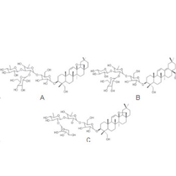
Breast cancer is a prevalent malignancy among women, especially in developing countries. A large number of anticancer agents with herbal origins have been reported. Hence, herbals may play an essential role in prevention and treatment of cancers. We investigated cytotoxic effects of dichloromethane fractions of Scrophularia oxysepala extract on the MCF-7 breast cancer cell line. The cytotoxic activity of Scrophularia oxysepala fractions on the MCF-7 cells was assessed using Trypan Blue dye exclusion and MTT (3-(4, 5-dimetylthiazol-2-yl) -2, 5-diphenyltetrazolium bromide) assays. In addition, apoptosis induction was determined using TUNEL (terminal deoxy transferase (TdT)-mediated dUTP nick- end labeling) assay and DNA fragmentation analysis. Quantitative Real-Time PCR was also used for analyzing the changes in Caspase-3, Caspase-9, and Bcl-2 genes’ expression. Results revealed an effective inhibition of growth and viability in MCF-7 cells treated with dichloromethane fractions. Cell death assay and DNA fragmentation analysis using the TUNEL test confirmed the induction of apoptosis in the MCF-7 cell line. Further, the fractions have resulted in an increased expression of Caspase-3and Caspase-9 mRNA, which highlights the possibility of apoptosis in the treatments. The expression study of Caspase-9 mRNA confirmed that, the fractions have triggered apoptosis via intrinsic mitochondrial pathway. In summary, fractions of Scrophularia oxysepala extract were found to be promising in growth inhibition and induction of apoptosis in MCF-7 breast cancer cells.

Downloads

Download data is not yet available.

References

Parkin DM, Bray F, Ferlay J, Pisani P. Estimating the world cancer burden: Globocan 2000. International journal of cancer. 2001;94(2):153-6.

http://dx.doi.org/10.1002/ijc.1440

Zhang X, Chen LX, Ouyang L, Cheng Y, Liu B. Plant natural compounds: targeting pathways of autophagy as anti‐cancer therapeutic agents. Cell proliferation. 2012;45(5):466-76.

http://dx.doi.org/10.1111/j.1365-2184.2012.00833.x

Cragg GM, Newman DJ. Plants as a source of anti-cancer agents. Journal of ethnopharmacology. 2005;100(1):72-9.

http://dx.doi.org/10.1016/j.jep.2005.05.011

Aghbali A, Hosseini SV, Delazar A, Gharavi NK, Shahneh FZ, Orangi M, et al. Induction of apoptosis by grape seed extract (Vitisvinifera) in oral squamous cell carcinoma. Bosn J Basic Med Sci 2013;13(3):186-91.

Pasdaran A, Nahar L, Asnaashari S, Sarker SD, Delazar A. GC-MS Analysis, Free-Radical-Scavenging and Insecticidal Activities of Essential Oil of Scrophularia oxysepala Boiss. 2013.

Shaw J, Lickey EB, Schilling EE, Small RL. Comparison of whole chloroplast genome sequences to choose noncoding regions for phylogenetic studies in angiosperms: the tortoise and the hare III. American Journal of Botany. 2007;94(3):275-88.

http://dx.doi.org/10.3732/ajb.94.3.275

Bhandari S, Babu U, Garg H. A triterpene glycoside from Scrophulariakoelzii. Phytochemistry. 1997;45(8):1717-9.

http://dx.doi.org/10.1016/S0031-9422(97)00256-2

Bhandari S, Roy R, Agrawal P, Garg H. Scrokoelziside A, A triterpene glycoside from Scrophulariakoelzii. Phytochemistry. 1996;41(3):879-82.

http://dx.doi.org/10.1016/0031-9422(95)00697-4

Giner RM, Villalba ML, RecioMdC, Má-ez S, Gray AI, Ríos JL. A New Iridoid from Scrophulariaauriculata ssp. p seudoauriculata. Journal of natural products. 1998;61(9):1162-3.

http://dx.doi.org/10.1021/np980067o

Li J, Huang X, Du X, Sun W, Zhang Y. Study of chemical composition and antimicrobial activity of leaves and roots of Scrophularianingpoensis. Natural product research. 2009;23(8):775-80.

http://dx.doi.org/10.1080/14786410802696247

Evan GI, Vousden KH. Proliferation, cell cycle and apoptosis in cancer. Nature. 2001;411(6835):342-8.

http://dx.doi.org/10.1038/35077213

Valiyari S. Dichloromethane and methanol extracts of Scrophularia oxysepala induces apoptosis in MCF-7 human breast cancer cells. Advanced pharmaceutical bulletin. 2012;2(2):223.

Harmon B, Corder A, Collins R, Gobe G, Allen J, Allan D, et al. Cell death induced in a murine mastocytoma by 42-47 C heating in vitro: evidence that the form of death changes from apoptosis to necrosis above a critical heat load. International journal of radiation biology. 1990;58(5):845-58.

http://dx.doi.org/10.1080/09553009014552221

Helmlinger G, Yuan F, Dellian M, Jain RK. Interstitial pH and pO2 gradients in solid tumors in vivo: high-resolution measurements reveal a lack of correlation. Nature medicine. 1997;3(2):177-82.

http://dx.doi.org/10.1038/nm0297-177

Kroemer G. The proto-oncogene Bcl-2 and its role in regulating apoptosis. Nature medicine. 1997;3(6):614-20.

http://dx.doi.org/10.1038/nm0697-614

Okamoto T, Coultas L, Metcalf D, van Delft MF, Glaser SP, Takiguchi M, et al. Enhanced stability of Mcl1, a prosurvival Bcl2 relative, blunts stress-induced apoptosis, causes male sterility, and promotes tumorigenesis. Proceedings of the National Academy of Sciences. 2014;111(1):261-6.

http://dx.doi.org/10.1073/pnas.1321259110

Alshatwi AA, Shafi G, Hasan TN, Syed NA, Khoja KK. Fenugreek induced apoptosis in breast cancer MCF-7 cells mediated independently by fas receptor change. Asian Pacific journal of cancer prevention : APJCP. 2013;14(10):5783-8. PubMed PMID: 24289578. Epub 2013/12/03. eng.

Gross A, McDonnell JM, Korsmeyer SJ. BCL-2 family members and the mitochondria in apoptosis. Genes & development. 1999;13(15):1899-911.

http://dx.doi.org/10.1101/gad.13.15.1899

Strasser A, O'Connor L, Dixit VM. Apoptosis signaling. Annual review of biochemistry. 2000;69(1):217-45.

http://dx.doi.org/10.1146/annurev.biochem.69.1.217

Deng X. Bcl2 Family Functions as Signaling Target in Nicotine-/NNK-Induced Survival of Human Lung Cancer Cells. Scientifica. 2014

Green DR, Reed JC. Mitochondria and apoptosis. Science-AAAS-Weekly Paper Edition. 1998;281(5381):1309-11.

Callagy GM, Pharoah PD, Pinder SE, Hsu FD, Nielsen TO, Ragaz J, et al. Bcl-2 is a prognostic marker in breast cancer independently of the Nottingham Prognostic Index. Clinical Cancer Research. 2006;12(8):2468-75.

http://dx.doi.org/10.1158/1078-0432.CCR-05-2719

Chang Z, Xing J, Yu X. Curcumin induces osteosarcoma MG63 cells apoptosis via ROS/Cyto-C/Caspase-3 pathway. Tumor Biology. 2014;35(1):753-8.

http://dx.doi.org/10.1007/s13277-013-1102-7

Livak KJ, Schmittgen TD. Analysis of relative gene expression data using real-time quantitative PCR and the 2(-Delta DeltaC(T)) Method. Methods. 2001;25(4):402-8. http://dx.doi.org/10.1006/meth.2001.1262

Hidding JT, Beurskens CH, van der Wees PJ, van Laarhoven HW, Nijhuis-van der Sanden MW. Treatment Related Impairments in Arm and Shoulder in Patients with Breast Cancer: A Systematic Review. PloS one. 2014;9(5):e96748.

http://dx.doi.org/10.1371/journal.pone.0096748

Liu R, Hua BJ, Li J. [Safety evaluation of Chinese medicine on tumor therapy]. ZhongguoZhongyaozazhi = China journal of Chinese materia medica. 2013 Dec;38(23):4181-4. PubMed PMID: 24791513. Epub 2014/05/06. chi.

Craig WJ. Health-promoting properties of common herbs. Am J ClinNutr. 1999 Sep;70(3 Suppl):491S-9S. PubMed PMID: 10479221. Epub 1999/09/09. eng.

Kuete V, Sandjo LP, Ouete JLN, Fouotsa H, Wiench B, Efferth T. Cytotoxicity and modes of action of three naturally occurring xanthones (8-hydroxycudraxanthone G, morusignin I and cudraxanthone I) against sensitive and multidrug-resistant cancer cell lines. Phytomedicine : international journal of phytotherapy and phytopharmacology. 2014;21(3):315-22.

http://dx.doi.org/10.1016/j.phymed.2013.08.018

Asadi H, Orangi M, Shanehbandi D, Babaloo Z, Delazar A, Mohammadnejad L, et al. Methanolic Fractions of Ornithogalumcuspidatum Induce Apoptosis in PC-3 Prostate Cancer Cell Line and WEHI-164 Fibrosarcoma Cancer Cell Line. Advanced Pharmaceutical Bulletin. 2014/12/30;4(5):455-8.

Kanchanapoom T, Noiarsa P, Ruchirawat S, Kasai R, Otsuka H. Triterpenoidal glycosides fromJusticiabetonica. Phytochemistry. 2004;65(18):2613-8.

http://dx.doi.org/10.1016/j.phytochem.2004.06.029

Bhandari S, Roy R, Agrawal P, Garg H. Scrokoelziside A, A triterpene glycoside fromScrophularia koelzii. Phytochemistry. 1996;41(3):879-82.

http://dx.doi.org/10.1016/0031-9422(95)00697-4

Trouillas P, Corbière C, Liagre B, Duroux J-L, Beneytout J-L. Structure–function relationship for saponin effects on cell cycle arrest and apoptosis in the human 1547 osteosarcoma cells: a molecular modelling approach of natural molecules structurally close to diosgenin. Bioorganic & medicinal chemistry. 2005;13(4):1141-9.

http://dx.doi.org/10.1016/j.bmc.2004.11.031

Kim S, Lee Y, Park J, Lee S. Ginsenoside-Rs 4, a new type of ginseng saponin concurrently induces apoptosis and selectively elevates protein levels of p53 and p21 WAF1 in human hepatoma SK-HEP-1 cells. European Journal of Cancer. 1999;35(3):507-11.

http://dx.doi.org/10.1016/S0959-8049(98)00415-8

Chae S, Kang KA, Chang WY, Kim MJ, Lee SJ, Lee YS, et al. Effect of compound K, a metabolite of ginseng saponin, combined with γ-ray radiation in human lung cancer cells in vitro and in vivo. Journal of agricultural and food chemistry. 2009;57(13):5777-82.

http://dx.doi.org/10.1021/jf900331g

Chwalek M, Lalun N, Bobichon H, Plé K, Voutquenne-Nazabadioko L. Structure–activity relationships of some hederagenindiglycosides: haemolysis, cytotoxicity and apoptosis induction. Biochimica et BiophysicaActa (BBA)-General Subjects. 2006;1760(9):1418-27.

http://dx.doi.org/10.1016/j.bbagen.2006.05.004

Azadmehr A, Hajiaghaee R, Mazandarani M. Induction of apoptosis and G2/M cell cycle arrest by Scrophulariastriata in a human leukaemia cell line. Cell proliferation. 2013;46(6):637-43.

http://dx.doi.org/10.1111/cpr.12074

Azadmehr A, Oghyanous KA, Hajiaghaee R, Amirghofran Z, Azadbakht M. Antioxidant and neuroprotective effects of scrophularia striata extract against oxidative stress-induced neurotoxicity. Cellular and molecular neurobiology. 2013;33(8):1135-41.

http://dx.doi.org/10.1007/s10571-013-9979-7

Valiyari S, Jahanban-Esfahlan R, Shahneh FZ, Yaripour S, Baradaran B, Delazar A. Cytotoxic and apoptotic activity of Scrophularia oxysepala in MCF-7 human breast cancer cells. Toxicological & Environmental Chemistry. 2013;95(7):1208-20.

http://dx.doi.org/10.1080/02772248.2013.854362

Thornberry NA, Lazebnik Y. Caspases: enemies within. Science. 1998;281(5381):1312-6.

http://dx.doi.org/10.1126/science.281.5381.1312

Akagi H, Higuchi H, Sumimoto H, Igarashi T, Kabashima A, Mizuguchi H, et al. Suppression of myeloid cell leukemia-1 (Mcl-1) enhances chemotherapy-associated apoptosis in gastric cancer cells. Gastric cancer. 2013;16(1):100-10.

http://dx.doi.org/10.1007/s10120-012-0153-6

Shore GC, Warr MR. Unique biology of Mcl-1: therapeutic opportunities in cancer. Current molecular medicine. 2008;8(2):138-47.

http://dx.doi.org/10.2174/156652408783769580

Karami H, Baradaran B, Esfahani A, Sakhinia M, Sakhinia E. Therapeutic Effects of Myeloid Cell Leukemia-1 siRNA on Human Acute Myeloid Leukemia Cells. Advanced pharmaceutical bulletin. 2014;4(3):243.

Adams JM, Cory S. The Bcl-2 protein family: arbiters of cell survival. Science. 1998;281(5381):1322-6.

http://dx.doi.org/10.1126/science.281.5381.1322

Karami H, Baradaran B, Esfahani A, Estiar MA, Naghavi-Behzad M, Sakhinia M, et al. siRNA-Mediated silencing of survivin inhibits proliferation and enhances etoposidechemosensitivity in acute myeloid leukemia cells. Asian Pac J Cancer Prev. 2013;14:7719-24.

http://dx.doi.org/10.7314/APJCP.2013.14.12.7719

Giessrigl B, Yazici G, Teichmann M, Kopf S, Ghassemi S, Atanasov AG, et al. Effects of Scrophularia extracts on tumor cell proliferation, death and intravasation through lymphoendothelial cell barriers. Int J Oncol. 2012;40(6):2063-74.

Dichloromethane fractions of Scrophularia oxysepala extract induce apoptosis in MCF-7 human breast cancer cells

Downloads

Additional Files

Published

2015-01-29

How to Cite

1.
Hosseini B-A, Pasdaran A, Kazemi T, Shanehbandi D, Karami H, Orangi M, Baradaran B. Dichloromethane fractions of Scrophularia oxysepala extract induce apoptosis in MCF-7 human breast cancer cells. Biomol Biomed [Internet]. 2015Jan.29 [cited 2023Sep.23];15(1):26-32. Available from: https://www.bjbms.org/ojs/index.php/bjbms/article/view/226

Issue

Section

Molecular Biology

INTRODUCTION

Breast cancer is one of the most prevalent types of cancer among women. Thus, searching for an effective treatment for this disease is a priority [1]. Using herbals for the treatment of malignancies is common in many cultures, because some herbals contain abundant anti-cancer compounds. In addition, useful compounds from these herbals are being used in the production of various modern drugs [2-4].

Scrophulariaceae is a family of flowering plants containing 3000 species and 280 genera that are widely distributed around the world, especially, in Asia and North America [5, 6]. Effects of S. oxysepala on cell death mechanisms have been assessed in previous studies. S. oxysepala fractions comprise ingredients such as verbascosaponin and scrokoelziside (Figure 1) [7-10]. Some herbal compounds are effective in the treatment of cancer by different mechanisms including apoptosis[11, 12].

FIGURE 1: Three saponins isolated from S. oxysepala DCM fractions A: verbascosaponin B: scrokoelziside and C: isolated from these fractions

Apoptosis is a biological process that maintains homeostasis in living beings without causing inflammation [13, 14]. Caspases are cysteine proteases that play a significant role in primary stages of executive phase of apoptosis. During the detection of Caspases, it was specified that Caspase-3 typically is activated by many death signals [15, 16]. Basically, there are two main signaling pathways for cellular apoptosis: a) the mitochondrial or intrinsic pathway that responds to intracellular stimuli and results in cytochrome c release from the mitochondria leading to the activation of Caspase-9; and b) the extrinsic death receptor pathway initiated by ligand binding to extracellular cell death receptors resulting in caspase-8 activation. Both pathways converge at downstream activation of caspase-3[17].

Caspase activation influences specific substrates, causing biochemical and morphological changes in the cells such as cell shrinkage, condensation of chromatin, and cleavage of DNA [18-20]. Thus, the caspase activity can be a biochemical marker for apoptosis. On the other hand, Bcl-2 is an anti-apoptosis protein playing an inhibitory role in apoptosis [21-23]. In the present study, we assessed the cytotoxic effects of dichloromethane (DCM) fractions of S. oxysepala extract on MCF-7 human breast cancer cells. In addition, the effect of fraction on the expression levels of Caspase-3 and Bcl-2 genes, as the main indicators of apoptosis, were evaluated. To determine whether the apoptosis was conducted via intrinsic or extrinsic pathway, changes in the expression of Caspase-9 mRNA was analyzed by quantitative real-time PCR.

MATERIALS AND METHODS

Preparation of fractions

S. oxysepala was collected from a 30 kilometer area in Kaleybar, Garehdagh Mountain during the flowering period. A voucher specimen (2821) was deposited at the Herbarium of the Researches Center for Agriculture and Natural Resources, East Azerbaijan, Iran.

Air-dried and powdered aerial parts of S. oxysepala (1800gr) were extracted with n-hexane, dichloromethane and methanol using a Soxhlet apparatus. All extracts were concentrated using a rotary evaporator at 45 ºC (Heildolph, Germany) under reduced pressure to obtain a powder or a viscous mass. Five grams of dichloromethane extract were dissolved in the minimum possible amount of methanol and loaded and fractionated in a Sephadex-LH20 column using an isocratic (CH2Cl2-MeOH, 1:1) elution. This method yielded fifteen fractions (F). These fractions were intermingled based on pattern similarities resulting from thin layer chromatography using a chloroform system (7:3). From 13 fractions, F3, F4, F6, and F13 fractions were monitored and assessed for their effects and compounds.

Cell culture

A human breast cancer cell line (MCF-7) and a mouse normal control cell line (L929) were purchased from the Iranian National Cell Bank (Pasteur Institute, Tehran, Iran). Cells were cultured in RPMI-1640 culture medium containing 10% fetal bovine serum (FBS), penicillin (100U/ml) and streptomycin (100 µg/ml) (all purchased from Sigma, Germany) and then incubated at 37 ºC at 95% humidity and 5 % CO2.

Cell viability assay

3-(4,5-Dimethylthiazol-2-yl)-2, 5-Diphenyltetrazolium Bromide (MTT) (Sigma, Germany) assay is based on tetrazolium salt breakage by mitochondrial succinate dehydrogenase within living cells. This results in the production of purple formazan crystals becoming dissolved by dimethyl sulfoxide (DMSO). Briefly, cancer cells as well as L929 cell (as a normal control cell) were cultured in 96 well plates and treated with various concentrations (0-300 µg/ml) of dichloromethane fractions for 24 h and 36 h. Untreated cells were also used as the control group. After incubations, MTT solution with a concentration of 5 mg/ml was added to the each well. After incubation in 37 ºC, 200 µl of DMSO and Sorenson buffer was added to each well. Thirty minute after incubation at room temperature, absorbance at 570 nm was measured using an ELISA reader (Awareness Technology, USA).

TUNEL assay

The TUNEL (Terminal deoxynucleotidyl transferase dUTP nick end labeling) (Roche, Germany) assay is based on the activity of TdT (Terminal deoxynucleotidyl transferase) (Roche, Germany) enzyme that adds probed nucleotides to the ends of broken DNA strands. Probed nucleotides bind to the free 3’-hydroxyl end of double- or single-stranded DNA. After 24 hours of treatment with fractions, cells were fixed using 4% paraformaldehyde diluted in PBS (pH, 7.4). After twice washing with PBS, cells were incubated in a solution containing 3% H2O2 in methanol for 10 minutes and then washed again with PBS. Next, cells were incubated in permeabilization solution on ice for 10 minutes. Next, 50 µl of the reaction mixture containing the TdT enzyme and nucleotides was added to the cells and incubated at 37°C for 1 hour. After washing with PBS three times, slides were incubated with 50 µl of POD (Roche, Germany) streptavidin HRP solution for 30 minutes. After another wash, the cells were incubated with DAB (Sigma, Germany) substrate and stained cells were then analyzed using a light microscope.

Cell death ELISA

In order to determine the type of the cell death induced by fractions, an ELISA kit (Roche Diagnostic GmbH, Germany) was used. First, MCF-7 and L929 cells were cultured in separate flasks. Cells (1×10⁴ cells/well) were then treated with DCM fractions of S. oxysepala in 75 µg/ml concentrations (IC50). After 24 hours, cells were lysed, and cell lysates were used for apoptosis assessment. Next, 80 µl of immune-reagent solution was added to wells and incubated for 2 hours at room temperature. 100 µl of stop solution was added to each well and absorbances at 405 nm were measured using the ELISA reader.

DNA fragmentation

DNA fragmentation is one of the most significant biochemical markers for apoptosis. Cells with a density of 7×10⁵ cells/well were exposed to a fractions extract of S. oxysepala. After 24 hours, DNA was extracted using the ­proteinase K method and sedimentation was done using cold isopropanol. Agarose gel (1.8%) electrophoresis and safe stain™ (Cinnagen, Iran) were used to visualize bands under UV light.

Quantitative real time-PCR

The total RNA was isolated using an RNX PLUS kit (Cinnagen, Iran) according to the manufacturer’s instructions. The quality and quantity of isolated RNA were assessed using a NANODROP 2000c spectrophotometer (Thermo Scientific, USA). The RNA was then transcribed to cDNA using a reverse transcriptase kit (Promega, USA). Quantitative RT-PCR (qRT-PCR) was performed using the Corbett Rotor-Gene 6000 system (Corbett Life Science, Australia). PCR was performed in a 20 µl reaction system containing 0.2 µM of each primer, 10 µl of SYBR green reagent (RR820L Takara Bio, Japan), 1 µl of cDNA template and 8.6 µl of nuclease-free water. The primer sequences were as follows: forward: 5’-TCCCTGGAGAAGAGCTACG-3’ and reverse: 5’-GTAGTTTCGTGGATGCCACA-3’, for β–actin, forward: 5’-ATGGTTTGAGCCTGAGCAGA-3’ and reverse: 5’-GGCAGCATCATCCACACATAC-3’ for Caspase-3, forward: GCAGGCTCTGGATCTCGGC and reverse: GCTGCTTGCCTGTTAGTTCGC for Caspase-9 and forward: 5’-CCTGGGATGACTGAGTACC-3’, and reverse: 5’-GGCAGCATCATCCACACATAC-3’ for Bcl-2. The PCR cycling was performed by initial denaturation step at 95 °C for 3 min followed by 45 cycles at 95°C for 10 seconds, 58°C for 30 seconds and 72°C for 20 seconds. Relative mRNA expression was measured using the 2 - (∆∆CT) method, using β-actin as a reference gene[24].

Statistical analysis

All experiments were assayed in triplicate (n = 3). Data are expressed as means ± S.D. Analysis of variance (ANOVA) followed by a two-tailed unpaired t-test was used to determine the significant differences between groups. The differences were considered to be statistically significant at p<0.05. All statistical analyzes were performed using GraphPad Prism 6.01 software (GraphPad Software Inc., San Diego, CA, USA).

RESULTS

Effects of the DCM fractions of S. oxysepala extract on cell viability

Cytotoxic effects of DCM fractions of S. oxysepala extract on cell viability were assessed using an MTT assay. As shown in Figures 2 and 3, all four DCM fractions (F3, F4, F6, and F13) decreased the viability of the both MCF-7 and L929 cells in a dose-dependent manner (relative to the blank control) after 24 and 36 hours. The F4 and F13 fractions were more cytotoxic compared to the F3 and F6 fractions (p<0.05). Therefore, we used F4 and F13fractions in the next experiments. Table 1 shows the IC50 (50% inhibitory concentrations) values of the DCM fractions on the cells. The F4 and F13 fractions had a lower IC50 values in comparison to the F3 and F6 fractions. In addition, the cytotoxic effects of DCM fractions of S. oxysepala extract on the MCF-7 cells were significantly higher than on the L929 cells (p<0.05).

FIGURE 2: Effects of S. oxysepala dichloromethane fractions on viability of MCF-7 cells, (A) (24) hr and (B) 36 hr. Values are presented as means (n = 3) ± S.E. *significant statistical difference (p < 0.05) relative to the blank control.
FIGURE 3: Effects of dichloromethane fractions of S. oxysepala on L929 cell line viability. Cells for (A) 24 hr and (B) 36 hr. Data are presented as means (n = 3) ± S.E. *sp< 0.05 normalized the control.
TABLE 1: (IC50) concentrations of DCM fractions of S. oxysepala for L929 and MCF-7 cell lines after 24 and 36 hr incubations.

Apoptotic effects of the DCM fractions of S. oxysepala

Apoptosis was monitored using TUNEL, DNA fragmentation, and ELISA cell death assays. In addition, expression levels of Caspase-3 and Bcl-2 apoptotic genes were measured using qRT-PCR.

DNA damage is a sign of apoptosis. First, apoptosis was evaluated using a TUNEL test. As shown in Figure 4, cell nuclei became brown after 24 hours of treatment with IC50 of the F13 fraction, while this did not occur in control cells. Moreover, treatment with all DCM fractions resulted in marked DNA fragmentation in the MCF-7 cells, although not in L929 cells, which revealed the specific apoptotic effect of the extract on tumor cells (Figure 5).

FIGURE 4: Apoptotic effects of DCM (fraction F4) of S. oxysepala on MCF-7 cells observed by TUNEL assay. (A) L929 control, (B) MCF-7 control, (C) treated L929 and (D) treated MCF-7. Arrows indicate representative apoptotic cells. A-D: 200× magnification.
FIGURE 5: DNA fragmentation assay in cells exposed to IC50 concentration (75 µg/mL)of S. oxysepala for 24 hr. (A) L929 Normal cells (left to right: Control, F3, F4, F6, F13 fractions) AND (B) MCF-7 breast cancer cell line (left to right: Control, F3, F4, F6, F13 fractions), M= size marker. DNA fragments were separated on 1.8% agarose gel electrophoresis.

Next, cell death ELISA assay was used to determine the type of cell death. Twenty-four hours after treatment, the cells were lysed and then cell lysates were assessed for the presence of DNA-nucleosome fragments. Results showed that a 24-hour exposure to F4 and F13 fractions led to 3.5- and 2.6-fold increases, respectively, in apoptosis in comparison with blank controls (p<0.05). However, DCM fractions had a less apoptotic effect on the L929 normal cells compared to the MCF-7 tumor cells (Figure 6).

FIGURE 6: Effects of dichloromethane fractions of S. oxysepala (IC50 = 75 µg/mL) on apoptosis of MCF-7 and L929 cells after 24 hr detected by cell death ELISA assay. Data are represented as the fold increase in apoptosis and expressed as means ± S.E. ­*signified (p < 0.05) relative to the control.

To further confirm the apoptotic effects of DCM fractions of S. oxysepala on the cells, the expression levels of Caspase-3 and Bcl-2 genes were examined. After a 24-hour treatment with IC50 concentrations of DCM fractions, relative mRNA expression was quantified using qRT-PCR.

Data showed that treatment of MCF-7 cells with F4 and F13 fractions caused 11.26 and 2.83fold increases in the expression level of Caspase-3 mRNA, respectively, while the expression level of Bcl-2 mRNA was increased, respectively, by 4.16- and 1.62-fold (compared to blank controls). However, in 24 hour exposures of the L929 cells with F4 and F13 fractions, the increase in expression levels of Caspase-3 mRNA were 1.12 and 1.33 fold, respectively, and the expression levels of Bcl-2 mRNA were 1.15 and 2.05, respectively, relative to blank controls (Figure 7).

FIGURE 7: Effects of S. oxysepala DCM fractions on (A) Caspase-3,(B) Caspase-9 and (C) Bcl-2 mRNA expression in MCF-7 and L929 cells at 24 hr. Relative expression was acquired by qRT-PCR using 2(-ΔΔCt) method. The results are presented as mean ± SD (n = 3); *p<0.05 versus control.

DISCUSSION

Breast cancer is one of the most frequent types of cancer among women all over the world [25]. Because of its high mortality and morbidity, there is a great deal of interest in finding an appropriate medication for the treatment of breast cancer. Among these treatments, medicinal plants are considered to be important sources of therapeutic substances and a variety of plants have been shown to contain bioactive and anti-cancer compounds [26-29]. For example, different species of the Scrophularia genus have bacteriostatic and anti-inflammatory properties [30, 31]. According to the literature, some Scrophularia spices contain high quantities of saponins[32-35], olean, epoxy olean, dammaraneand terpenoids[27, 28]. Many chemicals such as phenol glycosides and some phenilicacids extracted from Scrophularia species have significant cytotoxicity and cytostaticity. In a recent study, the inhibitory impacts of S. striata extract on leukemic cells by arresting G2/M phase and apoptosis has been reported[36, 37].

In our previous study, we assessed the anti-proliferative and cytotoxic effects of three S. oxysepala extracts (n-hexane, DCM, and methanol) on the MCF-7 breast cancer cell line. While the n-hexane extract did not exhibit any cytotoxic impacts, the DCM and methanol extracts significantly inhibited cancerous cells in a dose and time-dependent manner. At the same time, these had no significant adverse effects on human normal cells (HUVEC)[12, 38]. In the present study, we have further investigated the active components of the S. oxysepala DCM extract. For this purpose, we prepared four fractions of this extract: F3, F4, F6, and F13. We found that F4 and F13 fractions indicated more cytotoxic effects with no significant inhibitory effects on L929 cells, and nontoxic effects of these fractions on murine normal cells (L929) could be promising for potential use in future in vivo studies.

Besides the morphologic characteristics of the cells treated with these fractions, DNA fragmentation confirmed the occurrence of apoptosis. In addition, results of cell death ELISA showed that the fractions above have induced apoptosis in MCF-7 cells. Apoptosis, as the most acceptable form of cell death, depends on a Caspase proteolytic system. In other words, Caspase family members are required for the initiation and execution of apoptosis[39]. Caspase-3 is a major downstream effector in apoptosis and plays a crucial role in DNA fragmentation and chromatin condensation. Meanwhile, anti-apoptotic proteins such as Bcl-2 play important inhibitory roles in apoptotic processes and are shown to increase cell viability [40-44].

Results of quantitative RT-PCR indicated that S. oxysepala DCM fractions resulted in increased expression of Caspase-3 mRNA. Moreover, L929 normal cells showed no significant changes in Caspase gene expression patterns. In malignant cells, a lower rate of increase in Bcl-2 gene expression was evident 24 hours after exposure to the fractions. Giessrigl et al. also reported antineoplastic effects of Scrophularia components. They concluded that growth inhibition and cell death was due to the activation of Caspase-3 and down-regulation of cell cycle regulators, proto-oncogenes and NF-κB gene expression [45].

Results of an MTT assay indicated that an increased dose of fractions led to more intense cytotoxic effects. Nevertheless, major cytotoxic effects were detected in 24 hours rather than 36 hours, and extended time of incubation resulted in an increase in IC50 value. Consequently, it can be concluded that there is a half-life for the mentioned fractions, and repeated doses, at 24-hour intervals, are needed to re-establish the effects.

CONCLUSION

The present study demonstrates that, dichloromethane fractions of Scrophularia oxysepala induce apoptosis in MCF-7 breast cancer cells in a dose-dependent manner via mitochondrial intrinsic pathway.

DECLARATION OF INTERESTS

The authors declare no conflict of interest.

REFERENCES

  1. , , , (). Estimating the world cancer burden: Globocan 2000. International journal of cancer. http://dx.doi.org/10.1002/ijc.1440
  2. , , , , (). Plant natural compounds: targeting pathways of autophagy as anti-cancer therapeutic agents. Cell proliferation. http://dx.doi.org/10.1111/j.1365-2184.2012.00833.x
  3. , (). Plants as a source of anti-cancer agents. Journal of ethnopharmacology. http://dx.doi.org/10.1016/j.jep.2005.05.011
  4. , , , , , (). Induction of apoptosis by grape seed extract (Vitisvinifera) in oral squamous cell carcinoma. Bosn J Basic Med Sci.
  5. , , , , (). GC-MS Analysis, Free-Radical-Scavenging and Insecticidal Activities of Essential Oil of Scrophularia oxysepala Boiss.
  6. , , , (). Comparison of whole chloroplast genome sequences to choose noncoding regions for phylogenetic studies in angiosperms: the tortoise and the hare III. American Journal of Botany. http://dx.doi.org/10.3732/ajb.94.3.275
  7. , , (). A triterpene glycoside from Scrophulariakoelzii. Phytochemistry. http://dx.doi.org/10.1016/S0031-9422(97)00256-2
  8. , , , (). Scrokoelziside A, A triterpene glycoside from Scrophulariakoelzii. Phytochemistry. http://dx.doi.org/10.1016/0031-9422(95)00697-4
  9. , , , , , (). A New Iridoid from Scrophulariaauriculata ssp. p seudoauriculata. Journal of natural products. http://dx.doi.org/10.1021/np980067o
  10. , , , , (). Study of chemical composition and antimicrobial activity of leaves and roots of Scrophularianingpoensis. Natural product research. http://dx.doi.org/10.1080/14786410802696247
  11. , (). Proliferation, cell cycle and apoptosis in cancer. Nature. http://dx.doi.org/10.1038/35077213
  12. (). Dichloromethane and methanol extracts of Scrophularia oxysepala induces apoptosis in MCF-7 human breast cancer cells. Advanced pharmaceutical bulletin.
  13. , , , , , (). Cell death induced in a murine mastocytoma by 42-47 C heating in vitro: evidence that the form of death changes from apoptosis to necrosis above a critical heat load. International journal of radiation biology. http://dx.doi.org/10.1080/09553009014552221
  14. , , , (). Interstitial pH and pO2 gradients in solid tumors in vivo: high-resolution measurements reveal a lack of correlation. Nature medicine. http://dx.doi.org/10.1038/nm0297-177
  15. (). The proto-oncogene Bcl-2 and its role in regulating apoptosis. Nature medicine. http://dx.doi.org/10.1038/nm0697-614
  16. , , , , , (). Enhanced stability of Mcl1, a prosurvival Bcl2 relative, blunts stress-induced apoptosis, causes male sterility, and promotes tumorigenesis. Proceedings of the National Academy of Sciences. http://dx.doi.org/10.1073/pnas.1321259110
  17. , , , , (). Fenugreek induced apoptosis in breast cancer MCF-7 cells mediated independently by fas receptor change. Asian Pacific journal of cancer prevention: APJCP. PubMed PMID: 24289578. Epub: 2013/12/03.eng
  18. , , (). BCL-2 family members and the mitochondria in apoptosis. Genes & development. http://dx.doi.org/10.1101/gad.13.15.1899
  19. , , (). Apoptosis signaling. Annual review of biochemistry. http://dx.doi.org/10.1146/annurev.biochem.69.1.217
  20. (). Bcl2 Family Functions as Signaling Target in Nicotine-/NNK-Induced Survival of Human Lung Cancer Cells. Scientifica.
  21. , (). Mitochondria and apoptosis. Science-AAAS-Weekly Paper Edition.
  22. , , , , , (). Bcl-2 is a prognostic marker in breast cancer independently of the Nottingham Prognostic Index. Clinical Cancer Research. http://dx.doi.org/10.1158/1078-0432.CCR-05-2719
  23. , , (). Curcumin induces osteosarcoma MG63 cells apoptosis via ROS/Cyto-C/Caspase-3 pathway. Tumor Biology. http://dx.doi.org/10.1007/s13277-013-1102-7
  24. , (). Analysis of relative gene expression data using real-time quantitative PCR and the 2(-Delta DeltaC(T)) Method. Methods. http://dx.doi.org/10.1006/meth.2001.1262
  25. , , , , (). Treatment Related Impairments in Arm and Shoulder in Patients with Breast Cancer: A Systematic Review. PloS one. http://dx.doi.org/10.1371/journal.pone.0096748
  26. , , (). [Safety evaluation of Chinese medicine on tumor therapy]. ZhongguoZhongyaozazhi =China journal of Chinese materia medica. PubMed PMID: 24791513. Epub: 2014/05/06.chi
  27. (). Health-promoting properties of common herbs. Am J ClinNutr. PubMed PMID: 10479221. Epub 1999/09/09.eng
  28. , , , , , (). Cytotoxicity and modes of action of three naturally occurring xanthones (8-hydroxycudraxanthone G, morusignin I and cudraxanthone I) against sensitive and multidrug-resistant cancer cell lines. Phytomedicine: international journal of phytotherapy and phytopharmacology. http://dx.doi.org/10.1016/j.phymed.2013.08.018
  29. , , , , , (). Methanolic Fractions of Ornithogalumcuspidatum Induce Apoptosis in PC-3 Prostate Cancer Cell Line and WEHI-164 Fibrosarcoma Cancer Cell Line. Advanced Pharmaceutical Bulletin 2014/12/30.
  30. , , , , (). Triterpenoidal glycosides from<i>Justiciabetonica</i>. Phytochemistry. http://dx.doi.org/10.1016/j.phytochem.2004.06.029
  31. , , , (). Scrokoelziside A, A triterpene glycoside from<i>Scrophularia koelzii</i>. Phytochemistry. http://dx.doi.org/10.1016/0031-9422(95)00697-4
  32. , , , , (). Structure–function relationship for saponin effects on cell cycle arrest and apoptosis in the human 1547 osteosarcoma cells: a molecular modelling approach of natural molecules structurally close to diosgenin. Bioorganic & medicinal chemistry. http://dx.doi.org/10.1016/j.bmc.2004.11.031
  33. , , , (). Ginsenoside-Rs 4, a new type of ginseng saponin concurrently induces apoptosis and selectively elevates protein levels of p53 and p21 WAF1 in human hepatoma SK-HEP-1 cells. European Journal of Cancer. http://dx.doi.org/10.1016/S0959-8049(98)00415-8
  34. , , , , , (). Effect of compound K, a metabolite of ginseng saponin, combined with γ-ray radiation in human lung cancer cells in vitro and in vivo. Journal of agricultural and food chemistry. http://dx.doi.org/10.1021/jf900331g
  35. , , , , (). Structure–activity relationships of some hederagenindiglycosides: haemolysis, cytotoxicity and apoptosis induction. Biochimica et BiophysicaActa (BBA)-General Subjects. http://dx.doi.org/10.1016/j.bbagen.2006.05.004
  36. , , (). Induction of apoptosis and G2/M cell cycle arrest by Scrophulariastriata in a human leukaemia cell line. Cell proliferation. http://dx.doi.org/10.1111/cpr.12074
  37. , , , , (). Antioxidant and neuroprotective effects of scrophularia striata extract against oxidative stress-induced neurotoxicity. Cellular and molecular neurobiology. http://dx.doi.org/10.1007/s10571-013-9979-7
  38. , , , , , (). Cytotoxic and apoptotic activity of Scrophularia oxysepala in MCF-7 human breast cancer cells. Toxicological & Environmental Chemistry. http://dx.doi.org/10.1080/02772248.2013.854362
  39. , (). Caspases: enemies within. Science. http://dx.doi.org/10.1126/science.281.5381.1312
  40. , , , , , (). Suppression of myeloid cell leukemia-1 (Mcl-1) enhances chemotherapy-associated apoptosis in gastric cancer cells. Gastric cancer. http://dx.doi.org/10.1007/s10120-012-0153-6
  41. , (). Unique biology of Mcl-1: therapeutic opportunities in cancer. Current molecular medicine. http://dx.doi.org/10.2174/156652408783769580
  42. , , , , (). Therapeutic Effects of Myeloid Cell Leukemia-1 siRNA on Human Acute Myeloid Leukemia Cells. Advanced pharmaceutical bulletin.
  43. , (). The Bcl-2 protein family: arbiters of cell survival. Science. http://dx.doi.org/10.1126/science.281.5381.1322
  44. , , , , , (). siRNA-Mediated silencing of survivin inhibits proliferation and enhances etoposidechemosensitivity in acute myeloid leukemia cells. Asian Pac J Cancer Prev. http://dx.doi.org/10.7314/APJCP.2013.14.12.7719
  45. , , , , , (). Effects of Scrophularia extracts on tumor cell proliferation, death and intravasation through lymphoendothelial cell barriers. Int J Oncol.