The osteoplastic effectiveness of the implants made of mesh titanium nickelide constructs

Authors

  • Iurii Mikhailovich Irianov Laboratory of Morphology, Russian Ilizarov Scientific Center Restorative Traumatology and Orthopaedics (RISC RTO)
  • Olga Vladimirovna Diuriagina Laboratory of Purulent Osteology and Limb Defect Filling, RISC RTO
  • Tatiana Iurevna Karaseva Department of Orthopaedics, Russian Ilizarov Scientific Center Restorative Traumatology and Orthopaedics (RISC RTO)
  • Evgenii Anatolevich Karasev Department of Orthopaedics, Russian Ilizarov Scientific Center Restorative Traumatology and Orthopaedics (RISC RTO)

DOI:

https://doi.org/10.17305/bjbms.2014.2284

Keywords:

implant, titanium nickelide, bone defect, reparative osteogenesis

Abstract

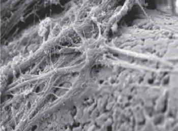
The purpose of the work was to study the features of reparative osteogenesis for filling the defect of tubular bone under implantation of meshtitanium nickelide constructs. Tibial fenestrated defect was modeled experimentally in 30 Wistar pubertal rats, followed by implant intramedullary insertion. The techniques of radiography, scanning electron microscopy and X-ray electron probe microanalysis were used. The mesh implant of titanium nickelide has been established to possess biocompatibility, osteoconductive and osteoinductive properties, the zone of osteogenesis and angiogenesis is created around it, bone cover is formed. Osteointegration of the implant occurs early, by 7 days after surgery, and by 30 days after surgery organotypical re-modelling of the regenerated bone takes place, as well as the defect is filled with lamellar bone tissue by the type of bone wound primary adhesion. By 30 days after surgery mineral content of the regenerated bone tissue approximates to the composition of intact cortex mineral phase.

Downloads

Download data is not yet available.
The osteoplastic effectiveness of the implants made of mesh titanium nickelide constructs

Downloads

Additional Files

Published

2014-05-20

How to Cite

1.
Mikhailovich Irianov I, Vladimirovna Diuriagina O, Iurevna Karaseva T, Anatolevich Karasev E. The osteoplastic effectiveness of the implants made of mesh titanium nickelide constructs. Biomol Biomed [Internet]. 2014May20 [cited 2023Mar.28];14(1):4-7. Available from: https://www.bjbms.org/ojs/index.php/bjbms/article/view/2284

Issue

Section

Biomaterials

INTRODUCTION

The development and experimental-and-clinical substantiation of implantation technologies for tissue restoration in the defect area is one of the most important trends of modern medicine [1, 2, 3, 4]. At present, intense development of medical technologies takes place – those technologies related to using the implants of titanium nickelide made in the form of mesh scaffolds with bioactive nano-structured surface [5]. Dense fibrous connective tissue filling the defect of abdominal wall muscular-and-aponeurotic layer was demonstrated to be formed during implantation [1]. Such constructs were not used for bone defect filling except our works, and the implants as an entire block of titanium nickelide were shown to have little osteoplastic effect [6]. The purpose of the work – to study the features of reparative osteogenesis for tubular bone defect filling by implanting mesh constructs of titanium nickelide.

MATERIAL AND METHODS

Animals

Experiments were performed on 30 Wistar pubertal rats, males and females, with body weight of 0.23-0.24 kg, in compliance with European Convention for the Protection of Vertebrate Animals (Strasbourg, 1986), they were approved by the Ethics Committee of RISC RTO.

Procedures

Defect of 3x4-mm extension was modeled in the proximal third of tibial shaft of animals by milling at low speed under general anesthesia (Rometar8 mg/kg and Zoletil 4 mg/kg IM). A sterile implant designed and manufactured at RISCRTO was placed into the zone of the defect immediately after surgery [7]. The implant presented fine-profile mesh constructs made of 90-μm thickness thread (mesh size 200-250 μm) which were spirally twisted and fastened in the form of coupling on titanium nickelide rod of 2-mm thickness, the end flanges of which were fixed in the medullary canal. The thread presented composite material including the core of nanostructured solid titanium nickelide, and the titanium oxide porous surface layer of 5-7-μm thickness. 7, 14, and 30 day safter surgery the animals were withdrawn from the experiment by intracardiac injection of 1 mL 10% Novocain solution. The operated bones were fixed in the mixture of 2% paraformaldehyde and glutaraldehyde solution, and 0.1% picric acid solution on 0.1 Mphosphate buffer at pH 7.4, after that they were embedded in araldit. The surface of araldite blocks was investigated using INCA-200 Energy x-ray electron probe microanalyser (OXFORDINSTRUMENTS, England) in calcium characteristic x-ray emission in order to reveal mineralized matrix and the evidences of osteogenic differentiation of the regenerate bone cells. Calcium and phosphorus content was determined in newly formed bone tissue. The block surface was pickled in 2 % sodium ethiolate solution. The obtained preparations were spray-coated with Platina and Palladium alloy using IB6 ion-and-vacuum sprayer (Eiko, Japan) at 6-mA ion current and 1.5-kV inter-electrode voltage. The objects were studied with JSM-840 scanning electron microscope (JEOL, Japan) in the mode of secondary electron registration at 20-kV accelerating voltage. Video image capturing and processing were made using INCA-200 Energy hardware-software system (OXFORD INSTRUMENTS, England).

RESULTS

The microrelief of the implant titanium nickelide thread surface has been established to be characterized by sharply marked roughness and nanostructuring, as well as by the presence of multiple macro- and micropores of irregular shape and different size, and some of them are of size below 100 nm (Figure 1a). Close contact of the regenerated bone tissue with the implant thread surface, and formation of osteointegrative connection without fibrous capsule forming is observed 7 days after surgery (Figure 1, b). Multiple trabeculae of newly formed bone tissue forming dense aggregations are arranged round the implant designs. Adhesion of capillary buds and perivascular osteogenic cells forming the layers of osteoid tissue is noted on the implant surface. The development of osteogenic differentiation of the perivascular cells on the implant surface and around the implant is proved due to specific calcified matrix formation by them (Figure 1, b). Thus, reparative osteogenesis occurs by the type of direct intramembranous osteogenesis and spreads throughout the defect volume. A network of thin collagen fibers oriented predominantly in the longitudinal direction with respect to the implant structures is detected in osteointegration zone osteoid, and large functionally active osteoblasts are localized secreting collagen and proteoglycans of bone matrix, as evidenced by pericellular fibrillogenesis near their surface (Figure 1, a). Osteoblasts are attached to the implant structures with focal contacts of cytoplasmic processes branching at the ends and forming specialized fixing structures. The presence of matrix vesicles – initial centers of mineralization, in the zone of interaction with the implant surface is a specific sign of calcifying activity of osteoblasts. The value of Ca/P coefficient in osteointegration zone is 1.33±0.05, and that in the regenerate bone tissue – 1.59±0.07. Zones of active apposition osteogenesis are found around the implant and on its surface 14 days after surgery (Figure 2, a). At this stage of the experiment intense neoangiogenesis is observed in the regenerated bone, as well as the phase of organogenesis and bone tissue remodeling, as evidenced by the reorganization of rough-fibred bone tissue trabeculae into primary osteon structures (Figure 2, b). The defect zone is partly filled with newly formed lamellar bone tissue having the signs of osteoclast resorption. Blood vessels of the microcirculatory bed, osteogeniccells, and bone structures are not only adjacent to the implant surface, but they grow into it, allowing the implant to acquire its osteoinductive and osteogenic properties. Tissue-specific regenerated bone growing deeply into the implant mesh constructs is formed in bone defect 30 days after surgery (Figure 3, a). The implant due to this is filled with vessels and perivascular osteogenic cells which form bone tissue by the type of interstitial (internal) osteogenesis. Primary osteons of rough-fibred bone tissue are replaced by organotypic secondary osteons of lamellar bone tissue.

FIGURE 1: Osteointegration of the implant in the tibial defect zone 7 days after surgery: a – adhesion of osteogenic cells on the thread surface, scanning electron microscopy; b – a bone cover around the implant threads, a map of electron probe microanalysis, image in calcium characteristic x-ray emission. Magnification: a – x 3700, b – x 310.
FIGURE 2: The regenerated bone in the zone of tibial defect 14 days after surgery: a – osteoblasts and afine-fibredosteoid on the implant surface; b – osteons of lamellar bone tissue being formed within the implant, scanning electron microscopy. Magnification: a – × 2700, b – × 400.

Osteocyte lacunae of specific structure are often found in the regenerated bone, and one of their walls is the surface of the implant elements. Empty osteocytelacunae are few. Lamellar bone tissue replaces the zone of defect over its greater extent (Figure 3, b), and this occurs by the type of primary adhesion of bone wounds. In this period of the experiment calcium and phosphorus content in the regenerated bone tissue is 20.3±1.1 % and 10.5±0.5 %, respectively.

FIGURE 3: Tibial defect filling by 30 days after implantation: a – lamellar bone tissue in the zone of cortical defect; b – bundles of collagen fibers of dense fibrous connective tissue in the zone of periosteal defect, scanning electron microscopy. Magnification: a – × 210, b – × 3500.

DISCUSSION

The mechanical properties of alloys based on nickel and titanium are known to approximate to the mechanical characteristics of bone tissue and possess biocompatibility [1, 5]. The studies performed by us couldn’t reveal the signs of forming inflammation foci pathomorphology of which was described before [8]. The implant titanium nickelide thr eadshave been established to have roughness, nanostructuring and high porosity of their surface. A layer of titanium oxide located on the thread surface prevents metal diffusion and provides adhesive properties and the most favorable conditions for functioning of perivascular osteogenic cells [1, 5]. Regenerated bone tissue grows into the implant three-dimensional structure, contributes to expression of osteogenic factors and to osteogenic differentiation of cells, and to mass accumulation of mineralized matrix. This activates osteogenesis in the pre-implantation zone and contributes to osteointegration of the implant in early periods. Osteoinductive properties of the implants specified by the presence of bone morphogenetic proteins and osteogenic growth factors [9] are known to be of them a in importance for the implant successive use, as well as its osteoconductive properties providing directed growing into the defect zone of blood vessels, surrounding them perivascular osteogenic cells and newly formed bone tissue [10]. The performed studies have demonstrated that a zone of active apposition osteogenesisis formed around the implantand within it, and a bone cover is produced having the properties of osteogenesis conductor and inductor which provides directed growth of bone tissue, prolonged stimulation of angiogenesis and reparative osteogenesis. Defect healing occurs early according to the primary type without cartilaginous and connective tissue formation in regenerated bone. Quantitative parameters of mineralization in the zone of osteointegration evidence that Ca/P coefficient value is less than that in crystal hydroxyapatite. This indicates the presence of mainly amorphous calcium phosphate in this area, that is consistent with the data of the literature which note the necessity of the presence of amorphous calcium phosphate surface layer for the implant osteointegration [11, 12]. Mineral composition of the regenerated bone tissue approximates to the composition of cortical tibial mineral phase in adult rats.

CONCLUSION

Thus, it has been established that the implant of mesh titanium nickelide constructs is not only an effective osteoconductor providing prolonged activation of reparative osteogenesis, but it acquires the properties of osteogenicity and osteoinductivity contributing to three-dimensionals patial development of bone tissue and fast bone filling with a unified regenerated bone due to inter growing of the bone tissue-containing osteoinductors (growth factors and bone morphogenetic proteins) releasing during osteoclast resorption. This points to the possibility of using the implant as an incubator and a carrier for cells of osteogenic differentiation. Ease of implant manufacturing technology, the relative atraumatic surgical intervention, the absence of biological reaction of rejection at tribute the implant studied to a number of the most optimal osteoplastic materials, and its use seems to be theoretically substantiated and promising, especially under reducing the individual osteogenetic potential in adult and elderly patients.

DECLARATION OF INTEREST

The authors declare no conflict of interest.

REFERENCES

  1. , , , (). Plastic efficiency of different implants used for repair of soft and bone tissue defects. Bull Exp Biol Med.
  2. , , , , , (). Evaluation of titanium implants placed into simulated ex-traction sockets: a study in dogs. Int J Oral Maxillofac Implants.
  3. , (). Bone graft materials. An overview of the basic science. Clin Orthop Relat Res.
  4. , , (). Biomaterials and osseous regeneration. Ann Chir Plast Esthet.
  5. , , , , , (). Tissue reaction to a titanium-nickelide mesh implant after plasty of postresection defects of anatomic structures of the chest. Bull Exp Biol Med.
  6. , , , , (). Application of biologically and mechanically compatible implants of Nitinol for surgical treatment of spine and spinal cord injuries and diseases. Genij Orthop.
  7. (). Implant to compensate for the defect in the bone transosseous osteo synthesis. Irianov YM. No 118554. Filed 06.04.2012. Bull.
  8. , , , (). Pathologic morphology of acute experimental osteomyelitis. Bosn J Basic Med Sci.
  9. , (). Ultrastructural study of direct bone formation induced by BMPs-collagen complex implanted into an ectopic site. Oral Dis.
  10. , , , , , (). Osteoconductive and hemostatic properties of apatite formed on/in agarose gel as a bone-grafting material. J Biomed Mater Res B Appl Biomater.
  11. , , , , , (). Study of biomimetic apatite formation on dentine surface. Stomatologiia (Mosk).
  12. (). Osteoconductive coatings for total joint arthroplasty. Clin Orthop Relat Res.