Calcitonin is expressed in the submaxillary glands of rats

Authors

  • Yongli Zhang Department of Endocrinology, Affiliated Hospital of Yanan University
  • Baoli Li Department of Pharmacology, Yanan University Medical College
  • Zhaoying Fu Department of Immunology, Yanan University Medical College
  • Sheli Li Department of Endocrinology, Affiliated Hospital of Yanan University

DOI:

https://doi.org/10.17305/bjbms.2014.2294

Keywords:

submaxillary gland, calcitonin, immunocytochemistry, nucleic acid hybridization

Abstract

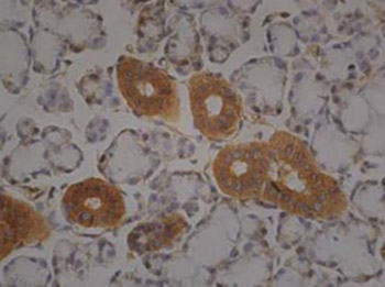
Calcitonin is usually produced by the parafollicular cells of the thyroid. However in an immunohistochemistry experiment we observed that the cells of the serous acini of rat submaxillary gland tissue were stained positive with calcitonin antibodies. We further used immunocytochemistry and nucleic acid hybridization to localize the distribution of calcitonin protein and calcitonin mRNA respectively in cultivated cells of rat submaxillary glands. The results showed that the cytoplasm of the epithelial cells of the submaxillary glands had positive staining in immunocytochemistry using calcitonin monoclonal antibody and positive reaction in nucleic acid hybridization using calcitonin mRNA complementary DNA probe. For the first time we found that the cells of the submaxillary glands of rats can produce the hormone calcitonin.

Downloads

Download data is not yet available.
Calcitonin is expressed in the submaxillary glands of rats

Downloads

Additional Files

Published

2014-05-20

How to Cite

1.
Zhang Y, Li B, Fu Z, Li S. Calcitonin is expressed in the submaxillary glands of rats. Biomol Biomed [Internet]. 2014May20 [cited 2023May30];14(1):35-9. Available from: https://www.bjbms.org/ojs/index.php/bjbms/article/view/2294

Issue

Section

Molecular Biology

INTRODUCTION

In recent years, researchers have discovered and extracted more than 30 various bioactive substances in the submaxillary glands [1-6]. These bioactive substances were either secreted directly into the blood or secreted first into the digestive tract with saliva and then enter the blood stream through gastrointestinal absorption; they exert regulatory roles on multiple tissues and cells to adjust their physiological activities [7, 8]. Some of these factors fully meet the standards of hormone, and it was put forward that they have dual functions of endocrine and exocrine [9, 10]. In doing an immunohistochemistry examination with calcitonin antiserum, we accidently observed a positive staining in the rat tissue of submaxillary glands, which suggested that the submaxillary glands might secrete calcitonin. In order to clarify this phenomenon, we repeated the immunohistochemistry experiment using calcitonin monoclonal antibody, and further cultivated rat submaxillary gland cells and performed immunocytochemistry and nucleic acid hybridization to localize the distribution of calcitonin protein and calcitonin mRNA respectively in the cultivated cells of the submaxillary glands. Our study for the first time showed that the cells of the submaxillary glands of rats can produce the hormone calcitonin. This paper reports our study and the results.

MATERIAL AND METHODS

Materials and Animals

DMEM/F12 medium was the product of Fisher’s Biological Chemical Products Co., LTD (Deputy in Beijing, China). Fetus bovine serum was the product of Lanzhou Rongye Biological Science and Technology Limited Company (Lanzhou, China). Rabbit anti-CK8 (rat keratin-8) monoclonal antibody was the product of Beijing BIOS Biological Technology Co., LTD (Beijing, China). Rabbit anti-calcitonin monoclonal antibody was the product of Thermo Fisher (NeoMarkers, Shanghai, China). Biotin labeled goat anti-rabbit IgG serum was the product of Zymed Laboratories (San Diego, California, United States). SP immunocytochemical staining kit and DAB color-display reagent was the product of Beijing Zhongshanjinqiao Biological Technology Co., LTD (Beijing, China). Nucleic acid hybridization detection kit was the product of Wuhan BOSTER Company (Wuhan, China). Digoxin labeled calcitonin DNA oligonucleotide was made by Beijing AUGCT Biological Technology Limited Liability Company (Beijing, China). SD rats were from Experimental Animal Center of Xi’an Jiaotong University Medical School (Xi’an, China).

Preparation of rat submaxillary gland tissue

Male and female SD rats, weigh 180-220 g, were used in the experiment. The rats were anesthetized with ether, and the submaxillary glands quickly taken, fixed in 10% formalin solution. After conventional paraffin embedding, the tissue blocks were cut into continuous slices of 5 μm thick. The experiment was performed in accordance to the international, national and institutional rules concerning animal experiments and rights.

Immunohistochemical staining of submaxillary gland tissue with anti-calcitonin antibody

After dehydration of paraffin wax, immunohistochemical staining SP (Streptavidin protein) method was performed [11]. 1:20 diluted rabbit anti-rat calcitonin monoclonal antibody (the first antibody) was applied and the slides were put into 4°C incubator and for the night. After rewarming at 37°C for 30 min, biotin labeled goat anti-rabbit serum (the second antibody) and horseradish peroxidase labeled Streptavidin were added in turn according to the instruction of the detection kits. The color was shown with DAB (3, 3,-diaminobenzidine). After conventional dehydration, transparent, and cementing, microscopic examination was done and photos were taken. For controls, PBS buffer was used instead of the first antibody.

Isolation and cultivation of epithelial cells of submaxillary glands

The rats were put to death by breaking the neck and the sub-maxillary glands were taken in sterile conditions. The newly removed submaxillary glands were put into 75% ethanol for 5 minutes. After careful removing of capsule, fat, blood vessels, and connective tissue, the obtained gland tissue was rinsed with PBS for three times. The submaxillary gland tissue was then cut into small masses of 0.1 mm~0.3 mm, rinsed with PBS for 3 times, and digested with 4 ml of 0.25% EDTA (trypsin plus Ethylenediamine tetraacetic acid) at 37°C for 20 min. The digestion was terminated by adding 4 ml culture medium (DMEM/F12 plus 10% calf bovine serum) and the cell suspension was collected and centrifuged at 800 rpm for 5 minutes. The pellet was taken and the process of digestion and centrifugation was repeated once more. Then the cells were re-suspended with 2 ml culture medium and the cell number was counted. After that, fibroblasts or fibrocytes were removed by differential attachment technique [12]. The cells were adjusted to 4 x 10⁵/ml concentration and cultivated at 37°C, saturated humidity and 5% CO2. Two days later, the original culture medium was changed and eight days later the cells were transferred to a new passage of cultivation (subculture).

Identification of cell type and demonstration of calcitonin distribution in the cultured submaxillary cells with immunocytochemistry

The sub-cultivated cells were used to do immunocytochemistry to show cell type as well as calcitonin localization with anti-cytokeratin 8 (CK8) antibody and anti-calcitonin antibody respectively. After 5 day cultivation on cover glass, cells were fixed with 4% formaldehyde for 30 min, saturated with 0.3% triton for 5 min, and incubated with hydrogen peroxide for 5 min to reduce nonspecific dyeing. Then 1:400 rabbit anti-rat CK8 monoclonal antibody and 1:25 rabbit anti-calcitonin monoclonal antibody were added and all of these study materials were put into 4°C refrigerator for the night. Next, biotin labeled goat anti-rabbit IgG serum was added and incubated at 37°C for 15 min. Following that the SP complex was added and after incubating at room temperature for 15 min, color was shown with DAB. After re-dyeing, dehydration, transparency, and slide sealing, microscopic examination was performed. For negative control, PBS was used to substitute rabbit anti-rat CK8 monoclonal antibody and rabbit anti-calcitonin monoclonal antibody.

Nucleic acid hybridization with calcitonin mRNA complementary probe

The cover glass treated with poly-lysine was used in cell climb cultivation. When cells grew to cover 95%-100% of the cover glass, it was removed and the cells were fixed with 4% poly-formaldehyde for 10 min. After gradient alcohol hydration, pepsin 20 μl was added followed with room temperature incubation for 20 min. Gradient alcohol dehydration was done again and this was followed with addition of 25 μl pre-hybrid solution and incubation at 40°C for 90 min. Then 25 μl hybrid solution containing the digoxin labeled calcitonin mRNA complementary DNA oligonucleotide probe (5-CCCATA-ATAGCCCAGAGAACAGCCAGAGAGG-3’) was applied and followed with 40°C incubation for 18 hours. After 3 times rinse with TTBS (Tris-Buffered Saline and Tween 20. Contents: 50 mM Tris, 150 mM NaCl, 0.05% Tween 20), 20-30 μl of 1% blockers was added, which was followed with 37°C incubation for 30 min. Again rinsed with TTBS for 3 times, enzyme labeled digoxin antibody 20μl was added and this was followed with 37°C incubation for 90 min. Then NBT/BCIP (Nitro-Blue-Tetrazolium/ 5-bromo-4-chloro-3-indolyl-phosphate) 30μl was added to show color with the reaction kept in dark place at 37°C for 2 hours. After rinsing with deionized water for 5 min, nuclear solid red 30μl was added and allowed to react for 5 min. Finally, after washing, dehydration, transparency, and slide sealing, a microscopic examination was performed. The hybrid solution used as negative control contained the same hybrid buffer but no probes.

RESULTS

Immunohistochemistry with anti-calcitonin antibody demonstrated calcitonin positive cells in rat submaxillary gland

The immunohistochemistry results of rat submaxillary gland tissue with anti-calcitonin monoclonal antibody revealed calcitonin positive staining cells in the submaxillary glands of the rats, which was manifested as brown color. The anti-calcitonin immune reactive staining was mainly distributed in the cells of the serous acini while the cells of the mucous acini were only slightly stained. No positive staining or brown color was seen in the cells of the negative control. The anti-calcitonin immune reactivity showed no sex differentiation in the cells of the sub-maxillary gland tissue of the rats. See Figure 1a, 1b and 1c.

FIGURE 1: The immunohistochemistry results of submaxillary gland tissue from male (A) and female (B) SD rats with anti-calcitonin monoclonal antibody. Calcitonin positive cells are stained brown. The positive staining is mainly distributed in the cells of the serous acini while the cells of the mucous acini are only slightly colored. The staining shows no sex difference. In the negative control (C), the cells are not stained by the antibody.

Anti-CK8 antibody staining indicated that the cells obtained by primary isolation/cultivation were secretory epithelial cells

In preparing primary isolation/culture of rat submaxillary gland cells, care was taken to remove capsule, fat, blood vessels, and connective tissue of the originally obtained glands; this guaranteed that the obtained cells were mostly secretory epithelial cells. Differential attachment technique utilized before the cultivation of the isolated cells removed fibroblasts or fibrocytes; this made the cell population more pure. Microscopic observation showed that the shapes of the cultured cells suggested epithelial cells rather than fibroblasts or fibrocytes (Figure 2a, 2b). Finally, immunocytochemistry with anti-CK8 monoclonal antibody showed that the cytoplasm of the cultured cells were positively stained (colored yellow or brown), indicating secretory epithelial cells (Figure 3a).

FIGURE 2a: Primary culture (8th days) of submaxillary gland cells observed with inverted microscope. x400.
FIGURE 2b: Subculture (6th days) of submaxillary gland cells observed with inverted microscope. x400.
FIGURE 3: Immunocytocheiriistry results showing the cell type and calcitonin distribution in the cultivated submaxillary gland cells; (A) The cytoplasm of the isolated cells is positively stained by anti-CK8 antibody, indicating secretory epithelial cells; (B) The cytoplasm of the cultivated submaxillary gland cells stains brown or yellow by anti-calcitonin antibody; (C) In the negative control, the cytoplasm is grey in color.

Immunocytochemistry showed the distribution of calcitonin staining in the cytoplasm of cultivated submaxillary gland cells

Immunocytochemical results in the cultivated cells of the submaxillary glands with anti-calcitonin monoclonal antibody staining showed that the cells were calcitonin positive as indicated by the easily recognizable yellow or brown color in their cytoplasm (Figure 3b). The cytoplasm was grey or unstained in the negative control (Figure 3c). The nuclei were blue in color for both antibody stained and control cells, although with different deepness.

Nucleic acid hybridization results showed that the cytoplasm of the cultivated submaxillary gland cells was calcitonin mRNA positive

The cytoplasm of the positive cells in the nucleic acid hybridization was blue or blue-violet in color and their nuclei were shallow red, which formed clear contrast with the control; the background was not colored (Figure 4a, 4b). The positive cells were very easy to identify and the mRNAs were widely spread in the cytoplasm of the submaxillary gland cells.

FIGURE 4: The nucleic acid hybridization result shows calcitonin mRNA positive cells in the submaxillary gland of rat; (A) The cytoplasm of the positive cells is blue in the hybridization result (The shallow red regions are the nuclei); (B) In the negative control, only the nuclei are seen (shallow red).

DISCUSSION

In recent years, it was found that the granular convoluted tubule cells of rat submaxillary gland can synthesize, store, and release nearly 30 kinds of bioactive peptides, such as epithelial growth factor, blood vessels relieve agent, insulin, endothelin, and liver cell growth factor [1-8]. Moreover, new bioactive substances were continually found in the submaxillary glands of rats. Fu et al. [13] found that submaxillary glands have the expression of follicle-stimulating hormone and luteinizing hormone. Chen et al. [11] found that submaxillary glands have the expression of luteinizing hormone and its receptors. And there were reports [14] showing that go-nadotrophin releasing hormone and its receptors can coexist in submaxillary glands. However, no paper has ever reported that the submaxillary glands could produce calcitonin. Calcitonin is a 32-amino acid linear polypeptide hormone that primarily maintains the homeostasis of calcium and phosphate; it also inhibits the activity of osteoclast cells and promotes the activity of osteoblast cells. Calcitonin is usually produced by the parafollicular cells (C-cells) of the thyroid. When doing an immunohistochemical experiment on rat thyroid with anti-calcitonin serum, we observed unexpectedly that the submaxillary glands of the animal contained positive staining cells. We carefully re-performed the immunohistochemistry examination of submaxillary gland tissue with anti-calcitonin monoclonal antibody using both male and female adult SD rats. Our results showed calcitonin positive staining in the submaxillary glands of the rats which was the most obvious in the cells of the serous acini and which showed no sex difference. The results indicated that the sub-maxillary glands of rats do can synthesis and release calcitonin. We further investigated the cellular localization of calcitonin protein and calcitonin mRNAs in the cultivated cells (primary culture) of the submaxillary glands. With rabbit anti-rat calcitonin monoclonal antibody as primary antibody, the immunocytochemical results showed that the cytoplasm of the cultivated cells of the submaxillary glands contained calcitonin. By doing nucleic acid hybridization with digoxin labeled calcitonin mRNA complementary DNA as probe, we observed positive reaction in the cytoplasm of the cells, indicating the existence of calcitonin mRNAs in the cells of the submaxillary glands.

CONCLUSION

In summary, by using submaxillary gland tissue and primary and secondary cell cultures and through detection of calcitonin protein and calcitonin mRNA, our investigation demonstrated that the cells of the submaxillary gland of rats could synthesize and secrete calcitonin. We speculate that the submaxillary glands of rats participate to a certain degree in the regulation of calcium and phosphate metabolism. Further studies are needed to address the problem of whether this is also the case in human beings.

DECLARATION OF INTEREST

The authors declare that there is no conflicts of interest with respect to the authorship or the publication of this article.

Acknowledgements

ACKNOWLEDGMENT

The study was funded by Shaanxi Education Department research fund (08JK506).

REFERENCES

  1. , , , , , (). Immunohistochemical expression and distribution of orexin, orphanin and leptin in the major salivary glands of some mammals. Folia Histochem Cytobiol.
  2. , , , (). Salivary gland derived peptides as a new class of anti-inflammatory agents: review of preclinical pharmacology of C-terminal peptides of SMR1 protein. J Inflamm (Lond).
  3. , , , , (). Molecular cloning and characterization of the neonatal rat and mouse submandibular gland protein SMGC. Gene.
  4. , , , (). A new member of the hor-monally regulated rodent submaxillary gland glycoprotein gene family: cDNA cloning and tissue specific expression. Mol Cell Endocrinol.
  5. , , (). Characterization of novel mRNAs encoding enzymes involved in peptide alpha-amidation. J Biol Chem.
  6. (). Biologically active polypeptides in submandibular glands. J Histochem Cytochem.
  7. , , , , , , (). Autonomic nervous system regulates secretion of anti-inflammatory prohormone SMR1 from rat salivary glands. Am J Physiol Cell Physiol.
  8. , , (). Natriuretic peptide system in the rat submaxillary gland. Jankowski M, Regul Pept.
  9. , , (). The research progress of bioactive substances in submaxilary gland. Sichuan Journal of Anatomy.
  10. , , , , (). The rodent granular convoluted tubule cell --an update. Eur J Morphol.
  11. , , , , , (). The distribution, cloning and analysis of partial sequence of LH and its receptor in submaxillary gland of rats. Acta Anatomica Sinica.
  12. , , (). Improvement on primary culture method of rat submaxillary gland cells. Medical Information.
  13. , , , (). Studies on distribution of FSH, LH and colocalization with GnRHR in rat submaxilary grands. Acta Anatomica Sinica.
  14. , , , , (). Studies of Localization and Distribution of Follicle-Stimulating Hormone Receptor in Rat Submandibular Gland and Stomach. Acta Anatomica Sinica.