Evaluation of bone mineral density (BMD) and indicators of bone turnover in patients with hemophilia

Authors

  • Mehmet Dagli Department of Internal Medicine, Faculty of Medicine, Selcuk University, Konya, Turkey
  • Ali Kutlucan Department of Internal Medicine, Faculty of Medicine, Selcuk University, Konya, Turkey
  • Sedat Abusoglu Department of Biochemistry, Faculty of Medicine, Selcuk University, Konya, Turkey
  • Abdulkadir Basturk Department of Internal Medicine, Faculty of Medicine, Selcuk University, Konya, Turkey
  • Mehmet Sozen Department of Internal Medicine, Faculty of Medicine, Selcuk University, Konya, Turkey
  • Leyla Kutlucan Department of Anesthesiology, Konya Training and Research Hospital, Konya, Turkey
  • Ali Unlu Department of Biochemistry, Faculty of Medicine, Selcuk University, Konya, Turkey
  • Farise Yilmaz Department of Nuclear Medicine, Faculty of Medicine, Selcuk University, Konya, Turkey

DOI:

https://doi.org/10.17305/bjbms.2018.2335

Keywords:

Hemophilia, osteoporosis, bone mineral density, BMD, procollagen type I N-terminal propeptide, PINP, urinary N-terminal telopeptide, uNTX, complete blood count, CBC, bone resorption, bone formation

Abstract

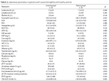
A decrease in bone mass is observed in hemophilic patients. The aim of this study was to evaluate bone mineral density (BMD), parathyroid hormone (PTH), 25-hydroxy vitamin D (vitamin D), and a bone formation and resorption marker, procollagen type I N-terminal propeptide (PINP) and urinary N-terminal telopeptide (uNTX) respectively, in hemophilic patients and healthy controls. Laboratory parameters related to the pathogenesis of bone loss such as neutrophil-lymphocyte ratio (NLR) and platelet-lymphocyte ratio (PLR) were also evaluated. Thirty-five men over 18 years of age, with severe hemophilia (A and B) and receiving secondary prophylaxis, were included in the study. The same number of age-, sex-, and ethnicity-matched healthy controls were evaluated. Anthropometric, biochemical, and hormonal parameters were determined in both groups. No significant difference in anthropometric parameters was found between the two groups. The BMD was low in 34% of hemophilic patients. Vitamin D, calcium, and free testosterone levels were significantly lower (p < 0.001, p = 0.011, p < 0.001, respectively), while PTH, PINP, and activated partial thromboplastin time (aPTT) levels were significantly higher (p < 0.014, p = 0.043, p < 0.001, respectively), in hemophilic patients compared to controls. There was no significant difference between the two groups in NLR, PLR, phosphorus, thyroid-stimulating hormone, and uNTX level. The reduction of bone mass in hemophilic patients may be evaluated using the markers of bone formation and resorption, enabling early detection and timely treatment.

Downloads

Download data is not yet available.

Author Biographies

Mehmet Dagli, Department of Internal Medicine, Faculty of Medicine, Selcuk University, Konya, Turkey

Department of Internal Medicine

Ali Kutlucan, Department of Internal Medicine, Faculty of Medicine, Selcuk University, Konya, Turkey

Department of Internal Medicine

Sedat Abusoglu, Department of Biochemistry, Faculty of Medicine, Selcuk University, Konya, Turkey

Department of Biochemistry

Abdulkadir Basturk, Department of Internal Medicine, Faculty of Medicine, Selcuk University, Konya, Turkey

Department of Internal Medicine

Mehmet Sozen, Department of Internal Medicine, Faculty of Medicine, Selcuk University, Konya, Turkey

Department of Internal Medicine

Leyla Kutlucan, Department of Anesthesiology, Konya Training and Research Hospital, Konya, Turkey

Department of Anesthesiology

Ali Unlu, Department of Biochemistry, Faculty of Medicine, Selcuk University, Konya, Turkey

Department of Biochemistry

Farise Yilmaz, Department of Nuclear Medicine, Faculty of Medicine, Selcuk University, Konya, Turkey

Department of Nuclear Medicine

References

Stephensen D, Drechsler W, Scott O. Comparison of muscle strength and in-vivo muscle morphology in young children with haemophilia and those of age-matched peers. Haemophilia 2012;18(3):e302-10. https://doi.org/10.1111/j.1365-2516.2011.02705.x.

Hoyer LW. Haemophilia A. N Engl J Med 1994;330(1):38-47. https://doi.org/10.1056/NEJM199401063300108.

Anagnostis P, Vakalopoulou S, Slavakis A, Charizopoulou M, Kazantzidou E, Chrysopoulou T, et al. Reduced bone mineral density in patients with haemophilia A and B in Northern Greece. Thromb Haemost 2012;107(3):545-51.

https://doi.org/10.1160/TH11-08-05563.

Gerstner G, Damiano ML, Tom A, Worman C, Schultz W, Recht M, et al. Prevalence and risk factors associated with decreased bone mineral density in patients with haemophilia. Haemophilia 2009;15(2):559-65. https://doi.org/10.1111/j.1365-2516.2008.01963.x.

Katsarou O, Terpos E, Chatzismalis P, Provelengios S, Adraktas T, Hadjidakis D, et al. Increased bone resorption is implicated in the pathogenesis of bone loss in hemophiliacs: Correlations with hemophilic arthropathy and HIV infection. Ann Hematol 2010;89(1):67-74. https://doi.org/10.1007/s00277-009-0759-x.

Wallny TA, Scholz DT, Oldenburg J, Nicolay C, Ezziddin S, Pennekamp PH, et al. Osteoporosis in haemophilia – an underestimated comorbidity? Haemophilia 2007;13(1):79-84. https://doi.org/10.1111/j.1365-2516.2006.01405.x.

Linari S, Montorzi G, Bartolozzi D, Borderi M, Melchiorre D, Benelli M, et al. Hypovitaminosis D and and osteopenia/osteoporosis in a haemophilia population: A study in HCV/HIVor HCV infected patients. Haemophilia 2013;19(1):126-33.

https://doi.org/10.1111/j.1365-2516.2012.02899.x.

Kovacs CS. Haemophilia, low bone mass, and osteopenia/osteoporosis. Transfus Apheres Sci 2008;38(1):33-40. https://doi.org/10.1016/j.transci.2007.12.003.

Lewiecki EM, Watts NB, McClung MR, Petak SM, Bachrach LK, Shepherd JA, et al. Official positions of the International Society for Clinical Densitometry. J Clin Endocrinol Metab 2004;89(8):3651-5. https://doi.org/10.1210/jc.2004-0124.

Gurevitch O, Slavin S. The hematological etiology of osteoporosis. Med Hypotheses 2006;67(4):729-35. https://doi.org/10.1016/j.mehy.2006.03.051.

Kaufman JM, Reginster JY, Boonen S, Brandi ML, Cooper C, Dere W, et al. Treatment of osteoporosis in men. Bone 2013;53(1):134-44. https://doi.org/10.1016/j.bone.2012.11.018.

Brennan SL, Henry MJ, Wluka AE, Nicholson GC, Kotowicz MA, Pasco JA. Socioeconomic status and bone mineral density in a population-based sample of men. Bone 2010;46(4):993-9. https://doi.org/10.1016/j.bone.2009.12.029.

Watts NB, Adler RA, Bilezikian JP, Drake MT, Eastell R, Orwoll ES, et al. Osteoporosis in men: An endocrine society clinical practice guideline. J Clin Endocrinol Metab 2012;97(6):1802-22. https://doi.org/10.1210/jc.2011-3045.

Hoch AZ, Pajewski NM, Moraski L, Carrera GF, Wilson CR, Hoffmann RG, et al. Prevalence of the female athlete triad in high school athletes and sedentary students. Clin J Sport Med 2009;19(5):421-8. https://doi.org/10.1097/JSM.0b013e3181b8c136.

Anagnostis P, Vakalopoulou S, Vyzantiadis TA, Charizopoulou M, Karras S, Goulis DG, et al. The clinical utility of bone turnover markers in the evaluation of bone disease in patients with haemophilia A and B. Haemophilia 2014;20(2):268-75. https://doi.org/10.1111/hae.12271.

Alioglu B, Selver B, Ozsoy H, Koca G, Ozdemir M, Dallar Y. Evaluation of bone mineral density in Turkish children with severe haemophilia A: Ankara hospital experience. Haemophilia 2012;18(1):69-74. https://doi.org/10.1111/j.1365-2516.2011.02587.x.

Tlacuilo-Parra A, Villela-Rodríguez J, Garibaldi-Covarrubias R, Soto-Padilla J, Orozco-Alcala J. Bone turnover markers and bone mineral density in children with haemophilia. Haemophilia 2011;17(4):657-61. DOI: 10.1111/j.1365-2516.2010.02439.x.

Brown JP, Albert C, Nassar BA, Adachi JD, Cole D, Davison KS, et al. Bone turnover markers in the management of postmenopausal osteoporosis. Clin Biochem 2009;42(10-11):929-42. https://doi.org/10.1016/j.clinbiochem.2009.04.001.

Öztürk ZA, Yesil Y, Kuyumcu ME, Bilici M, Öztürk N, Yeşil NK, et al. Inverse relationship between neutrophil lymphocyte ratio (NLR) and bone mineral density (BMD) in elderly people. Arch Gerontol Geriatr 2013;57(1):81-5. https://doi.org/10.1016/j.archger.2013.02.005.

Huang C, Li S. Association of blood neutrophil lymphocyte ratio in the patients with postmenopausal osteoporosis. Pak J Med Sci 2016;32(3):762-5. https://doi.org/10.12669/pjms.323.10292.

Koseoglu SB. Bone loss & platelet-to-lymphocyte ratio. Biomark Med 2017;11(1):5-10. https://doi.org/10.2217/bmm-2016-0188.

Li XS, Zhang JR, Meng SY, Li Y, Wang RT. Mean platelet volume is negatively associated with bone mineral density in postmenopausal women. J Bone Miner Metab 2012;30(6):660-5. https://doi.org/10.1007/s00774-012-0362-4.

Hakala M, Kröger H, Valleala H, Hienonen-Kempas T, Lehtonen-Veromaa M, Heikkinen J, et al. Once-monthly oral ibandronate provides significant improvement in bone mineral density in postmenopausal women treated with glucocorticoids for inflammatory rheumatic diseases: A 12-month, randomized double-blind, placebo-controlled trial. Scand J Rheumatol 2012;41(4):260-6. https://doi.org/10.3109/03009742.2012.664647.

Azab B, Bhatt VR, Phookan J, Murukutla S, Kohn N, Terjanian T, et al. Usefulness of the neutrophil-to-lymphocyte ratio in predicting short- and long-term mortality in breast cancer patients. Ann Surg Oncol 2012;19(1):217-24.

https://doi.org/10.1245/s10434-011-1814-0.

Gomez D, Farid S, Malik HZ, Young AL, Toogood GJ, Lodge JP, et al. Preoperative neutrophil-to-lymphocyte ratio as a prognostic predictor after curative resection for hepatocellular carcinoma. World J Surg 2008;32(8):1757-62.

https://doi.org/10.1007/s00268-008-9552-6.

Papa A, Emdin M, Passino C, Michelassi C, Battaglia D, Cocci F. Predictive value of elevated neutrophil-lymphocyte ratio on cardiac mortality in patients with stable coronary artery disease. Clin Chim Acta 2008;395(1-2):27-31. https://doi.org/10.1016/j.cca.2008.04.019.

Tamhane UU, Aneja S, Montgomery D, Rogers EK, Eagle KA, Gurm HS. Association between admission neutrophil to lymphocyte ratio and outcomes in patients with acute coronary syndrome. Am J Cardiol 2008;102(6):653-7. https://doi.org/10.1016/j.amjcard.2008.05.006.

Evaluation of bone mineral density (BMD) and indicators of bone turnover in patients with hemophilia

Downloads

Additional Files

Published

2018-05-20

How to Cite

1.
Dagli M, Kutlucan A, Abusoglu S, Basturk A, Sozen M, Kutlucan L, Unlu A, Yilmaz F. Evaluation of bone mineral density (BMD) and indicators of bone turnover in patients with hemophilia. Biomol Biomed [Internet]. 2018May20 [cited 2023Jun.6];18(2):206-10. Available from: https://www.bjbms.org/ojs/index.php/bjbms/article/view/2335

Issue

Section

Short Communication

INTRODUCTION

Hemophilia is an inherited bleeding disorder characterized by hemorrhagic events, such as intra-articular and intramuscular hemorrhage [1,2]. Recent studies have shown a reduction in bone mass in patients with hemophilia [3-5]. Due to recurrent hemorrhage and decreased mobility, hemophilic patients are unable to maintain bone mineral density (BMD) observed in healthy children. In addition, 25-hydroxy vitamin D (vitamin D) deficiency, presence of plasma-derived factor VIII (FVIII) and factor IX (FIX) inhibitors, degree of hemophilia, low body weight, hepatitis C virus (HCV) and human immunodeficiency virus (HIV) infections, as well as treatment with antiviral drugs were also associated with osteoporosis and reduction in bone mass in patients with hemophilia [3,4,6,7]. However, the pathophysiological mechanism leading to reduced bone mass, other causes of osteoporosis, and markers that can predict the reduction in bone mass in hemophilic patients have not been fully identified.

Osteoporosis is disease characterized by bone loss, increased bone fragility and bone fracture risk [8]. Risk factors for the development of osteoporosis include: age, use of glucocorticoids for more than three months, fracture history in first-degree relatives, low body mass index (BMI), cigarette smoking, nutritional factors, work conditions, exercise amount, and low socioeconomic status [9-12]. Dual energy X-ray absorptiometry (DXA) is the recommended test for the evaluation of osteoporosis [13]. Diagnosis is made based on hip and lumbar (central region) DXA scans and the results are presented as bone mineral content (BMC, gr/cm2) of the specific region. BMD is reported as a T score compared to a healthy, young adult of the same sex and as a Z score compared to an individual of the same sex, weight, and ethnic or racial group. In males over 50 years old, osteoporosis and osteopenia can be diagnosed based on the T score obtained with central DXA. For men under 50 years of age, according to the recommendations of the International Society for Clinical Densitometry, the Z score adjusted for race and age should be used instead of T score. A Z score below −2.0 is reported as “low BMD according to chronological age” or “below expected range according to age.” The score above −2.0 is reported as “in the expected range according to age” [14].

Biochemical markers of bone turnover reflect the metabolic activity of the bone and are classified into markers of bone formation and resorption. Up to now, a number of bone turnover markers have been identified. A few studies showed that vitamin D, parathyroid hormone (PTH), serum N-terminal telopeptide of type I collagen (NTX), C-terminal telopeptide of type I collagen (CTX), bone alkaline phosphatase (BALP), and osteocalcin (OC) are the markers of bone turnover in patients with hemophilia [15-17]. On the other hand, procollagen type I N-terminal propeptide (PINP), a bone formation marker released from osteoblasts, and urinary N-terminal telopeptide (uNTX), a marker of bone resorption detected in urine [18], have not been investigated in hemophilic patients with osteoporosis.

Moreover, several recent studies have found a significant association between neutrophil-lymphocyte ratio (NLR), platelet-lymphocyte ratio (PLR), and mean platelet volume (MPV), with low BMD [19-22]. However, this association has not been evaluated in hemophilic patients.

The aim of this prospective study was to measure BMD in hemophilic patients and to compare markers of bone formation and resorption between patients with hemophilia and controls. Laboratory parameters such as NLR and PLR, which may be related to the pathogenesis of bone loss, were also evaluated.

MATERIALS AND METHODS

Patients

The study was conducted between January 2016 and July 2016 in hemophilic patients who had been followed up at the adult hematology clinic of the Konya Selcuk University Faculty of Medicine and volunteered to participate in the study. Ethics approval was obtained from the Selcuk University Faculty of Medicine (Ethics no: 2015/228). The participants were informed and written consents were obtained. We included 35 men with severe hemophilia (29 with hemophilia A and 6 with hemophilia B). All patients had been receiving secondary prophylaxis at the beginning of the study, for 8.1 ± 0.3 years. The therapy consisted of 1500 daily units of FVIII and FIX concentrates, 3 days per week, for patients with hemophilia A and B, respectively. None of the patients were positive for the FVIII and FIX inhibitor.

The control group consisted of healthy individuals of the same age, sex, and ethnicity. Patients undergoing treatment with vitamin D, calcium, antiepileptic drugs and long-term corticosteroids, alcohol users, as well as patients with previous malignancies, malabsorption, morbid obesity, hypogonadism, thyroid and parathyroid diseases were not included in the study.

A detailed history was obtained and physical examination was performed in patients and controls. Anthropometric measurements including height (m2) and weight (kg) were also performed. The BMI was calculated as weight per height squared. The standard deviation score of height, weight, and BMI was determined for each group. Joint deformities in hemophilic patients were evaluated by physical examination. Flat graphs of joints of lower extremities were evaluated.

BMD measurement

BMD measurement was performed only in the patients with hemophilia. The BMD of lumbar vertebra and femoral neck was measured with a DXA device (Lunar DPX Prodigy-Tech, General Electric Company, Madison, WI, USA).

Laboratory assessment

All participants underwent the following laboratory assessments: complete blood count (CBC), FVIII or FIX activity, level of FVIII/IX inhibitor, glucose, serum creatinine, calcium (Ca), albumin (Alb), phosphorus (P), alkaline phosphatase (ALP), total bilirubin, direct bilirubin, aspartate aminotransferase (AST), alanine aminotransferase (ALT), activated partial thromboplastin time (aPTT), international normalized ratio (INR), C-reactive protein (CRP), hepatitis B surface antigen (HBsAg), antibodies against HCV (anti-HCV) and HIV (anti-HIV), vitamin D, free testosterone, PTH, and thyroid-stimulating hormone (TSH). The HCV viral load of hemophilic patients was not determined. The bone turnover markers, uNTX and PINP, were measured in all participants.

CBC was determined on a Coulter LH 780 hematology analyzer (Beckman Coulter, Inc., CA, USA). aPTT, INR, FVIII, and FIX were analyzed with a coagulation analyzer (Siemens Healthineers, Erlangen, Germany). Glucose, serum creatinine, CRP, Ca, Alb, P, ALP, total bilirubin, direct bilirubin, and liver function tests were determined on an AU analyzer (Beckman Coulter, Inc.). Hepatitis B surface antigen (HBsAg), anti-HCV and anti-HIV were analyzed using an Abbott analyzer (Abbott, Abbott Park, IL, USA). Free testosterone and vitamin D were quantified with liquid chromatography tandem-mass spectrometry [LC-MS] (ABSCIEX, USA). PTH and TSH were analyzed with Roche Modular E170 system (Roche Diagnostics, Basel, Switzerland). Urinary NTX and serum PINP were analyzed using commercially available enzyme linked immunosorbent assay (ELISA) kits (Elabscience, TX, USA).

Statistical analysis

Statistical analysis was performed with IBM SPSS Statistics for Windows, Version 22.0. (IBM Corp., Armonk, NY). Neutrophil count, NLR, PLR, hemoglobin, mean corpuscular volume (MCV), erythrocyte sedimentation rate (ESR), CRP, urea, creatinine, AST, ALT, Alb, total bilirubin, direct bilirubin, vitamin D, PTH, free testosterone, TSH, uNTX, PINP, aPTT, and MPV were analyzed with the Kolmogorov–Smirnov and Mann–Whitney U test. Age, years of diagnosis, height, weight, BMI, white blood count (WBC), lymphocyte count, platelet count, Ca, P, ALP, INR, and glucose were assessed with independent sample t-test. Univariate logistic regression analyses were conducted to identify potential predictors of BMD. The relationship between joint deformity and biochemical parameters was assessed by Spearman’s correlation analysis. Values of p < 0.05 were considered statistically significant.

RESULTS

The mean age of patients with hemophilia was 29.9 ± 8.07 years and it was 31.8 ± 12.0 years in the control group. There was no significant difference in the age, height, weight, and BMI between the two groups (Table 1). The hemophilic patients had an average of 2.4 ± 1.6 bleeding events per year. Two patients had a bone fracture. HCV was detected in 7 patients, but none of the patients received anti-HCV therapy. There were no HIV- or HBsAg-positive patients.

TABLE 1: Anthropometric measurements in patients with severe hemophilia and healthy controls

Approximately 85% of hemophilic patients had one or more joint deformities. Three patients had 1, 11 had 2, 8 had 3, and 8 patients had more than 3 joint deformities. Five patients did not have joint deformities. The number of joint deformities was not correlated with biochemical test results, bone turnover parameters and BMD in patients with hemophilia.

Twelve of 35 hemophilic patients (34%) had low BMD. Osteoporosis of the femur neck was detected in 1 of 2 hemophilic patients over the age of 50. According to the age, BMD was within the expected range in 22 patients out of 33 patients under the age of 50, while low BMD was detected in 11/33 patients. Seven of these 11 patients had low BMD of the femur neck, 2 patients had low BMD of the lumbar vertebrae, and 2 patients had low BMD of both the femur neck and lumbar vertebrae. There was no significant correlation between the low BMD and biochemical parameters, bone turnover parameters, and HCV infection in patients with hemophilia.

Vitamin D, Ca, and free testosterone levels were significantly lower (p < 0.001, p = 0.011, and p < 0.001, respectively) in patients with hemophilia compared to control group. PTH, PINP, and aPTT levels were significantly higher (p = 0.014, p = 0.043, and p < 0.001, respectively) in hemophilic patients compared to controls (Table 2). There were no significant differences in the blood parameters, NLR, PLR, ESR, CRP, creatinine, AST, ALT, Alb, total bilirubin, ALP, glucose, P, TSH, and uNTX values between the two groups.

TABLE 2: Laboratory parameters in patients with severe hemophilia and healthy controls

DISCUSSION

A decrease in bone mass is observed in hemophilic patients. In our study, a significant proportion (34%) of patients with severe hemophilia had low BMD. This percentage is significantly higher compared to healthy population (7.5%). The inability to perform regular physical activity because of joint deformities, and immobility due to bleeding events, probably lead to low BMD. Unfortunately we could not analyze the BMD in control group because of ethical constraints. In addition, we observed significantly lower levels of vitamin D in hemophilic patients, which might be the result of inadequate sunlight exposure due to immobility. Free testosterone and Ca levels were also significantly lower, while PTH was significantly higher in hemophilic patients compared to control group. However, the mean values of these parameters were within the normal range in both groups, and probably do not have a significant impact on BMD in hemophilic patients. Similarly, other studies did not show a significant relationship of PTH and total testosterone levels with bone turnover in hemophiliacs [15-16].

Previous studies evaluated several bone formation and resorption markers in hemophilic patients, including serum NTX-I, CTX-I, TRAP-5b, BALP, and OC [15-17]. In one of these studies, a significant negative correlation was found between BALP and BMD [15]. In the second study, OC was significantly lower in hemophilic patients compared to control group [16]. The third study evaluated OC and CTX-I levels in hemophilic children with low and normal BMD. While the level of OC was significantly lower in the group with low BMD, there was no relationship between CTX-I level and BMD [17]. On the contrary, PINP and uNTX have not been evaluated in hemophilic patients previously. An important finding of our study was that the serum PINP, which is a marker of bone formation, was significantly higher in hemophilic patients. On the other hand, the level of uNTX, a bone resorption marker, was not significantly different between the two groups. PINP can be used to monitor the progress of antiresorptive therapies as it is associated with the processes of bone remodeling. A decrease in PINP levels by more than 60% was reported one month after the start of antiresorptive treatment [23]. In patients with low BMD, PINP could be used for the initial diagnostic evaluation.

We found no significant differences in CBC, ESR, CRP, liver and renal functions, ALP, Alb, P, total bilirubin, and fasting blood glucose between the two groups. A few studies reported an association of NLR, PLR, and MPV with osteoporosis [19-22]. However, no previous study evaluated the association between low BMD and NLR, PLR and MPV in hemophiliacs.

NLR as a simple and inexpensive marker, which can be rapidly assessed, has been used as an indicator of inflammation in many diseases [24-27]. Öztürk et al. [19] found that NLR is an independent marker for osteoporosis and suggested that inflammation is present during bone remodeling [19]. Huang and Li [20] demonstrated a significant association between BMD and NLR in osteoporotic postmenopausal women [20]. In our study, there was no significant relationship between low BMD and NLR in patients with hemophilia. Koseoglu et al. [21] reported that PLR in postmenopausal women might be predictive of low BMD [21]. In this study, although the PLR was higher in hemophilic patients compared to controls, this result was not statistically significant.

MPV is an early indicator of platelet activity and recent studies have shown that MPV plays an important role in bone remodeling [22]. In a study evaluating 410 postmenopausal women with osteoporosis, a significant negative correlation was found between MPV and the BMD of lumbar vertebrae and femoral neck after correction for other risk factors [22]. In our study, no significant relationship between low BMD and MPV was observed in patients with hemophilia.

CONCLUSION

We showed that BMD and vitamin D levels were significantly lower in patients with severe hemophilia. PINP was significantly higher in hemophilic patients compared to control group. There was no significant difference between the two groups in uNTX, NLR, PLR, and MPV values. Our results form a basis for future studies which should comprehensively investigate a higher number of bone turnover markers in a larger sample of hemophilic patients.

Acknowledgements

ACKNOWLEDGMENTS

This work was supported by Selcuk University Scientific Research Project (Project No: 15401129).

DECLARATION OF INTERESTS

The authors declare no conflict of interests.

REFERENCES

  1. , , (). Comparison of muscle strength and in-vivo muscle morphology in young children with haemophilia and those of age-matched peers. Haemophilia. https://doi.org/10.1111/j.1365-2516.2011.02705.x
  2. (). Haemophilia A. N Engl J Med. https://doi.org/10.1056/NEJM199401063300108
  3. , , , , , (). Reduced bone mineral density in patients with haemophilia A and B in Northern Greece. Thromb Haemost. https://doi.org/10.1160/TH11-08-05563
  4. , , , , , (). Prevalence and risk factors associated with decreased bone mineral density in patients with haemophilia. Haemophilia. https://doi.org/10.1111/j.1365-2516.2008.01963.x
  5. , , , , , (). Increased bone resorption is implicated in the pathogenesis of bone loss in hemophiliacs: Correlations with hemophilic arthropathy and HIV infection. Ann Hematol. https://doi.org/10.1007/s00277-009-0759-x
  6. , , , , , (). Osteoporosis in haemophilia – an underestimated comorbidity?. Haemophilia. https://doi.org/10.1111/j.1365-2516.2006.01405.x
  7. , , , , , (). Hypovitaminosis D and osteopenia/osteoporosis in a haemophilia population: A study in HCV/HIVor HCV infected patients. Haemophilia. https://doi.org/10.1111/j.1365-2516.2012.02899.x
  8. (). Haemophilia, low bone mass, and osteopenia/osteoporosis. Transfus Apheres Sci. https://doi.org/10.1016/j.transci.2007.12.003
  9. , , , , , (). Official positions of the International Society for Clinical Densitometry. J Clin Endocrinol Metab. https://doi.org/10.1210/jc.2004-0124
  10. , (). The hematological etiology of osteoporosis. Med Hypotheses. https://doi.org/10.1016/j.mehy.2006.03.051
  11. , , , , , (). Treatment of osteoporosis in men. Bone. https://doi.org/10.1016/j.bone.2012.11.018
  12. , , , , , (). Socioeconomic status and bone mineral density in a population-based sample of men. Bone. https://doi.org/10.1016/j.bone.2009.12.029
  13. , , , , , (). Osteoporosis in men: An endocrine society clinical practice guideline. J Clin Endocrinol Metab. https://doi.org/10.1210/jc.2011-3045
  14. , , , , , (). Prevalence of the female athlete triad in high school athletes and sedentary students. Clin J Sport Med. https://doi.org/10.1097/JSM.0b013e3181b8c136
  15. , , , , , (). The clinical utility of bone turnover markers in the evaluation of bone disease in patients with haemophilia A and B. Haemophilia. https://doi.org/10.1111/hae.12271
  16. , , , , , (). Evaluation of bone mineral density in Turkish children with severe haemophilia A: Ankara hospital experience. Haemophilia. https://doi.org/10.1111/j.1365-2516.2011.02587.x
  17. , , , , (). Bone turnover markers and bone mineral density in children with haemophilia. Haemophilia. DOI: 10.1111/j.1365-2516.2010.02439.x
  18. , , , , , (). Bone turnover markers in the management of postmenopausal osteoporosis. Clin Biochem. https://doi.org/10.1016/j.clinbiochem.2009.04.001
  19. , , , , , (). Inverse relationship between neutrophil lymphocyte ratio (NLR) and bone mineral density (BMD) in elderly people. Arch Gerontol Geriatr. https://doi.org/10.1016/j.archger.2013.02.005
  20. , (). Association of blood neutrophil lymphocyte ratio in the patients with postmenopausal osteoporosis. Pak J Med Sci. https://doi.org/10.12669/pjms.323.10292
  21. (). Bone loss & platelet-to-lymphocyte ratio. Biomark Med. https://doi.org/10.2217/bmm-2016-0188
  22. , , , , (). Mean platelet volume is negatively associated with bone mineral density in postmenopausal women. J Bone Miner Metab. https://doi.org/10.1007/s00774-012-0362-4
  23. , , , , , (). Once-monthly oral ibandronate provides significant improvement in bone mineral density in postmenopausal women treated with glucocorticoids for inflammatory rheumatic diseases: A 12-month, randomized double-blind, placebo-controlled trial. Scand J Rheumatol. https://doi.org/10.3109/03009742.2012.664647
  24. , , , , , (). Usefulness of the neutrophil-to-lymphocyte ratio in predicting short- and long-term mortality in breast cancer patients. Ann Surg Oncol. https://doi.org/10.1245/s10434-011-1814-0
  25. , , , , , (). Preoperative neutrophil-to-lymphocyte ratio as a prognostic predictor after curative resection for hepatocellular carcinoma. World J Surg. https://doi.org/10.1007/s00268-008-9552-6
  26. , , , , , (). Predictive value of elevated neutrophil-lymphocyte ratio on cardiac mortality in patients with stable coronary artery disease. Clin Chim Acta. https://doi.org/10.1016/j.cca.2008.04.019
  27. , , , , , (). Association between admission neutrophil to lymphocyte ratio and outcomes in patients with acute coronary syndrome. Am J Cardiol. https://doi.org/10.1016/j.amjcard.2008.05.006