The effect of 131I-induced hypothyroidism on the levels of nitric oxide (NO), interleukin 6 (IL-6), tumor necrosis factor alpha (TNF-α), total nitric oxide synthase (NOS) activity, and expression of NOS isoforms in rats

Authors

  • Jing Zhou Department of Nuclear Medicine, The Fuling Central Hospital of Chongqing, Chongqing, China
  • Gang Cheng Department of Nuclear Medicine, The First Affiliated Hospital of Chongqing Medical University, Chongqing, China
  • Hua Pang Department of Nuclear Medicine, The First Affiliated Hospital of Chongqing Medical University, Chongqing, China
  • Qian Liu Department of Nuclear Medicine, The First Affiliated Hospital of Chongqing Medical University, Chongqing, China
  • Ying Liu Department of Nuclear Medicine, The First Affiliated Hospital of Chongqing Medical University, Chongqing, China

DOI:

https://doi.org/10.17305/bjbms.2018.2350

Keywords:

Hypothyroidism, I-131, radioactive iodine, aorta, nitric oxide, NO, nitric oxide synthase, NOS, eNOS, nNOS, iNOS, inflammatory mediators, interleukin, IL-6, TNF-α

Abstract

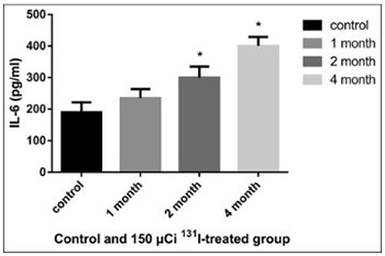
Accumulating evidence has shown that hypothyroidism affects the cardiovascular system, significantly increasing the incidence of cardiovascular diseases. In the present study we investigated the effect of radioactive iodine (I-131)-induced hypothyroidism on several parameters of vascular function, such as nitric oxide (NO), total nitric oxide synthase (NOS) activity and expression of NOS isoforms, as well as on interleukin 6 (IL-6) and tumor necrosis factor alpha (TNF-α) as indicators of inflammation, in rats. A dose of 150 µCi of 131-I was determined as optimal for establishing the model of hypothyroidism in rats. After administration of 131-I, at the end of month 1, 2, and 4 (n = 3 for each time point), NO, IL-6, and TNF-α in the serum and total NOS activity in the aorta were determined in 150 µCi group, compared to controls. The mRNA and protein expression of endothelial, neuronal, and inducible NOS (eNOS, nNOS, and iNOS) in the rat aorta was also estimated, using quantitative reverse transcription polymerase chain reaction and Western blot, respectively. The levels of IL-6 and TNF-α increased in 150 µCi group; the results were significant at the end of month 2 and 4 for IL-6, and at all time points for TNF-α. The levels of NO decreased significantly at the end of month 2 and 4 in 150 µCi group. The total NOS activity increased significantly in 150 µCi group, at all three time points. Significant changes in the mRNA and protein expression of all three NOS isoforms were observed in 150 µCi group compared to controls. NO, IL-6, TNF-α levels and NOS activity and expression are altered in hypothyroid state, and the underlying mechanism should be further investigated.

Downloads

Download data is not yet available.

Author Biographies

Jing Zhou, Department of Nuclear Medicine, The Fuling Central Hospital of Chongqing, Chongqing, China

Department of Nuclear Medicine

Gang Cheng, Department of Nuclear Medicine, The First Affiliated Hospital of Chongqing Medical University, Chongqing, China

Department of Nuclear Medicine

Hua Pang, Department of Nuclear Medicine, The First Affiliated Hospital of Chongqing Medical University, Chongqing, China

Department of Nuclear Medicine

Qian Liu, Department of Nuclear Medicine, The First Affiliated Hospital of Chongqing Medical University, Chongqing, China

Department of Nuclear Medicine

Ying Liu, Department of Nuclear Medicine, The First Affiliated Hospital of Chongqing Medical University, Chongqing, China

Department of Nuclear Medicine

References

Lu ZY, Zhong NS. Internal Medicine. 7th ed. Beijing: People's Medical Publishing House; 2008.

Aizawa Y, Yoshida K, Kaise N, Fukazawa H, Kiso Y, Sayama N, et al. The development of transient hypothyroidism after iodine-131 treatment in hyperthyroid patients with Graves' disease: Prevalence, mechanism and prognosis. Clin Endocrinol (Oxf) 1997;46(1):1-5. https://doi.org/10.1046/j.1365-2265.1997.d01-1737.x.

Alexander EK, Larsen PR. High dose of (131)I therapy for the treatment of hyperthyroidism caused by Graves' disease. J Clin Endocrinol Metab 2002;87(3):1073-7. https://doi.org/10.1210/jcem.87.3.8333.

Gomez N, Gomez JM, Orti A, Gavalda L, Villabona C, Leyes P, et al. Transient hypothyroidism after iodine-131 therapy for Grave's disease. J Nucl Med 1995;36(9):1539-42.

Canaris GJ, Manowitz NR, Mayor G, Ridgway EC. The Colorado thyroid disease prevalence study. Arch Intern Med 2000;160(4):526-34. https://doi.org/10.1001/archinte.160.4.526.

Biondi B, Palmieri EA, Lombardi G, Fazio S. Effects of subclinical thyroid dysfunction on the heart. Ann Intern Med 2002;137(11):904-14.

https://doi.org/10.7326/0003-4819-137-11-200212030-00011.

Cappola AR, Ladenson PW. Hypothyroidism and atherosclerosis. J Clin Endocrinol Metab 2003;88(6):2438-44. https://doi.org/10.1210/jc.2003-030398.

Klein I, Ojamaa K. Thyroid hormone and the cardiovascular system. N Engl J Med 2001;344(7):501-9. https://doi.org/10.1056/NEJM200102153440707.

Vargas F, Moreno JM, Rodriguez-Gomez I, Wangensteen R, Osuna A, Alvarez-Guerra M, et al. Vascular and renal function in experimental thyroid disorders. Eur J Endocrinol 2006;154(2):197-212. https://doi.org/10.1530/eje.1.02093.

Flammer AJ, Luscher TF. Three decades of endothelium research: From the detection of nitric oxide to the everyday implementation of endothelial function measurements in cardiovascular diseases. Swiss Med Wkly 2010;140:w13122. https://doi.org/10.4414/smw.2010.13122.

Luscher TF, Vanhoutte PM. The Endothelium: Modulator of Cardiovascular function. Florida, USA: CRC press; 1990.

Förstermann U, Sessa WC. Nitric oxide synthases: regulation and function. Eur Heart J 2012;33(7):829-37,837a-837d. https://doi.org/10.1093/eurheartj/ehr304.

Tsutsui M, Shimokawa H, Otsuji Y, Ueta Y, Sasaguri Y, Yanagihara N. Nitric oxide synthases and cardiovascular diseases: Insights from genetically modified mice. Circ J 2009; 73(6):986-93. https://doi.org/10.1253/circj.CJ-09-0208.

Abbas AM, Sakr HF. Effect of magnesium sulfate and thyroxine on inflammatory markers in a rat model of hypothyroidism. Can J Physiol Pharmacol 2016;94(4):426-32. https://doi.org/10.1139/cjpp-2015-0247.

Hajje G, Saliba Y, Itani T, Moubarak M, Aftimos G, Fares N. Hypothyroidism and its rapid correction alter cardiac remodeling. PLoS One 2014;9(10):e109753. https://doi.org/10.1371/journal.pone.0109753.

Diez JJ, Hernanz A, Medina S, Bayon C, Iglesias P. Serum concentrations of tumour necrosis factor-alpha (TNF-alpha) and soluble TNF-alpha receptor p55 in patients with hypothyroidism and hyperthyroidism before and after normalization of thyroid function. Clin Endocrinol (Oxf) 2002;57(4):515-21. https://doi.org/10.1046/j.1365-2265.2002.01629.x.

Johnson C, Olivier NB, Nachreiner R, Mullaney T. Effect of 131I-induced hypothyroidism on indices of reproductive function in adult male dogs. J Vet Intern Med 1999;13(2):104-10. https://doi.org/10.1892/0891-6640(1999)013<0104:EOIHOI>2.3.CO;2.

Pasteur I, Tronko N, Drozdovich I, Donich S, Voitenko L. Xenotransplantation of cultured newborn pig thyroid tissue for the treatment of post-radioiodine hypothyroidism in rats. Cytotechnology 2000;33(1-3):89-92. https://doi.org/10.1023/A:1008185701106.

Reilly CP, Symons RG, Wellby ML. A rat model of the 131I-induced changes in thyroid function. J Endocrinol Invest 1986;9(5):367-70. https://doi.org/10.1007/BF03346944.

Torlak V, Zemunik T, Modun D, Capkun V, Pesutic-Pisac V, Markotic A, et al. 131 I-induced changes in rat thyroid gland function. Braz J Med Biol Res 2007;40(8):1087-94. https://doi.org/10.1590/S0100-879X2006005000127.

Hosseini M, Dastghaib SS, Rafatpanah H, Hadjzadeh MA, Nahrevanian H, Farrokhi I. Nitric oxide contributes to learning and memory deficits observed in hypothyroid rats during neonatal and juvenile growth. Clinics (Sao Paulo) 2010;65(11):1175-81. https://doi.org/10.1590/S1807-59322010001100021.

McAllister RM, Albarracin I, Price EM, Smith TK, Turk JR, Wyatt KD. Thyroid status and nitric oxide in rat arterial vessels. J Endocrinol 2005;185(1):111-9. https://doi.org/10.1677/joe.1.06022.

Quesada A, Sainz J, Wangensteen R, Rodriguez-Gomez I, Vargas F, Osuna A. Nitric oxide synthase activity in hyperthyroid and hypothyroid rats. Eur J Endocrinol 2002;147(1): 117-22. https://doi.org/10.1530/eje.0.1470117.

Engin AB, Sepici-Dincel A, Gonul II, Engin A. Oxidative stress-induced endothelial cell damage in thyroidectomized rat. Exp Toxicol Pathol 2012;64(5):481-5. https://doi.org/10.1016/j.etp.2010.11.002.

Dizdarevic-Bostandic A, Burekovic A, Velija-Asimi Z, Godinjak A. Inflammatory markers in patients with hypothyroidism and diabetes mellitus type 1. Med Arch 2013;67(3):160-1. https://doi.org/10.5455/medarh.2013.67.160-161.

Marfella R, Ferraraccio F, Rizzo MR, Portoghese M, Barbieri M, Basilio C, et al. Innate immune activity in plaque of patients with untreated and L-thyroxine-treated subclinical hypothyroidism. J Clin Endocrinol Metab 2011;96(4):1015-20. https://doi.org/10.1210/jc.2010-1382.

Guida MS, Abd El-Aal A, Kafafy Y, Salama SF, Badr BM, Badr G. Thymoquinone rescues T lymphocytes from gamma irradiation-induced apoptosis and exhaustion by modulating pro-inflammatory cytokine levels and PD-1, Bax, and Bcl-2 signaling. Cell Physiol Biochem 2016;38(2):786-800. https://doi.org/10.1159/000443034.

Koukkunen H, Penttila K, Kemppainen A, Halinen M, Penttila I, Rantanen T, et al. C-reactive protein, fibrinogen, interleukin-6 and tumour necrosis factor-alpha in the prognostic classification of unstable angina pectoris. Ann Med 2001;33(1):37-47. https://doi.org/10.3109/07853890109002058.

Gao C, Li T, Liu J, Guo Q, Tian L. Endothelial functioning and hemodynamic parameters in rats with subclinical hypothyroid and the effects of thyroxine replacement. PLoS One 2015;10:e0131776. https://doi.org/10.1371/journal.pone.0131776.

Ibuki Y, Goto R. Ionizing radiation-induced macrophage activation: Augmentation of nitric oxide production and its significance. Cell Mol Biol (Noisy-le-grand) 2004;50 Online Pub:OL617-626.

Lacoste-Collin L, Jozan S, Pereda V, Courtade-Saidi M. Influence of a continuous very low dose of gamma-rays on cell proliferation, apoptosis and oxidative stress. Dose Response 2015;13(1): dose-response.14-010.Lacoste-Collin.

https://doi.org/10.2203/dose-response.14-010.Lacoste-Collin.

Nowosielska EM, Wrembel-Wargocka J, Cheda A, Lisiak E, Janiak MK. Enhanced cytotoxic activity of macrophages and suppressed tumor metastases in mice irradiated with low doses of X- rays. J Radiat Res 2006;47(3-4):229-36. https://doi.org/10.1269/jrr.0572.

Sarati LI, Martinez CR, Artes N, Arreche N, Lopez-Costa JJ, Balaszczuk AM, et al. Hypothyroidism: Age-related influence on cardiovascular nitric oxide system in rats. Metabolism 2012;61(9):1301-11. https://doi.org/10.1016/j.metabol.2012.01.022.

Fellet AL, Arza P, Arreche N, Arranz C, Balaszczuk AM. Nitric oxide and thyroid gland: Modulation of cardiovascular function in autonomic-blocked anaesthetized rats. Exp Physiol 2004;89(3):303-12. https://doi.org/10.1113/expphysiol.2004.027201.

Rodriguez-Gomez I, Moliz JN, Quesada A, Montoro-Molina S, Vargas-Tendero P, Osuna A, et al. L-Arginine metabolism in cardiovascular and renal tissue from hyper- and hypothyroid rats. Exp Biol Med (Maywood) 2016;241(5):550-6. https://doi.org/10.1177/1535370215619042.

The effect of 131I-induced hypothyroidism on the levels of nitric oxide (NO), interleukin 6 (IL-6), tumor necrosis factor alpha (TNF-α), total nitric oxide synthase (NOS) activity, and expression of NOS isoforms in rats

Downloads

Additional Files

Published

2018-11-07

How to Cite

1.
Zhou J, Cheng G, Pang H, Liu Q, Liu Y. The effect of 131I-induced hypothyroidism on the levels of nitric oxide (NO), interleukin 6 (IL-6), tumor necrosis factor alpha (TNF-α), total nitric oxide synthase (NOS) activity, and expression of NOS isoforms in rats. Biomol Biomed [Internet]. 2018Nov.7 [cited 2023Sep.23];18(4):305-12. Available from: https://www.bjbms.org/ojs/index.php/bjbms/article/view/2350

Issue

Section

Biochemistry

INTRODUCTION

In many countries, radioactiveiodine (131I or I-131) has been utilized for more than 60 years; for example in China, I-131 treatment has been in use since 1958 [1]. Moreover, I-131 is the first choice for the treatment of hyperthyroidism in adults in European countries and the USA. Hypothyroidism is a major complication of I-131 treatment. The cumulative incidence of hypothyroidism, in I-131 treated patients with hyperthyroidism caused by Graves’ disease, has been gradually increasing each year, reaching around 80% in adults and 95% among children and adolescents [2-4]. Hypothyroidism is a relatively common endocrine disease. In a general population, the occurrence of hypothyroidism is 10% [5]; more than 95% of the affected individuals have primary hypothyroidism, most commonly caused by I-131 treatment of hyperthyroidism. Accumulating evidence has shown that hypothyroidism seriously affects the cardiovascular system, significantly increasing the incidence of cardiovascular diseases [6, 7]. Both subclinical hypothyroidism and hypothyroidism decrease the cardiac output, increase the peripheral resistance, and impair the endothelial function [6,8]. Impaired endothelial function is associated with decreased vasodilation [9]. The vascular endothelium, as the interface between the blood and vessel walls, plays a major role in regulating vascular structure and function [10].

Impaired nitric oxide (NO) bioavailability, due to reduced production of NO by nitric oxide synthase (NOS) or its increased breakdown by reactive oxygen species (ROS), is commonly linked to endothelial dysfunction [11]. Other studies confirmed that NO is one of the most important factors of vasodilation. NO biological half-life is only 4 to 8 seconds, and it participates in the function of cardiovascular system in the following ways: regulation of vascular tension, anti-oxidative, anti-inflammatory, and anticoagulant effect, fibrinolytic action, inhibition of leukocyte adhesion and migration, inhibition of proliferation and migration of smooth muscle cells, and inhibition of platelet aggregation and adhesion [10].

Three isoforms of NOS are known in mammals, encoded by different genes: 1) endothelial NOS (eNOS or NOS3) is mainly present in the endothelium, and is encoded by the NOS3 gene located on 7q35-36; 2) neuronal NOS (nNOS or NOS1), encoded by the NOS1 gene located on 12q24.2, is expressed predominantly in nervous tissue and skeletal muscle; and 3) inducible NOS (iNOS or NOS2) encoded by the NOS2 gene (17q11.2) has a role in the immune and cardiovascular system. Increased NO production is induced by intracellular antigens, some tumor cells, and microbial products as well as in abnormal physiological conditions, such as heart failure and inflammation. Moreover, all three NOS isoforms are expressed in other tissue/cell types, and many tissues express more than one isoform. For example, nNOS has also been identified in the spinal cord, sympathetic ganglia, adrenal glands, epithelial cells of different organs, kidney macula densa cells, pancreatic islet cells, and vascular smooth muscle. eNOS was also detected in platelets, some neurons in the brain, human placenta, and in kidney tubular epithelial cells. In addition, the three NOS isoforms are expressed in myocardial cells and play a critical role in the cardiovascular system [12,13].

Inflammatory mediators, such as interleukin 6 (IL-6) and tumor necrosis factor alpha (TNF-α), have been associated with hypothyroidism [14]. IL-6, a pleiotropic cytokine released by fibroblasts, T lymphocytes, endothelial cells, and monocytes, has multiple biological activities in different target cells. Furthermore, IL-6 has a crucial role in vascular inflammation. Also, a previous study showed that the concentration of IL-6 is increased in hypothyroid rats [14,15]. TNF-α, a cytokine mainly expressed by monocyte-macrophage cells, is activated in patients with thyroid dysfunction; a high concentration of TNF-α has been demonstrated in patients with hypothyroidism [16].

In the present study, we investigated the effect of 131I-induced hypothyroidism on NO, total NOS activity and expression of NOS isoforms as parameters of vascular function, and on IL-6 and TNF-α as indicators of inflammation, in rats. Rats were intraperitoneally (i.p.) injected with 131I to establish a model of hypothyroidism. According to the literature, the dosage of 131I ranged from 50-450 µCi and the longest exposure lasted 5 months [17-20]. In our experiments, the rats in different groups were i.p. injected with 75 µCi, 150 µCi, 300 µCi, or 450 µCi 131I. The weight of the animals, thyroid uptake (counts per minute [CPM]), and thyroid function at different time points were compared between the groups, to determine the optimal 131I dose for the model. In the selected rat model of hypothyroidism, we assessed the level of NO, IL-6, and TNF-α in the serum and total NOS activity in the aorta at different time points. Finally, we estimated the mRNA and protein expression of eNOS, nNOS, and iNOS in the rat aorta, using qRT-PCR and Western blot, respectively.

MATERIALS AND METHODS

Reagents

The enzyme-linked immunosorbent assay (ELISA) kits for IL-6 and TNF-α were obtained from Neobioscience Co., Ltd. (Guangdong, China). Kits for the detection of total NOS activity and NO were obtained from Nanjing Jiancheng Bioengineering Institute (Nanjing, China). Anti-eNOS, anti-iNOS, and anti-nNOS antibodies were obtained from Bioss Biotechnology Co., Ltd. (Beijing, China). TRIzol reagent was obtained from Tiangen Biotechnology Co., Ltd. (Beijing, China). PrimeScript™ RT reagent kit with gDNA Eraser, SYBR® Premix Ex Taq™ II (Tli RNaseH Plus), ROX plus, and DL2000 DNA marker were obtained from TaKaRa Biotechnology Co., Ltd. (Dalian, China). Primers for NOS genes (Table 1) were synthesized by Invitrogen (Shanghai, China). Rat anti-NOS monoclonal antibody and horseradish peroxidase (HRP)-conjugated anti-rat secondary antibody were obtained from Zhongshan Golden Bridge Biotechnology Co., Ltd (Beijing, China). Triiodothyronine (T3), thyroxine (T4), and thyroid stimulating hormone (TSH) kits were obtained from Beijing kit-guide Bo High Biological Technology Co., Ltd. (Beijing, China).

TABLE 1: Primers sequences for nitric oxide synthase (NOS) genes

Experimental groups and treatment

Forty-five male Sprague-Dawley rats, aged 4 weeks and weighing 80-100 g, were obtained from the Department of Laboratory Animal Center of the Chongqing Medical University. The guidelines of Chongqing Medical University for the care and use of animals, followed in this study, were approved by the Chongqing science and technology commission (SYXK2007-0001, SCXK2007-0001, and SCXK2007-0002). The animals were housed and fed for 14 days under a 12-hour light/dark cycle at an appropriate temperature and humidity. The rats were randomly assigned to 5 groups (n = 9 divided at three time points for each 131I dose): control group, and groups with 75 µCi, 150 µCi, 300 µCi, and 450 µCi of 131I injected i.p. Following the administration of 131I, the rats were euthanized (at the end of month 1, 2, and 4; n = 3 for each time point) for biochemical analysis of serum, aorta, and thyroid glands.

Weight measurement

Before the euthanization, the rats were weighed to determine the difference in weight between different time points in each group.

Blood and tissue samples

At each time point, an appropriate amount of chloral hydrate was injected i.p. in rats as an anesthetic. The blood was collected by puncture of the left ventricle, left for 30 minutes without anticoagulant and followed by centrifugation for 15 minutes at 3000 r/min to obtain the serum. The serum samples were stored at -80°C. After collecting the blood, the right atrium was punctured to clean immediately the blood. Then, the aortas (from the ascending to the abdominal aorta) were collected and stored at -80°C. Finally, the thyroid glands were collected.

Measurement of thyroid radioactivity

Following the collection of samples, the thyroid radioactivity was detected using a scintillator detector. The distance between the detector and tissue was 10 cm and the detection time was 1 minute. The results were recorded as CPM.

Assessment of thyroid function, IL-6, TNF-α, and NO

The levels of T3 and T4 were determined in the plasma using the radioimmunoassay kits. TSH was measured by a solid-phase competitive chemiluminescent enzyme immunoassay using the IMMULITE 2000 analyzer (Siemens Healthcare Diagnostics, USA). Serum IL-6, TNF-α, and NO were measured by ELISA, according to the kit instructions. All analyses were performed in triplicate, and the mean of the three measurements was used for the statistical analysis.

Total NOS activity

The total NOS activity was measured by the detection kit. The tissue from aorta (0.15 g) was homogenized in 1.35 g normal saline to make 10% tissue homogenate, and centrifuged at 2500 r/min for 10 minutes. The 50-µl supernatant was used to measure the NOS activity, protein content, and total NOS activity, according to the kit instructions.

Quantitative reverse transcription polymerase chain reaction (qRT-PCR)

The aorta tissue was ground to powder in a pre-cooled mortar. The total cellular RNA was extracted from the cells using TRIzol reagent. The samples were then purified with 75% ethanol, and the purity and concentration of RNA were determined at 260 nm with an ND-2000 NanoDrop spectrophotometer (Thermo Fisher Scientific, USA). The first-strand cDNA was synthesized using a PrimeScript™ RT reagent Kit with gDNA Eraser for RT-PCR. qRT-PCR was performed in a 20-µL reaction, including 0.5 µL forward primers and 0.5 µL reverse primers, 2 × SYBR Green reaction mix, and 2 µL cDNA. The PCR protocol was as follows: initial denaturation at 94°C for 30 seconds; 40 cycles of denaturation at 95°C for 15 seconds, annealing at 60°C for 30 seconds, and extension at 72°C for 30 seconds; 1 cycle at 72°C for 10 minutes. The melting curves were measured at the end of the amplification. All target genes (eNOS, nNOS, and iNOS) and β-actin gene were amplified in triplicate in a 96-well plate. Data were analyzed using the 2-ΔΔCT method.

Western blot analysis

The protein expression of eNOS, nNOS, and iNOS was assayed at the three time points (n = 3 for each time point). Briefly, the aorta was dissected, total protein was isolated using RIPA buffer, and the protein concentration was estimated. The proteins (30 µg) were denatured by a standard method, resolved by sodium dodecyl sulfate polyacrylamide gel electrophoresis (SDS-PAGE), and transferred to a polyvinylidene difluoride (PVDF) membrane. The membranes were blocked for 1 hour at room temperature with gentle agitation. Subsequently, they were incubated overnight at 4°C with monoclonal antibodies against nNOS, eNOS, and iNOS at a dilution of 1:1000. Then, the membranes were washed and incubated with the secondary antibody at 1:5000, for 1 hour at room temperature in the dark, with gentle agitation. Lastly, the immunoreactive bands were developed using detection reagents.

Statistical analysis

Statistical analyses were performed with IBM SPSS Statistics for Windows, Version 19.0. (IBM Corp., Armonk, NY). Quantitative data were expressed as mean ± standard deviation (SD). The statistical significance of the treatments was evaluated using Student’s t-test and two-way ANOVA. The differences were considered statistically significant at p < 0.05.

RESULTS

Rat body weight

As shown in Table 2, no differences were observed in the body weight of rats between control and 75 µCi group. Compared to control group, the body weight of rats in 150 µCi group was significantly increased (p < 0.05), while the weight was decreased in 300 µCi and 450 µCi groups (p < 0.05).

TABLE 2: Body weight of rats treated with 131I (n=3 per each time point, mean±SD)

Thyroid radioactivity in 131I-treated rats

The thyroid radioactivity gradually increased from the low-dose to high-dose group after the 131I administration at the end of month 1, and was markedly decreased in each dose group at the end of month 2 and 4; most of the radioactivity values being similar to the natural background value (Table 3).

TABLE 3: Thyroid radioactivity (counts per minute [CPM]) of rats treated with 131I (n=3 per each time point, mean±SD)

Thyroid function in 131I-treated rats

The thyroid function did not differ between control and 75 µCi group. In the other groups, serum T3 and T4 levels decreased gradually with increasing doses of 131I, and the results were statistically significant compared to control group (p < 0.05). On the contrary, serum TSH significantly increased with the increasing doses of 131I, compared to control group [p < 0.05] (Table 4). According to our results, 150 µCi dose of 131I was optimal and used for further experiments.

TABLE 4: Thyroid function of rats treated with 131I (n=3 per each time point, mean±SD)

Serum IL-6 and TNF-α levels in 131I-treated hypothyroid rats

Figures 1 and 2 indicate the serum levels of IL-6 and TNF-α, respectively, of hypothyroid rats treated with 150 µCi 131I. Compared to control group, the increase in IL-6 levels was significant in the treated hypothyroid rats (p < 0.05), at the end of month 2 and 4; in addition, the prolonged exposure to 131I led to higher levels of IL-6. The levels of TNF-α were also significantly increased in the treated hypothyroid rats compared to control group (p < 0.05), at all three time points; the levels of TNF-α at the end of month 2 and 4 were higher compared to those at the end of the 1st month.

FIGURE 1: Levels of serum interleukin 6 (IL-6) in hypothyroid rats treated with 150 µCi 131I. Compared to control group, the increase in IL-6 levels was significant in the treated hypothyroid rats, at the end of month 2 and 4; moreover, the prolonged exposure to 131I led to higher levels of IL-6. *p < 0.05 vs. control group.
FIGURE 2: Levels of serum tumor necrosis factor alpha (TNF-α) in hypothyroid rats treated with 150 µCi 131I. The levels of TNF-α were significantly increased in the treated hypothyroid rats compared to control group, at all three time points; the levels of TNF-α at the end of month 2 and 4 were higher compared to the levels at the end of month 1. *p < 0.05 vs. control group.

Serum NO levels and total NOS activity in the aorta of 131I-treated hypothyroid rats

Figure 3 shows that the serum levels of NO decreased significantly at the end of month 2 and 4 in hypothyroid rats treated with 150 µCi 131I, compared to control group (p < 0.05); however, at the end of month 1, the NO levels were distinctly higher in the treated hypothyroid rats compared to control group (p < 0.05). Figure 4 indicates that the total NOS activity in the aorta increased significantly in hypothyroid rats treated with 150 µCi 131I, compared to control group (p < 0.05), at all three time points. The total NOS activity was higher with the prolonged exposure to 131I.

FIGURE 3: Serum levels of nitric oxide (NO) in hypothyroid rats treated with 150 µCi 131I. The levels of NO decreased significantly at the end of month 2 and 4 in the treated hypothyroid rats, compared to control group; however, at the end of month 1, the NO levels were significantly higher in the treated hypothyroid rats compared to controls. *p < 0.05 vs. control group.
FIGURE 4: Total nitric oxide synthase (NOS) activity in the aorta of hypothyroid rats treated with 150 µCi 131I. The total NOS activity in the treated hypothyroid rats increased significantly compared to control group. *p < 0.05 vs. control group.

eNOS, nNOS, and iNOS gene expression in the aorta of 131I-treated hypothyroid rats

Compared to control group (all p < 0.05), the mRNA expression of eNOS increased in 150 µCi group at the end of month 1 (Figure 5A), but decreased at the end of month 2 and 4; the eNOS gene expression was the lowest after 4 months. However, we obtained completely the opposite results for nNOS (Figure 5B). The nNOS mRNA expression was decreased at the end of month 1 and increased at the end of month 2 and 4 in 150 µCi compared to control group (all p < 0.05). After 4 months, the expression of nNOS was the highest. The expression of iNOS increased with the prolonged exposure to 131I (Figure 5C), and it was significantly higher in 150 µCi group at the end of month 2 and 4, compared to control group (all p < 0.05).

FIGURE 5: Quantitative reverse transcription polymerase chain reaction (qRT-PCR) analysis of mRNA expression of endothelial nitric oxide synthase [eNOS] (A), neuronal NOS [nNOS] (B), and inducible NOS [iNOS] (C) in the aorta of hypothyroid rats treated with 150 µCi 131I at different time points. Compared to control group, the mRNA expression of eNOS increased in 150 µCi group at the end of month 1, but decreased at the end of month 2 and 4; the eNOS gene expression after 4 months was the lowest. The nNOS mRNA expression was decreased at the end of month 1 and increased at the end of month 2 and 4 in 150 µCi compared to control group. After 4 months, the expression of nNOS was the highest. The expression of iNOS increased with the prolonged exposure to 131I, and it was significantly higher in 150 µCi group at the end of month 2 and 4, compared to control group. *p < 0.05 vs. control group.

eNOS, nNOS, and iNOS protein levels in the aorta of 131I-treated hypothyroid rats

The results of Western blot analysis of eNOS, nNOS, and iNOS in hypothyroid rats treated with 150 µCi 131I are shown in Figure 6A. The eNOS protein levels decreased in 150 µCi group compared to control group (Figure 6B); the results were significant except for the group at the end of month 1 (p > 0.05). Compared to control group, the protein levels of nNOS in 150 µCi group were markedly decreased at the end of month 1 (Figure 6C). The levels of nNOS were gradually increased at the end of month 2 and 4, compared to the 1st month, and the differences were significant at the end of month 1 and 4 in relation to control group (p < 0.05). As shown in Figure 6D, the protein levels of iNOS in 150 µCi group were significantly increased compared to control group at all time points (p < 0.05).

FIGURE 6: Western blot analysis of endothelial nitric oxide synthase [eNOS], neuronal NOS [nNOS], and inducible NOS [iNOS] in the aorta of hypothyroid rats treated with 150 μCi 131I at different time points (A). The eNOS protein levels decreased in 150 μCi compared to control group (B). The results were significant except for the group at the end of month 1. Compared to control group, the protein levels of nNOS in 150 μCi group were markedly decreased at the end of month 1 (C). The levels of nNOS were gradually increased at the end of month 2 and 4, compared to the 1st month, and the differences were significant at the end of month 1 and 4 in relation to control group. The protein levels of iNOS in 150 μCi group were significantly increased compared to control group, at all three time points (D). The NOS protein expressions were compared to β-actin. The results are presented as mean ± SD (n = 3 per group). *p < 0.05 vs. control group.

DISCUSSION

In previous studies, chronic hypothyroidism was induced using antithyroid drugs (e.g., methimazole [MMI] and propylthiouracil [PTU]), which reduce the production of thyroid hormones [21-23], while acute hypothyroidism was induced by thyroid resection [24]. Comparable to the above-mentioned chronic model, in this study, we induced hypothyroidism in rats by 131I, and a similar approach is used in clinical setting in the treatment of hyperthyroidism with 131I. The radioactive iodine is taken up by thyroid cells which leads to their death and decreased levels of thyroid hormone. However, hypothyroidism as well as radiation from 131I also affect the expression of NOS in those patients. Several studies reported the dosage of 131I ranging from 50 µCi to 450 µCi, and the most effective doses ranged between 75-277 µCi [17-20]. In this study, we tested 75 µCi, 150 µCi, 300 µCi, and 450 µCi dose of 131I, and 150 µCi was selected as the optimal dose for establishing the model of hypothyroidism in rats.

Similar to our results, a correlation between hypothyroidism and low-grade inflammation was indicated previ ously [14,25]. Levothyroxine (L-T4) treatment of hypothyroid rats markedly decreased the elevated serum levels of TNF-α and IL-6 [1]. Marfella et al. [26] also observed significantly lower plasma TNF-α and IL-6 levels in patients with subclinical hypothyroidism treated with L-T4 compared to the untreated individuals [26]. However, only a few studies observed those changes over a prolonged period. As shown in Figures 1 and 2, compared to control group, the levels of IL-6 and TNF-α increased with longer duration of the 131I treatment. Although an increase of TNF-α and IL-6 was also induced by the exposure of rats to gamma radiation [27], we showed that at the end of month 2 and 4, when the CPM was similar to the natural background value, the levels of TNF-α and IL-6 were still continually increasing, indicating that the elevated levels of the pro-inflammatory cytokines are probably associated with hypothyroidism. In addition, increased levels of TNF-α and IL-6 have been suggested as a risk factor for adverse cardiovascular events [14,28].

In our study, the hypothyroid rats had lower serum NO levels at the end of month 2 and 4 compared to controls, while the NOS activity in the aorta was increased at all three time points; moreover, the prolonged exposure to 131I led to higher NOS activity. Lower NO levels in hypothyroid rats were also shown in another study [29], while other authors reported that radiation could increase the level of NO [30-32]. A markedly higher NO level observed in our study after 1 month of 131I treatment may be due to the radiation injury induced by 131I. Namely, the effect of 131I is stronger at 1 month and it gradually weakens in the subsequent period. In line with our results, higher NOS activity in the aorta of hypothyroid rats was demonstrated in another study [33], but several other studies showed lower NOS activity in hypothyroid rats [22-24]. This discrepancy may, at least partially, be related to the age and strain of animals, methodological differences, changes in the expression of the NOS isoforms, or to the altered NOS activity in subcellular fractions [23,33,34]. Nevertheless, in this study, we did not find a significant correlation between NO levels and NOS activity.

After the administration of 131I in our study, the gene expression of eNOS decreased in hypothyroid rats, except in the first month, the expression of nNOS was the opposite to eNOS, while the gene expression of iNOS continuously increased over the three time points. The protein levels of eNOS decreased significantly in the hypothyroid rats treated with 150 µCi 131I, compared to control group. The nNOS protein levels in the treated hypothyroid rats were drastically decreased at the end of month 1, then gradually increased at the end of month 2 and 4. The protein levels of iNOS in 150 µCi group were significantly increased compared to control group, at all three time points. The few differences between our qRT-PCR and Western blot results may be attributable to the method of hypothyroidism induction, hypothyroid state, or radiation effect. Only a few other studies investigated the eNOS, iNOS, and nNOS levels in the aorta of hypothyroid rats, using qRT-PCR and Western blot, with variable results. In agreement with our results, two studies showed decreased levels of eNOS in the aorta of hypothyroid rats [22,35]. However, one of those studies [35] showed the opposite results for iNOS levels. In the study of Sarati et al. [33] adult hypothyroid rats had higher eNOS and iNOS protein levels compared to control animals, while no difference was observed in nNOS protein levels between the two groups [33]. Moreover, McAllister et al. observed decreased protein expression of nNOS in their hypothyroid rats [22], contrary to our results at the end of month 2 and 4.

Overall, we observed a similar trend for NO and NOS levels based on qRT-PCR results in hypothyroid rats treated with 150 µCi 131I, but some differences were noticed between the NO and eNOS at protein level, and this should be further investigated.

CONCLUSION

Previous studies indicated the effect of thyroid hormones on the cardiovascular system. Some of our results agree with that observation, while other differ. The underlying mechanisms of the effect of hypothyroidism on the cardiovascular system need further clarification.

DECLARATION OF INTERESTS

The authors declare no conflict of interests.

REFERENCES

  1. , (). . Internal Medicine.
  2. , , , , , (). The development of transient hypothyroidism after iodine-131 treatment in hyperthyroid patients with Graves’ disease: Prevalence, mechanism and prognosis. Clin Endocrinol (Oxf). https://doi.org/10.1046/j.1365-2265.1997.d01-1737.x
  3. , (). High dose of (131)I therapy for the treatment of hyperthyroidism caused by Graves’ disease. J Clin Endocrinol Metab. https://doi.org/10.1210/jcem.87.3.8333
  4. , , , , , (). Transient hypothyroidism after iodine-131 therapy for Grave’s disease. J Nucl Med.
  5. , , , (). The Colorado thyroid disease prevalence study. Arch Intern Med. https://doi.org/10.1001/archinte.160.4.526
  6. , , , (). Effects of subclinical thyroid dysfunction on the heart. Ann Intern Med. https://doi.org/10.7326/0003-4819-137-11-200212030-00011
  7. , (). Hypothyroidism and atherosclerosis. J Clin Endocrinol Metab. https://doi.org/10.1210/jc.2003-030398
  8. , (). Thyroid hormone and the cardiovascular system. N Engl J Med. https://doi.org/10.1056/NEJM200102153440707
  9. , , , , , (). Vascular and renal function in experimental thyroid disorders. Eur J Endocrinol. https://doi.org/10.1530/eje.1.02093
  10. , (). Three decades of endothelium research: From the detection of nitric oxide to the everyday implementation of endothelial function measurements in cardiovascular diseases. Swiss Med Wkly. https://doi.org/10.4414/smw.2010.13122
  11. , (). . The Endothelium: Modulator of Cardiovascular function.
  12. , (). Nitric oxide synthases: regulation and function. Eur Heart J. 837a-837dhttps://doi.org/10.1093/eurheartj/ehr304
  13. , , , , , (). Nitric oxide synthases and cardiovascular diseases: Insights from genetically modified mice. Circ J. https://doi.org/10.1253/circj.CJ-09-0208
  14. , (). Effect of magnesium sulfate and thyroxine on inflammatory markers in a rat model of hypothyroidism. Can J Physiol Pharmacol. https://doi.org/10.1139/cjpp-2015-0247
  15. , , , , , (). Hypothyroidism and its rapid correction alter cardiac remodeling. PLoS One. https://doi.org/10.1371/journal.pone.0109753
  16. , , , , (). Serum concentrations of tumour necrosis factor-alpha (TNF-alpha) and soluble TNF-alpha receptor p55 in patients with hypothyroidism and hyperthyroidism before and after normalization of thyroid function. Clin Endocrinol (Oxf). https://doi.org/10.1046/j.1365-2265.2002.01629.x
  17. , , , (). Effect of 131I-induced hypothyroidism on indices of reproductive function in adult male dogs. J Vet Intern Med. https://doi.org/10.1892/0891-6640 (1999)013<0104:EOIHOI> 2.3.CO;2
  18. , , , , (). Xenotransplantation of cultured newborn pig thyroid tissue for the treatment of post-radioiodine hypothyroidism in rats. Cytotechnology. https://doi.org/10.1023/A:1008185701106
  19. , , (). A rat model of the 131I-induced changes in thyroid function. J Endocrinol Invest. https://doi.org/10.1007/BF03346944
  20. , , , , , (). 131 I-induced changes in rat thyroid gland function. Braz J Med Biol Res. https://doi.org/10.1590/S0100-879X2006005000127
  21. , , , , , (). Nitric oxide contributes to learning and memory deficits observed in hypothyroid rats during neonatal and juvenile growth. Clinics (Sao Paulo). https://doi.org/10.1590/S1807-59322010001100021
  22. , , , , , (). Thyroid status and nitric oxide in rat arterial vessels. J Endocrinol. https://doi.org/10.1677/joe.1.06022
  23. , , , , , (). Nitric oxide synthase activity in hyperthyroid and hypothyroid rats. Eur J Endocrinol. https://doi.org/10.1530/eje.0.1470117
  24. , , , (). Oxidative stress-induced endothelial cell damage in thyroidectomized rat. Exp Toxicol Pathol. https://doi.org/10.1016/j.etp.2010.11.002
  25. , , , (). Inflammatory markers in patients with hypothyroidism and diabetes mellitus type 1. Med Arch. https://doi.org/10.5455/medarh.2013.67.160-161
  26. , , , , , (). Innate immune activity in plaque of patients with untreated and L-thyroxine-treated subclinical hypothyroidism. J Clin Endocrinol Metab. https://doi.org/10.1210/jc.2010-1382
  27. , , , , , (). Thymoquinone rescues T lymphocytes from gamma irradiation-induced apoptosis and exhaustion by modulating pro-inflammatory cytokine levels and PD-1, Bax, and Bcl-2 signaling. Cell Physiol Biochem. https://doi.org/10.1159/000443034
  28. , , , , , (). C-reactive protein, fibrinogen, interleukin-6 and tumour necrosis factor-alpha in the prognostic classification of unstable angina pectoris. Ann Med. https://doi.org/10.3109/07853890109002058
  29. , , , , (). Endothelial functioning and hemodynamic parameters in rats with subclinical hypothyroid and the effects of thyroxine replacement. PLoS One. https://doi.org/10.1371/journal.pone.0131776
  30. , (). Ionizing radiation-induced macrophage activation: Augmentation of nitric oxide production and its significance. Cell Mol Biol (Noisy-le-grand).
  31. , , , (). Influence of a continuous very low dose of gamma-rays on cell proliferation, apoptosis and oxidative stress. Dose Response. dose-response.14-010.Lacoste-Collin. https://doi.org/10.2203/dose-response.14-010.Lacoste-Collin
  32. , , , , (). Enhanced cytotoxic activity of macrophages and suppressed tumor metastases in mice irradiated with low doses of X- rays. J Radiat Res. https://doi.org/10.1269/jrr.0572
  33. , , , , , (). Hypothyroidism: Age-related influence on cardiovascular nitric oxide system in rats. Metabolism. https://doi.org/10.1016/j.metabol.2012.01.022
  34. , , , , (). Nitric oxide and thyroid gland: Modulation of cardiovascular function in autonomic-blocked anaesthetized rats. Exp Physiol. https://doi.org/10.1113/expphysiol.2004.027201
  35. , , , , , (). L-Arginine metabolism in cardiovascular and renal tissue from hyper- and hypothyroid rats. Exp Biol Med (Maywood). https://doi.org/10.1177/1535370215619042