Solitary intracranial tuberculoma mimicking a malignant tumor in a patient without tubercular lesions or a history of disease: a case report

Authors

  • Javier A. Bustamante-Rengifo Population-Based Cali Cancer Registry, Department of Pathology, School of Medicine, Universidad del Valle Biotechnology and Bacterial Infections Group, Department of Microbiology, Universidad del Valle
  • Luz F. Sua Pathology Unit, Fundación Valle del Lili-Universidad ICESI
  • Miryam Astudillo Biotechnology and Bacterial Infections Group, Department of Microbiology, Universidad del Valle
  • Luis E. Bravo Population-Based Cali Cancer Registry, Department of Pathology, School of Medicine, Universidad del Valle

DOI:

https://doi.org/10.17305/bjbms.2013.2396

Keywords:

tuberculosis, intracranial tuberculoma, tomography x-ray computed, magnetic resonance imaging, polymerase chain reaction

Abstract

Cerebral tuberculoma is a rare cause of intracranial mass. In Latin America and Colombia where tuberculosis is endemic, it represents between 5 and 30% of brain tumours. A 53-year-old Colombian woman was admitted to a third-level hospital in Cali, Colombia, after reporting loss of consciousness, headache, paresthesia, and flight of ideas for a two-week period. Imaging studies showed a left frontal mass of malignant appearance whose first possible diagnosis was metastatic neoplasia or glioma. With the initial results, absence of history of chronic infectious diseases and a history of thyroidectomy, a surgical procedure was carried out and a histopathological and molecular evaluation was conducted. The pathology report noted necrotizing granulomatous inflammation and tissue staining and molecular tests for detection of M. tuberculosis were positive and the patient was managed with anti-tubercular treatment. Intracranial masses are frequently targeted as a malignant neoplastic disease for surgical treatment. Considering an infectious etiology must be a diagnostic option.

Downloads

Download data is not yet available.
Solitary intracranial tuberculoma mimicking a malignant tumor in a patient without tubercular lesions or a history of disease: a case report

Downloads

Additional Files

Published

2013-05-20

How to Cite

1.
Bustamante-Rengifo JA, F. Sua L, Astudillo M, E. Bravo L. Solitary intracranial tuberculoma mimicking a malignant tumor in a patient without tubercular lesions or a history of disease: a case report. Biomol Biomed [Internet]. 2013May20 [cited 2023Dec.4];13(2):129-33. Available from: https://www.bjbms.org/ojs/index.php/bjbms/article/view/2396

Issue

Section

Case Reports

INTRODUCTION

The World Health Organization (WHO) estimated that at least one-third of the world’s population is infected with M. tuberculosis. Although, in recent years efforts have been made in developing control programs, as well as providing antibiotics to which the bacterium is sensitive, tuberculosis (TB) remains a major cause of mortality worldwide and a public health challenge for all countries [1]. In Colombia, the epidemiological profile of TB disease ranks it as a priority problem for public health in the country with an annual incidence rate ranging from 25 to 49 cases per 100,000 inhabitants. However, in our midst it is a latent problem that seems not to obtain much attention, but is gaining in importance every day. The tuberculosis bacillus can affect the central nervous system (CNS) in various ways and cause meningitis, serous meningitis, tuberculoma, brain abscesses, and leptomeninx. Tubercular meningitis and tuberculomas are the two most important clinical manifestations, the result of the hematogenous dissemination of M. tuberculosis [2]. Intracranial tuberculoma is a potentially lethal event, it is difficult to diagnose, and in most cases clinically and radio-logically indistinguishable from other expansive lesions [3]. It is for this reason that the concern arises to review this particular case because the clinical, imaging, and the direct intra-operative evaluation strongly suggested that this is an expansive process of malignancy, which changed the pathological findings and the subsequent molecular corroboration.

CASE REPORT

The patient was a 53-year-old Afro-Colombian woman from Buenaventura in Colombia’s Pacific coast. The reason for consultation stemmed from her progressively severe headaches. The present illness was two weeks in duration and began with an episode of loss of consciousness for 30 minutes. She recovered completely and showed no signs of the illness being confined to a specific area. After the episode, she continued with intermittent mild headaches, which did not respond to the administration of analgesics and were located in the frontal region accompanied by paresthesia and flight of ideas. The local hospital requested a simple computerized axial tomography (CAT) and referred her to a third-level hospital in Cali, a reference center for the Southwestern Colombian region with a diagnosis of a cerebral mass to study. Important antecedents included a history of arterial hypertension that was under treatment and the history of a thyroidectomy without a clear etiology. On physical examination, she was found in generally good condition, hemodynamically stable, and afebrile, and normal in the remaining aspects of the physical examination.

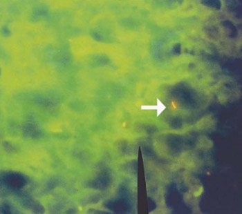
Concerning the neurological examination, the patient was alert, oriented to place and person, with a Glasgow of 15/15, isochoric and isoreactive pupils with no evidence of focalization, muscle strength of 5/5, preserved sensitivity and with a global ROT 2/4. The results of the simple CT showed a left frontal lesion with diffused limits associated to vasogenic edema with mass effect and midline displacement. She was hospitalized with a diagnosis of intracranial hypertension syndrome and frontal lesion under study. Laboratory and radiological studies were requested (Chest X-ray, CT contrast scan and magnetic resonance imaging of the brain). Medical treatment was given with amlodipine 5 mg and enalapril 20 mg every 12 hours and hydrochlorothiazide 25 mg and lovastatin 20 mg once daily. From the imaging studies, the chest X-ray was normal and a singular intra-cerebral neoplastic lesion was evidenced located in the left frontal lobe, associated with a large vasogenic edema, with mass effect, expansive growth, and displacement from the midline (Figure 1a). The blood cell profile, electrolytes, prothrombin and thromboplastin time, calcium, phosphorus, glucose, and creatinine were within normal limits, showing no signs of infection. She had no documented history of tuberculosis and for her history of a thyroidectomy of unknown etiology, the neurosurgery service considered a metastatic lesion or a high-grade glioma as diagnostic possibilities. During the craniotomy, a cortical-subcortical tumor was found and a left frontal lobectomy was performed to resect the entire tumoral lesion. The pathology service received a fragment of tissue fixed in formaldehyde 6.5 x 4 x 2.5 cm, with a vascularized surface and edematous. The sectioning was soft, light brown and with the presence of a rounded, pearl-colored, compact lesion of 2.5 cm in diameter. Four representative sections were taken for routine inclusion in paraffin and histopathologic evaluation with staining of hematoxylin/eosin. Edematous brain tissue was observed with chronic granulomatous inflammation, accompanied by abundant giant cells of the Langerhans type and extensive central caseous necrosis (Figure 1b). The initial evaluation to detect acid-fast bacilli (AFB) by using the Ziehl Neelsen (ZN) stain was negative. With the Auramine/Rhodamine stain, a scant number (3) of bacilli were identified (Figure 2a). A second examination of the slides for ZN confirmed this finding. Genomic DNA was obtained for molecular studies from the paraffin-embedded specimens. The characterization of the genus Mycobacterium was obtained by using a nested PCR that amplified a conserved region of the gene encoding the 65-kDa heat-shock protein. To identify the species, we amplified the insertion sequence IS6110, specific for the tuberculosis complex (Figure 2b). Staining with periodic acid of Schiff (PAS) for fungi was negative. Postoperative progress was satisfactory and antituberculous therapy was carried on for six months by using rifampicin, isoniazid, pyrazinamide, and ethambutol for the first two months. Subsequently, rifampicin and isoniazid were administered until the treatment was completed. The patient had complete resolution of symptoms during the seven-month follow-up period. All sputum smears during this time were negative.

FIGURE 1: (a) Cerebral Nuclear Magnetic Resonance Image where one observes the single left frontal mass lesion (white arrow) with vasogenic edema, which captures the contrast medium. (b) Formation of granulomas with surrounding lymphocytic infiltration and giant Langhans-type cells (white arrow) (hematoxylin-eosin, magnification X200).
FIGURE 2: (a) Staining with fluorochrome auramine/rhodamine in a sample of the cerebral lobe (magnification X1000). (b) Agarose gel electrophoresis of products amplified by PCR nested for IS6//0 specific for the tuberculosis complex (123 bp). MP line, molecular weight marker (100 bp); line +, positive control DNA from a strain of M. tuberculosis isolated from a TB patient; line N, negative control DNA from Bacillus cereus. Line 1-4, DNA extracted from cerebral lobe biopsies embedded in paraffin.

DISCUSSION

Cerebral tuberculoma is a rare cause of intracranial mass; clinically, it is one of the deadliest forms arising from an infection by M. tuberculosis to the CNS level in spite of having a low incidence in developed countries from 0.15 to 0.18% [4]. In Latin America and in Colombia, where tuberculosis is a priority public health problem, it accounts for 5 - 30% of brain tumours [3-8]. The course of the disease is sub-acute or chronic and the duration is from weeks to months. Its clinical manifestations are varied and non-specific, resulting from the effect of the mass of tuberculomas that can cause a variety of neurological manifestations depending on the location of the brain lesions [4-7]. Therefore, it can easily be confused with other diseases [6, 8], making the diagnosis more difficult. However, some of the most common signs and symptoms of tuberculomas in adults include headache (intracranial hypertension), seizures, altered mental status, and papilledema [3-5, 9]. This is the case of the patient in this study who consulted after experiencing an episode of momentary loss of consciousness and subsequent headache and paresthesia. No other symptoms were documented, which required looking at organicity in the genesis of syncope that demanded imaging studies (CT and cerebral MRI). Neuro-imaging studies found a solitary lesion with mass effect, which obligated making a differential diagnosis prior to surgery. As suggested by Castillo et al. [6], one should consider two main groups of processes that produce such images of the brain: tumours and infectious or parasitic processes. Of the first group, metastases, primary lymphoma of the CNS and glioblastoma multiform, and with the second group, granulomas from tuberculosis, cysticercosis and toxoplasmosis top the list. Initially, it was thought that the cause of the disease might be due to an infectious process, especially tuberculosis, because the patient came from an area of high prevalence of tuberculosis and high burden of drug-resistant TB. The absence of symptoms typical of tuberculosis with normal chest radiograph, blood tests without any alteration, absence of a history of tuberculosis and presence of a history of thyroidectomy without clear aetiology, quickly led to doubt an infectious process and considered as diagnostic possibilities a metastatic lesion or a high-degree glioma. When faced with diagnostic doubt, a pathologic diagnosis was necessary, which is the gold standard [6]. Within this context, surgery was planned to resection the lesion and for it to undergo pathologic examination. It was then that the definitive diagnosis was achieved and anti-tubercular treatment was established. This case illustrates the nonspecific presentation of cerebral tuberculoma. The lesion at the level of cerebral parenchyma or meninx may become active years after the infection [8], suggesting that to diagnose cerebral tuberculosis one must consider it, especially in countries like Colombia with a high incidence of tuberculosis. The rate at which it is diagnosed and treated is the main controlling factor preventing serious neurological sequelae.

Currently, the diagnostic tools based on clinical features - abnormal cerebrospinal fluid (CSF), imaging and biopsy characteristics - allow an approach to the diagnosis of tuberculoma [7]. Among the diagnostic methods, cytochemical study in CSF, staining of Ziehl-Neelsen in samples of CSF and brain tissue, culture studies, and chest radiograph are perhaps most valuable to diagnose this condition [9]. However, these methods have limitations worth mentioning, and in the presence of normal cellularity and suspicion of cerebral tuberculosis, consideration should be given to performing a cerebral biopsy [9], as the Ziehl-Neelsen staining has low sensitivity detecting bacilli in 25% of the cases. Given that culturing is slow due to the prolonged reproduction time for the mycobacteria [10] and its sensitivity is very low in biological samples containing a small number of microorganisms, as in CSF, such that 20% of patients with a clinical diagnosis have negative cultures. However, culturing is the most-recommended reference method [11]. In our case, it was not possible for the type of sample. Moreover, only 30% of patients with brain tuberculoma have a positive chest radiograph. The absence of features of tuberculosis on chest X-rays should, therefore, not rule out the possible existence of brain tuberculomas [12]. The chest X-ray of our patient was normal without evidence of tuberculosis. In recent decades, several methods have been developed to improve their direct detection in clinical samples, such as using fluorescent dyes, antibody detection, antigen detection and PCR-based methods [10, 13], ideal for clinical cases where the bacterial load is low. In our case, staining with auramine/rhodamine fluorescent dyes and PCR were very useful to detect the presence of mycobacteria in the brain biopsy and allowed the latter to specifically identify the infecting mycobacterial species. Increased use of computerized tomography and magnetic resonance imaging (MRI) has been of great diagnostic aid for this serious complication of tuberculosis [4, 7]. However, the radiological images are not pathognomonic and vary in number, size, and location [7]. As to imaging studies based on cerebral tomography, some controversy exists. For some, this method permits showing the typical image of tuberculomas [5, 7], while for others it suggests that its appearance is variable [8], coinciding with those appearing as lesions with increased uptake of the contrast medium in a ring, though it can also be seen as hyper-dense lesions [5] or hypo-dense [8]. However, its use alone in diagnosing tuberculoma remains questionable. Similarly, nuclear magnetic resonance imaging has a variable appearance [4, 5], although it is more sensitive than CT to show the extent of the lesions [5]. The ’target sign’ has been described as a pathognomonic finding of tuberculomas, consisting of a ring-enhancing lesion with an additional central area of enhancement or calcification [12], but it has now been shown that it is a nonspecific finding and may lead to erroneous diagnosis of CNS tuberculoma [14]. Despite increasing technological advances in imaging studies and of the antibiotic era, diagnosis and treatment of intracranial tuberculoma remains a challenge for clinicians. Given that the absence of clinical findings of systemic tubercular infection is common, tubercular lesions or a history of disease is present in only about half of the large series of patients undergoing surgery for tuberculoma [4]. As if that were not enough, tubercle bacilli is not always obvious in CSF or in the extracted mass; therefore, the negativity of the results does not rule out the possibility of tuberculosis infection [5], coupled with the low specificity of radiological examinations [8]. The combination of diagnostic tests constitutes an approach to diagnosis of intracranial tuberculoma of increasing sensitivity. Before the development of chemotherapy, intracranial tuberculoma mortality was very high [4]. It has now been shown that brain tuberculomas are curable with medical treatment [2, 8]. The standard treatment consists of an isoniazid drug of choice for good penetration into the CNS with rifampicin, streptomycin, and pyrazinamide [4-6, 9], but it is necessary to have a high suspicion in its diagnosis to justify chemotherapy [4, 10]. Although some authors like Krivoy et al. [9] justify its use for any suspicion without waiting for laboratory confirmation, which is particularly highly regarded in tuberculosis-endemic areas. This method avoids the need for a histological diagnosis of tuberculoma, although taking a biopsy is imposed when faced with diagnostic doubt or difficulty [2, 7, 9]. In this regard, the histology rules out many neoplastic aetiologies and confirms the presence of granulomas and, in any event, surgical resection is reserved for when pharmacological therapy fails, when there is increased intracranial pressure, or when there is a large, life-threatening mass effect [5, 7, 9].

CONCLUSIONS

Intracranial masses are frequently targeted as a malignant neoplastic pathology with surgical treatment in most cases. The consideration of an infectious aetiology should be an important diagnostic option in a country like Colombia, where increased frequency of tuberculosis, both pulmonary and extra-pulmonary, makes it a priority that in presence of fever, severe headache, late onset crisis and single or multiple lesions on brain scan, consideration be given to tuberculosis granulomas. Finally, we recommend a thorough evaluation of the patient, taking into account the existence of risk factors for tuberculosis, along with results from thoracic X-ray, CT and MRI brain scans, PCR and Ziehl-Neelsen staining of CSF and culturing, which together allow early identification of cerebral tuberculoma and timely initiation of antitubercular treatment, avoiding unnecessary surgical intervention and reducing the associated morbidity.

Acknowledgements

ACKNOWLEDGEMENTS

This work was carried out with resources from the Bacteriology Section of the Microbiology Department and from the Research Group Cancer Population Registry of the city of Cali (RPCC) at Universidad del Valle. We also thank the support by COLCIENCIAS and their Young Researchers program.

DECLARATION OF INTEREST

The authors report no conflicts of interest present in the development and execution of this manuscript.

REFERENCES

  1. , (). Tuberculosis en Colombia: análisis de la situación epidemiológica, año 2006. Infectio.
  2. (). Infeccion por micobacterias del sistema nervioso central. Bol Med Hosp Infant Mex.
  3. , , , , (). Tuberculomas Intracraneales: Caracteristicas tomográficas y por resonancia magnética correlación histopatológica. Revista Peruana de Radiología.
  4. , , , (). Intracranial tuberculoma: Clinical and MRI findings. Turgut Ozal Tip Merkezi Dergisi.
  5. , , , (). Tuberculoma cerebral con clÍnica pseudotumoral. Reporte de un caso clÍnico y revisión de la literatura. Rev Chil Neurocirug.
  6. , , , , , (). Tuberculoma cerebral, ¿Una patologia infrecuente?Reporte de caso. Revista de Especialidades Médico-Quirúrgicas.
  7. , , , , , (). Tuberculomas intracraneales múltiples en la infancia. Rev Neurol.
  8. , , , , (). Tuberculoma del seno cavernoso. Arch Neurocien (Mex, DF).
  9. , , (). Tuberculosis del sistema nervioso central. Experiencia del Hospital de Niños “J M de Los Rios“. Caracas 1990-2008. Gac Méd Caracas.
  10. , (). Tuberculomas cerebrales: reporte de caso. Univ Med Bogota Colombia.
  11. , , , , (). Two-laboratory collaborative study on identification of mycobac-teria: molecular versus phenotypic methods. J Clin Microbiol.
  12. , , (). Cerebral tuberculoma mimicking high grade glial tumor. Turk Neurosurg.
  13. , , , , (). Central nervous system tuberculosis: pathogenesis and clinical aspects. Clin Microbiol Rev.
  14. , , , , , (). The “target sign”:is it a specific sign of CNS tuberculoma?. Neuroradiology.