Expression of phosphorylated extracellular signal-regulated kinase in rat kidneys exposed to high +Gz

Authors

  • Hyun-Soo Kim Molecular Biology Laboratory, Aerospace Medical Center
  • Youn Wha Kim Department of Pathology, School of Medicine

DOI:

https://doi.org/10.17305/bjbms.2012.2453

Keywords:

Gz acceleration, rat, kidney, extracellular signal-regulated kinase, immunohistochemistry

Abstract

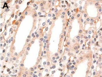
Exposure to high gravitational acceleration forces acting along the body axis from the head to the feet (+Gz) severely reduces blood flow to the visceral organs, including the kidneys. Extracellular signal-regulated kinase (ERK) figures predominantly in mediating kidney cell responses to a wide variety of stress-related stimuli. Though previous studies have shown the activation of ERK in some experimental models, the regulation of ERK associated with +Gz exposure has not yet been investigated. The aim of this study was to examine the effect of high +Gz exposure on ERK activation in the kidneys. Using a small animal centrifuge, eight male Sprague-Dawley rats were exposed to +10Gz or +13Gz three times for 3 minutes each. The bilateral kidneys were obtained from each rat, and the expression levels of phosphorylated ERK (p-ERK) were evaluated using immunohistochemistry. In the control group, the collecting duct epithelium displayed faint cytoplasmic staining with no nuclear staining of p-ERK. By contrast, rats exposed to +10Gz showed strong nuclear staining intensity for p-ERK. In the renal papilla, the epithelial cells of collecting ducts and thin segments of the loop of Henle exhibited strong nuclear immunoreactivity for p-ERK. Rats exposed to +13Gz also showed the same staining intensity and distribution of p-ERK expression as that of rats exposed to +10Gz. This study is the first to describe +Gz exposure-induced alteration in the expression of p-ERK in the kidneys. Our finding suggests that high +Gz exposure leads to the activation of ERK in the renal papilla.

Downloads

Download data is not yet available.
Expression of phosphorylated extracellular signal-regulated kinase in rat kidneys exposed to high +Gz

Downloads

Additional Files

Published

2012-11-20

How to Cite

1.
Kim H-S, Kim YW. Expression of phosphorylated extracellular signal-regulated kinase in rat kidneys exposed to high +Gz. Biomol Biomed [Internet]. 2012Nov.20 [cited 2023Jun.6];12(4):269-72. Available from: https://www.bjbms.org/ojs/index.php/bjbms/article/view/2453

Issue

Section

Biochemistry

INTRODUCTION

Mitogen-activated protein kinases (MAPKs) comprise a family of protein serine/threonine kinases which participate in signal transduction pathways that control intracellular events including acute responses to extracellular stimuli [1]. The MAPK family utilizes three parallel signaling pathways: extracellular signal-regulated kinase (ERK), stress-activated protein kinase/c-Jun N-terminal kinase and p38 MAPK [2]. In particular, activation of the ERK pathway plays an important role in the response of many cell types to a wide variety of physiological and stress-related stimuli, including ultraviolet light, heat shock, ischemia, oxygen free radicals and hyperosmolarity [3]. After binding of ligands to their respective receptors on the cell surface, a cascade of phosphorylation events leads to the activation of ERK: Ras directly interacts with and activates Raf, and Raf phosphorylates and activates MAPK/ERK kinase (MEK), which in turn phosphorylates and activates ERK [4].

The phosphorylated form of ERK (p-ERK) is then an active kinase and is able to phosphorylate a number of transcription factor targets and thus alter the pattern of gene transcription. A high gravitational acceleration force acting along the body axis from the head to the feet (+Gz) causes considerable strain on several organ systems, including the brain, heart, liver and kidneys. Exposure to high +Gz has been shown to severely decrease blood flow to the visceral organs, including the spleen, pancreas, liver and kidneys, in an apparent effort to maintain blood flow to the brain and heart [5, 6]. It is likely that the changes in the visceral blood flow are the result of some combination of +Gz-induced cardiovascular reflex responses and emotional stress that cause sympathetic vasoconstriction and an increase in the total peripheral resistance of the visceral vascular beds. Acute or chronic kidney injury results from various insults and pathological conditions, and is accompanied by the activation of compensatory repair mechanisms. Both insults and repair mechanisms are initiated by circulating factors whose cellular effects are mediated by the activation of selective signal transduction pathways. The ERK pathway is one of the main signaling pathways that are activated during these processes. For example, ERK activation plays a crucial role in the response of cultured mesangial cells to a variety of stimuli such as growth factors, angiotensin II, high glucose and mechanical stretch [7-10]. In cisplatin-induced nephrotoxicity, ERK activation mediates the renal inflammation and tubular epithelial cell apoptosis [11, 12]. In vitro studies have identified an important role for the activation of the ERK pathway in the proliferation of cultured mesangial cells, tubular epithelial cells and fibroblasts [13-15]. Furthermore, many studies have demonstrated an increase in ERK activation in diseased kidneys. There was a marked increase in tubular ERK activation after unilateral ureteral obstruction [16]. A study using renal biopsy specimens demonstrated that ERK activation in human glomerulopathies was associated with cell proliferation, histological lesions and renal dysfunction [17]. In addition, Bokemeyer et al. reported that glomerular ERK activation occurred in the rat model of mesangioproliferative glomerulonephritis and the blockade of the ERK pathway resulted in a significant reduction in mesangial cell proliferation in this disease model [18, 19]. However, the regulation of p-ERK during +Gz exposure, a condition that can adversely affect the kidneys, has not yet been described. The purpose of this study was to examine the effects of high +Gz exposure on the expression of p-ERK in the kidneys using immunohistochemical staining.

MATERIALS AND METHODS

Experimental Animals and +Gz Exposure Protocol

Ten male Sprague-Dawley rats, seven to eight weeks of age and weighing between 200 and 230 g, were purchased from Samtako Bio Korea Co., Ltd. (Osan-si, Gyeonggi-do, Republic of Korea). Throughout the experimental period, animals were fed standard laboratory rat chow, provided with free access to water and maintained on a 12:12-hour light-dark cycle with temperature controlled at 20 to 25°C in pathogen-free conditions. Rats were randomly assigned to three experimental groups. Eight of 10 rats were exposed to +10GZ (4 rats) or +13GZ (4 rats) three times for 3 minutes each, and the remaining 2 rats were assigned to the control group (+1Gz). Rats were placed inside a cylindrical plastic restraint device which, when mounted in a centrifuge, allowed +Gz to be delivered along their rostro-caudal axes. After the rats were secured, the restraint device was placed onto a small animal centrifuge. During the intervals between centrifuge runs, rats were allowed to move freely in the cage.

Histochemistry

At the end of the experiment, animals were anesthetized with sodium pentobarbital (45 mg/kg) and laparotomized via midline incision. The bilateral kidneys obtained from the control and centrifuged animals were immediately preserved in a 10% formaldehyde (formalin) solution. After 48 to 72 hours of formalin fixation, the entire kidney was dissected. Subsequently, the kidney tissues were embedded in paraffin and processed for routine histology staining, including hematoxylin-eosin, periodic acid-Schiff, Masson trichrome and periodic acid-methenamine-silver, and immunohistochemical staining.

Immunohistochemistry

p-ERK expression was assessed by immunostaining using the Bond Polymer Intense Detection System (Vision BioSystems, Mount Waverley, Victoria, Australia), following the manufacturer’s instructions. To summarize, 4-μm sections of formalin-fixed, paraffin-embedded tissue were deparaffinized with Bond Dewax Solution (Vision BioSystems), and an antigen retrieval procedure was performed using Bond ER Solution (Vision BioSystems) for 30 minutes at 100°C. Endogenous peroxidases were quenched by incubation with hydrogen peroxide for 5 minutes. The sections were incubated for 15 minutes at ambient temperature with a rabbit polyclonal anti-phospho-p44/42 MAPK (ERK1/2) (Thr202/Tyr204) antibody (1:200, Cell Signaling Technology, Incorporated, MA, USA). The biotin-free polymeric horseradish peroxidase-linker antibody conjugate system was used in the Bond-maX™ automatic slide stainer (Vision BioSystems), and visualization was performed by using a 3.3-diaminobenzidine (DAB) solution (1 mM DAB, 50 mM Tris-HCl buffer [pH 7.6] and 0.006% H2O2). Nuclei were counterstained with hematoxylin. Slides were subsequently dehydrated following a standard procedure and sealed with coverslips. In order to minimize interassay variation, positive and negative control samples were included in each run. The positive control sample was lung cancer tissue. The negative control was prepared by substituting non-immune serum for the primary antibody; no detectable staining was evident. Because the nuclear translocation of ERK may be necessary for ERK-activated transcription, strong nuclear staining was considered positive for p-ERK.

RESULTS

Upon gross inspection, the bilateral kidneys of all rats appeared normal. No discoloration, hemorrhage, nodularity, shrinkage or scarring was detected on the capsular surfaces. The centrifuged rats showed no significant alterations in the size or weight of their kidneys. All parts of the kidneys cut with great ease. In the renal cut surface, the centrifuged rats revealed no pathologic abnormalities; no evidence of infarction, hemorrhage, mass or cystic lesion was identified. The pelvicalyceal system remained intact. Histologically, the centrifuged rats showed no significant morphological changes in comparison to those of the control group; no evidence of glomerular capillary wall lesion, mesangial hypercellularity, tubular atrophy or interstitial fibrosis was detected.

Immunohistochemically, in the control group, the collecting duct (CD) epithelium displayed faint cytoplasmic p-ERK staining (Figure 1A). No nuclear p-ERK immunoreactivity was detected in the kidneys of the control group. By contrast, rats exposed to +10Gz showed strong nuclear staining intensity for p-ERK. In the renal papilla, the epithelial cells of CDs and thin segments of the loop of Henle (TLHs) exhibited strong nuclear and cytoplasmic immunoreactivity for p-ERK (Figure 1B). Rats exposed to +13Gz showed the same staining intensity and distribution of p-ERK expression as that of rats exposed to +10Gz; intense p-ERK staining was observed in the nuclei and cytoplasm of epithelial cells lining CDs and TLHs (Figure 1C). The glomeruli and cortical tubules and interstitium demonstrated no significant immunoreactivity.

FIGURE 1: Representative photomicrographs showing p-ERK immunoreactivity in rat kidneys exposed to high +Gz. (A) The control group shows faint p-ERK staining in the cytoplasm of collecting duct epithelium. The nuclear staining is absent. (B) By contrast, rats exposed to +10Gz show a significantly increased nuclear p-ERK staining intensity compared to the control group. The epithelial cells of collecting ducts and thin segments of the loop of Henle (arrow) in the renal papilla exhibit strong nuclear immunoreactivity for p-ERK. (C) Rats exposed to +13Gz show the same staining intensity and distribution of renal p-ERK expression as that of +10Gz-exposed rats. (Polymer method. Original magnification, A to C, ×400)

DISCUSSION

In this study, the subcellular localization of renal p-ERK after centrifugation was determined using immunohistochemical staining. A significantly increased level of nuclear p-ERK expression was identified in the CD and TLH epithelium of the renal papilla after exposure to both +10Gz and +13Gz. This finding suggests that high +Gz exposure up-regulates renal p-ERK. Our study is the first to describe +Gz exposure-induced alteration to p-ERK expression, while the precise mechanism by which +Gz exposure alters the expression of p-ERK in the renal papilla remains unknown. The renal papilla is particularly vulnerable to hypoxia/ischemia because of the peculiar arrangement of their blood supply and the hypertonic environment. The vasa recta form wide and plentiful vascular bundles at the base of the medullary pyramid, but the bundles taper as they continue distally toward the apex. As a result, the papilla receives only a marginal blood supply, a predisposing factor for ischemia and the subsequent development of renal papillary necrosis. On the basis of a previous study demonstrating a marked reduction in blood flow to the kidneys during +Gz exposure [5, 6], we hypothesized that hypoxia due to high +Gz-induced inadequate renal blood flow to the renal papilla might be attributable to the up-regulation of p-ERK. This hypothesis is supported by a cell culture study indicating that hypoxia led to an increase in ERK phosphorylation [20]. Ko et al. [21] also provided evidence for hypoxia-induced ERK activation in their study demonstrating that caspase-3 activation and apoptosis was accompanied by increased phosphorylation of ERK in renal epithelial cells cultured in a hypoxic chamber. Thus, it is reasonable to assume that high +Gz exposure, which causes displacement of blood towards lower extremities and a subsequent severe reduction in blood flow to the kidneys, would exacerbate hypoxia in the renal papilla and lead to the activation of ERK. The possibility also that the increased expression of renal p-ERK might be caused by repeated +Gz exposure-induced reperfusion injury cannot be excluded. We hypothesized that reperfusion injury might occur when the blood supply returned to the kidneys after a period of hypoxia/ischemia due to +Gz exposure (i.e., during the intervals between centrifuge runs). This hypothesis is supported by previous data showing that ERK was transiently activated in renal epithelial cells during the reperfusion after ischemia [22]. Previous studies have reported that ERK activation was enhanced after renal ischemia [23, 24], and the ERK pathway has been implicated as a modulator of ischemia/reperfusion injury in the kidneys [25, 26]. Reperfusion after ischemia has been documented to cause the accumulation of reactive oxygen species, which then play a role as intracellular signaling molecules. Increasing evidence supports the idea that reactive oxygen species cause phosphorylation of growth factor receptors, leading to downstream activation of the Raf/MEK/ERK cascade [27, 28]. Further studies are necessary to clarify the relationship between the expression of p-ERK and accumulation of reactive oxygen species in kidneys exposed to high +Gz.

CONCLUSION

In conclusion, this study is the first to report the altered expression of renal p-ERK associated with high +Gz exposure. We demonstrated significantly increased p-ERK immunoreactivity in the papilla of rat kidneys exposed to +Gz, suggesting that after high +Gz exposure, the activation of ERK occurs in the kidneys.

Acknowledgements

ACKNOWLEDGEMENTS

The views and opinions expressed in this article are those of the author and do not reflect the official policy or position of the Republic of Korea Air Force. The author thanks histotechnician Jung Hee Park for sharing her expertise in immunohistochemistry and medical librarian Ja Ok Kim for her assistance searching for literature and establishing key words.

DECLARATION OF INTEREST

The author declared that he had no conflicts of interest with respect to his authorship or the publication of this article.

REFERENCES

  1. , , , , , (). Mitogen-activated protein (MAP) kinase pathways: regulation and physiological functions. Endocr Rev.
  2. , (). Mitogen and stress response pathways: MAP kinase cascades and phosphatase regulation in mammals and yeast. Curr Opin Cell Biol.
  3. , , (). MAPK signaling and the kidney. Am J Physiol Renal Physiol.
  4. , , , , , (). Roles of the Raf/MEK/ERK pathway in cell growth, malignant transformation and drug resistance. Biochim Biophys Acta.
  5. , , (). Regional distribution of cardiac output in unanesthetized baboons during +Gz stress with and without an anti-G suit. Aviat Space Environ Med.
  6. , , (). Renal blood flow in miniature swine during +GZ stress and anti-G suit inflation. J Appl Physiol.
  7. , , , , , (). Mitogen-activated protein kinase cascade is activated in glomeruli of diabetic rats and glomerular mesangial cells cultured under high glucose conditions. Diabetes.
  8. , , , (). Platelet-derived growth factor and angiotensin II stimulate the mitogen-activated protein kinase cascade in renal mesangial cells: comparison of hypertrophic and hyperplastic agonists. Biochem J.
  9. , , , , (). Activation of mesangial cell signaling cascades in response to mechanical strain. Kidney Int.
  10. , , (). Sustained ERK-2 activation in rat glomerular mesangial cells: differential regulation by protein phosphatases. Am J Physiol.
  11. , , , , (). Cisplatin-induced cell death is EGFR/src/ERK signaling dependent in mouse proximal tubule cells. Am J Physiol Renal Physiol.
  12. , (). Cisplatin nephrotoxicity: mechanisms and reno-protective strategies. Kidney Int.
  13. , , (). Isoproterenol inhibits fibroblast growth factor-2-induced growth of renal epithelial cells. Pediatr Nephrol.
  14. , , , , , (). Differential contribution of three mitogen-activated protein kinases to PDGF-BB-induced mesangial cell proliferation and gene expression. J Am Soc Nephrol.
  15. , , , , , (). Hepatocyte growth factor regulates proteoglycan synthesis in interstitial fibroblasts. Kidney Int.
  16. , , , , , (). Activation of the ERK pathway precedes tubular proliferation in the obstructed rat kidney. Kidney Int.
  17. , , , , , (). Activation of the extracellular-signal regulated protein kinase pathway in human glomerulopathies. J Am Soc Nephrol.
  18. , , , , , (). Differential activation of mitogen-activated protein kinases in experimental mesangioproliferative glomerulonephritis. J Am Soc Nephrol.
  19. , , , , , (). In vivo identification of the mitogen-activated protein kinase cascade as a central pathogenic pathway in experimental mesangioproliferative glomerulonephritis. J Am Soc Nephrol.
  20. , , , , , (). Endothelial exposure to hypoxia induces Egr-1 expression involving PK- Calpha-mediated Ras/Raf-1/ERK1/2 pathway. J Cell Physiol.
  21. , , , , , (). Requirement of ERK activation in hypoxia-induced caspase activation and apoptosis of cultured tubular cells. Korean J Nephrol.
  22. , , , , , (). Signal transduction of MEK/ERK and PI3K/Akt activation by hypoxia/reoxygenation in renal epithelial cells. Eur J Cell Biol.
  23. , , (). Prevention of kidney ischemia/reperfusion-induced functional injury and JNK, p38, and MAPK kinase activation by remote ischemic pretreatment. J Biol Chem.
  24. , , , , (). Prevention of kidney ischemia/reperfusion-induced functional injury, MAPK and MAPK kinase activation, and inflammation by remote transient ureteral obstruction. J Biol Chem.
  25. , , , , , (). Renal ischemia/reperfusion and ATP depletion/repletion in LLC-PK(1) cells result in phosphorylation of FKHR and FKHRL1. Kidney Int.
  26. , , (). MAPK activation determines renal epithelial cell survival during oxidative injury. Am J Physiol.
  27. , , , , , (). EGF-Receptor phosphorylation and signaling are targeted by H2O2 redox stress. Am J Respir Cell Mol Biol.
  28. , , , , (). Activation of mitogen-activated protein kinase by H2O2. Role in cell survival following oxidant injury. J Biol Chem.