Persistent submacular fluid diagnosed with Optical Coherence Tomography after successful scleral buckle surgery for macula-off retinal detachment
Abstract
The objective of our study was to analyze the presence of persistent submacular fluid after apparently successful scleral buckle surgery for macula-off retinal detachment, using Optical Coherence Tomography (OCT), and to correlate these findings to postoperative visual acuity. Prospective study of consecutive patients hospitalized for macula-off retinal detachment, between February 2008 and April 2010, was carried out at the single referral centre for vitreoretinal surgery in Serbia. Outcomes were evaluated 1, 3, 6 and 12 months postoperatively, using OCT and best corrected visual acuity (BCVA). All 48 patients with macula-off retinal detachment had undergone clinically successful scleral buckle surgery, 3 to 30 days (mean 14.2 ± 6.9) after the onset of symptoms. Reattachment was achieved and one month later, patients were divided in two groups, according to the presence of submacular fluid assessed by OCT group A with detectable residual submacular fluid (40%) and group B without (60%).
The mean BCVA one month postoperatively was significantly lower (p<0.01) in the group A (0.11 ± 0.03) opposed to the group B (0.51 ± 0.11).
However, after a 12-month-follow-up, BCVA was similar in both groups (0.52 ± 0.17 and 0.54 ± 0.15 respectively, p=0.541), due to the restitution of normal macular architecture confirmed by OCT.
The prolonged recovery of visual acuity in patients with macula-off retinal detachment who had undergone successful scleral buckle surgery could be explained with presence of submacular fluid, diagnosed by OCT. However, final visual outcome was favourable, due to the gradual resorption of submacular fluid in a 12-month-period.
Keywords
macula-off retinal detachment scleral buckle surgery optical coherence tomography submacular fluidFull text
Introduction
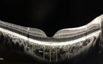
Optical Coherence Tomography (OCT) is a non-invasive, high-resolution, real-time imaging technique that enables the visualization of the retina and optic nerve head and analysis of their changes in vivo. Furthermore, Spectral domain Optical Coherence Tomography (SOCT) is a highly sensitive method for visualization of retinal layers with axial resolution of 4 to 6 μm [1]. Many factors are identified that might influence the final outcome of scleral buckle surgery (SBS) for rhegmatogenous retinal detachment and include preoperative findings such as: preoperative visual acuity [2-4], patient age [5], anatomical features such as whether macula is attached or detached, duration of macular detachment [2,3], extent of retinal detachment [6], position and number of retinal breaks [2, 4], refraction (myopia, as a predisposing factor for retinal breaks) [7] and formation of proliferative vitreoretinopathy [4]. Besides the duration of macular detachment (DMD), many ultrastructural intraretinal abnormalities that occur in affected macula may compromise the final outcome following successful SBS for macula-off retinal detachment. Although postoperatively macula appears clinically reattached, development of cystoid macular oedema, epiretinal membrane or persistent submacular fluid (SMF) may affect the visual recovery and all of those changes can be visualized by OCT [8-10]. In case of persistent SMF, postoperative best corrected visual acuity (BCVA) can vary depending on the amount of the persistent SMF, but at the course of time, as SMF fluid resolves, BCVA can improve. The aim of this prospective observational study is to evaluate the foveal architecture by OCT, with focus on persistent SMF and its effect on postoperative visual acuity, after clinically successful SBS.
Materials and methods
Patients
A prospective study of the consecutive patients hospitalized for macula-off retinal detachment who underwent scleral buckle surgery, between 01 February 2008 and 30 April 2010, at the Department for vitreoretinal surgery of the Clinic for Eye Diseases in Belgrade, was performed. This Department is the single referral centre for vitreoretinal surgery in Serbia.
Procedures
All patients underwent complete ophthalmological examination including medical history, visual acuity assessment (measured by Snellen chart at 6 m and converted to decimal notation), applanation tonometry, slit lamp examination, indirect ophthalmoscopy with 90D lens, fundus photography and OCT. Outcomes were evaluated 1, 3, 6 and 12 months postoperatively, using OCT (SOCT Copernicus, Optopol Technology S.A., Zawiercie, Poland) and best corrected visual acuity (BCVA). Inclusion criteria of the study: all patients with uncomplicated retinal detachment with macular involvement (macula-off), who had a scleral buckle surgery performed. Exclusion criteria were: 1) duration of the retinal detachment longer than one month, 2) complicated retinal detachment in which SBS could not be performed (multiple retinal tears localized in more than 2 quadrants, gigantic tear, posterior tear, unreachable for scleral buckling, extensive proliferative vitreoretinopathy), therefore indicated for vitrectomy, 3) associated ocular pathology (amblyopia, cataract, glaucoma, corneal or macular diseases) or a history of a previous eye surgery or ocular trauma, 4) systemic diseases such as: diabetes, neurological diseases. In all cases, SBS was performed by the same surgeon. The surgical procedure included: transscleral cryopexy of the retina using a cryo-probe, stitching of an encircling silicone band (2.5 mm) and a silicone explant to the sclera (one or more, corresponding in size and localisation to the position of the retinal tears). External drainage of subretinal fluid was not performed due to the potential complications. Fundus photography and OCT were done at the discharge from the hospital (3-5 days postoperatively), as well as during follow-up 1, 3, 6 and 12 months after surgery. The study has followed the tenets of the Declaration of Helsinki, and was approved by the Ethics Committee of the Clinical Centre of Serbia. All patients were informed about the study and had signed the informed consent.
Statistical analysis
Data were analyzed using SPSS 15.0. Methods used included Student's t-test for comparison of numerical variables, and X2 or Mann-Whitney U test (as appropriate) for categorical variables. The level of statistical significance was 0.05.
Results
In the study period, 48 patients (48 eyes) had undergone scleral buckle surgery for macula-off retinal detachment. All patients were symptomatic and the onset of symptoms lasted between 3 and 30 days (mean 14 days) prior the SBS, which was considered as the time of duration of retinal (macular) detachment. The average age of patients was 44.15 ± 16.07 years. Among them, 44% were males and 56% females (Table 1). Preoperatively, BCVA varied from light perception to 0.1. One month postoperatively, on control fundus photography retina was reattached in all cases (Figure 1 and 2). However, OCT examination revealed differences in the amount of residual submacular fluid: while in some cases retina was completely reattached (Figure 1), in others residual submacular fluid persisted (Figure 2). Patients were therefore divided in two groups depending on the OCT findings: group A, patients with residual SMF (40%) and group B, patients with macula reattached (60%) (Table 1). Groups were similar in respect to the gender (X2=0.002, p=0.963), age distribution (p=0.067) and mean duration of retinal detachment (p=0.582) (Table 1). One month postoperatively there was an obvious and significant difference in BCVA between groups (p<0.01), whereas a minimal recovery was observed in group A (0.11 ± 0.03) comparing to the group B (0.51 ± 0.11). However, at 12 months after surgery, significant improvement of visual acuity (p<0.01) was registered in group A, with final BCVA 0.52 ± 0.17 and mean increase in visual acuity of 0.41. On the other hand, final visual acuity (0.54 ± 0.15) didn’t differ much in group B comparing to the one-month-postoperative control, with insignificant (p=0.388) mean increase in visual acuity of 0.03 (Table 1). At examinations after 3 and 6 months postoperatively, the trend of VA improvement could be observed in both groups, but was significant only in group A (Figure 3). Finally, visual outcome after a 12 month-follow-up was similar in both groups (p=0.541) (Table 1).
Discussion
Scleral buckle surgery is an excellent surgical procedure for the repair of uncomplicated retinal detachment. Success rate of this type of surgery is 85-95% [3, 6]. The results of the surgery depend mainly on the duration of the retinal detachment, especially on the time lapse between the separation of the sensory retina from the retinal pigment epithelium (RPE) in the macular area and the surgical repair (duration of macular detachment, DMD). Studies have shown that duration of macular detachment less than 7 days has a better prognosis and better final visual outcome comparing to cases with longer DMD [3, 4]. However, despite the excellent postoperative anatomical results, in cases with macula-off retinal detachment repair, functional recovery is limited. The major reason for that is a permanent damage to the macula during detachment from the RPE. Development of OCT technology allowed the investigation of those pathoanatomical changes and several studies have shown that after clinically successful scleral buckle surgery for macula-off RD, SMF persists in almost half of the cases (47-66.6 %) [1, 11-14]. Benson et al. [13] identified 3 patterns of submacular fluid: confluent fluid, a single discrete bleb of fluid and multiple blebs of fluid. However even fine microstructural changes within the photoreceptor layer such as disruption of the inner segment/outer segment junction, irregular hyporeflectivity in the photoreceptor outer segments, discontinuation of the external limiting membrane and hyperreflective spots in the outer nuclear layer could be detected and are associated with lower postoperative visual acuity [1, 10, 15, 16]. In addition, cystoid macular oedema and epiretinal membranes may also develop and could be visualized by OCT [15, 17, 18]. All those abnormalities are usually accompanied by symptoms such as prolonged, incomplete visual recovery [13, 19, 20] and metamorphopsia [16, 21]. Both studies of Gharbiya et al. [10] and Cavallini et al. [14] found that presence of persistent SMF correlated with postoperative visual function and with final visual outcome. In most of the studies, SMF was fully reabsorbed within the period of 6 to 12 months after scleral buckle surgery [8, 9, 12], or could be detected at 12 months postoperatively in a small percentage (6-9 %) in the studies of Hagimura et al. [11], Wolfensberger et al. [20] and Gibran et al. [22]. In our study, SMF was completely reabsorbed in all cases after a 12-month-follow-up and thus yield a good visual recovery irrespectively to the fact that one-month-postoperatively in 40% of cases presence of residual SMF deteriorated visual acuity (to only 0.11 ± 0.03). Therefore, mean final best corrected visual acuity was 0.5 in all cases (Table 1). Prolonged visual recovery in some patients after successful SBS for macula-off retinal detachment could be explained by postoperative presence of persistent SMF detected by OCT, which can last even up to 12 months. In the vast majority of those cases, gradual resorption of SMF in the period of 12 months results in the gradual recovery of visual function. Similarly to our results, Benson et al. [13], Wolfensberger et al. [20] and Hagimura et al. [11] found that improvement of visual acuity can last up to 12 months after surgery and that it correlates with the amount of residual SMF. On the contrary, in the study of Baba et al. [23] the amount of residual SMF didn’t correlate with the final visual acuity. This may be due to the shorter follow up period in the latter study, of only 6 months. Finally, as in our study, Lecleire-Collet et al. [24] concluded that presence of postoperative “infraclinical” foveal detachment did not influence the final visual acuity. Those findings are confirmed by Seo et al. [12], who furthermore stated that the amount of SMF present one month after surgery did not correlate with the time necessary for it to absorb. However, in the study of Lecleire-Collet et al. [19] final visual acuity negatively correlated with height of the detachment at the central fovea and distance from the central fovea to the nearest undetached retina.
Conclusion
In conclusion, presence of SMF, diagnosed with OCT, can explain the extended recovery of visual acuity in patients after successful scleral buckle surgery for retinal detachment involving macula. Nevertheless, SMF does not influence the final visual acuity and it withdraws completely during the period of 12 months after surgery. Additional studies are needed to evaluate the effects of submacular fluid not only on the final visual acuity, but on the other visual function tests as well, such as contrast sensitivity, colour vision, microperimetry and multifocal electroretinography.
Declaration of interest
The authors declare no conflict of interest.
References
- Delolme MP, Dugas B, Nicot F, Muselier A, Bron AM, Creuzot-Garcher C. Anatomical and functional macular changes after rhegmatogenous retinal detachment with macula off. Am J Ophthalmol 2012;153(1):128–136.
- Heussen N, Hilgers RD, Heimann H, Collins L, Grisanti S. Scleral buckling versus primary vitrectomy in rhegmatogenous retinal detachment study (SPR study): multiple-event analysis of risk factors for reoperations. SPR Study report no. 4. Acta Ophthalmol 2011;89(7):622–628.
- Oshima Y, Yamanishi S, Sawa M, Motokura M, Harino S, Emi K. Two-year follow-up study comparing primary vitrectomy with scleral buckling for macula-off rhegmatogenous retinal detachment. Jpn J Ophthalmol 2000;44(5):538–549.
- Doyle E, Herbert EN, Bunce C, Williamson TH, Laidlaw DA. How effective is macula-off retinal detachment surgery. Might good outcome be predicted?. Eye (Lond) 2007;21(4):534–540.
- Pastor JC, Fernández I, Rodríguez de la Rúa E, Coco R, Sanabria-Ruiz Colmenares MR, Sánchez-Chicharro D, et al. Surgical outcomes for primary rhegmatogenous retinal detachments in phakic and pseudophakic patients: the Retina 1 Project--report 2. Br J Ophthalmol 2008;92(3):378–382.
- Thelen U, Amler S, Osada N, Gerding H. Outcome of surgery after macula-off retinal detachment - results from MUSTARD, one of the largest databases on buckling surgery in EuropeResults from a large German case series. Acta Ophthalmol 2010. DOI: 10.1111/j.1755-3768.2010.01939.x
- Alimanovic EH. Correlation between bulbar axis length and retinal ruptures in case of myopia eye. Bosn J Basic Med Sci 2009;9(3):187–190.
- Abouzeid H, Becker K, Holz FG, Wolfensberger TJ. Submacular fluid after encircling buckle surgery for inferior macula-off retinal detachment in young patients. Acta Ophthalmol 2009;87(1):96–99.
- Gibran SK, Alwitry A, Cleary PE. Foveal detachment after successful retinal reattachment for macula on rhegmatogeneous retinal detachment: an ocular coherence tomography evaluation. Eye (Lond) 2006;20(11):1284–1287.
- Gharbiya M, Grandinetti F, Scavella V, Cecere M, Esposito M, Segnalini A, et al. Correlation between spectral-domain optical coherence tomography findings and visual outcome after primary rhegmatogenous retinal detachment repair. Retina 2012;32(1):43–53.
- Hagimura N, Iida T, Suto K, Kishi S. Persistent foveal retinal detachment after successful rhegmatogenous retinal detachment surgery. Am J Ophthalmol 2002;133(4):516–520.
- Seo JH, Woo SJ, Park KH, Yu YS, Chung H. Influence of persistent submacular fluid on visual outcome after successful scleral buckle surgery for macula-off retinal detachment. Am J Ophthalmol 2008;145(5):915–922.
- Benson SE, Schlottmann PG, Bunce C, Xing W, Charteris DG. Optical coherence tomography analysis of the macula after scleral buckle surgery for retinal detachment. Ophthalmology 2007;114(1):108–112.
- Cavallini GM, Masini C, Volante V, Pupino A, Campi L, Pelloni S. Visual recovery after scleral buckling for macula-off retinal detachments: an optical coherence tomography study. Eur J Ophthalmol 2007;17(5):790–796.
- Wakabayashi T, Oshima Y, Fujimoto H, Murakami Y, Sakaguchi H, Kusaka S, et al. Foveal microstructure and visual acuity after retinal detachment repair: imaging analysis by Fourier-domain optical coherence tomography. Ophthalmology 2009;116(3):519–528.
- Rossetti A, Doro D, Manfre A, Midena E. Long-term follow-up with optical coherence tomography and microperimetry in eyes with metamorphopsia after macula-off retinal detachment repair. Eye (Lond) 2010;24(12):1808–1813.
- Schocket LS, Witkin AJ, Fujimoto JG, Ko TH, Schuman JS, Rogers AH, et al. Ultrahigh-resolution optical coherence tomography in patients with decreased visual acuity after retinal detachment repair. Ophthalmology 2006;113(4):666–672.
- Abouzeid H, Wolfensberger TJ. Macular recovery after retinal detachment. Acta Ophthalmol Scand 2006;84(5):597–605.
- Lecleire-Collet A, Muraine M, Menard JF, Brasseur G. Predictive visual outcome after macula-off retinal detachment surgery using optical coherence tomography. Retina 2005;25(1):44–53.
- Wolfensberger TJ, Gonvers M. Optical coherence tomography in the evaluation of incomplete visual acuity recovery after maculaoff retinal detachments. Graefes Arch Clin Exp Ophthalmol 2002;240(2):85–89.
- Wang Y, Li SY, Zhu M, Chen SJ, Liu Y, Men XH, et al. Metamorphopsia after successful retinal detachment surgery: an optical coherence tomography study. Acta Ophthalmol Scand 2005;83(2):168–171.
- Gibran SK, Cleary PE. Ocular coherence tomographic examination of postoperative foveal architecture after scleral buckling vs vitrectomy for macular off retinal detachment. Eye (Lond) 2007;21(9):1174–1178.
- Baba T, Hirose A, Moriyama M, Mochizuki M. Tomographic image and visual recovery of acute macula-off rhegmatogenous retinal detachment. Graefes Arch Clin Exp Ophthalmol 2004;242(7):576–581.
- Lecleire-Collet A, Muraine M, Menard JF, Brasseur G. Evaluation of macular changes before and after successful retinal detachment surgery using stratus-optical coherence tomography. Am J Ophthalmol 2006;142(1):176–179.
About this article
Correspondence
*Corresponding author: Aleksandra Radosavljević, Clinic for Eye Diseases, Clinical Centre of Serbia, Pasterova 2, 11000 Belgrade, Serbia Telephone: +381 66 83 01 210; Fax: +381 11 26 88 164 e-mail: [email protected]
Article history
Received: 2012-02-16 · Accepted: 2012-04-23
Copyright
Copyright: © Association of Basic Medical Sciences of FBIH. All rights reserved
Downloads
References
Delolme MP, Dugas B, Nicot F, Muselier A, Bron AM, Creuzot-Garcher C. Anatomical and functional macular changes after rhegmatogenous retinal detachment with macula off. Am J Ophthalmol. 2012;153(1):128-136. DOI: https://doi.org/10.1016/j.ajo.2011.06.010
Heussen N, Hilgers RD, Heimann H, Collins L, Grisanti S, SPR Study group. Scleral buckling versus primary vitrectomy in rhegmatogenous retinal detachment study (SPR study): multiple-event analysis of risk factors for reoperations. SPR Study report no. 4. Acta Ophthalmol. 2011;89(7):622-628. DOI: https://doi.org/10.1111/j.1755-3768.2009.01766.x
Oshima Y, Yamanishi S, Sawa M, Motokura M, Harino S, Emi K. Two-year follow-up study comparing primary vitrectomy with scleral buckling for macula-off rhegmatogenous retinal detachment. Jpn J Ophthalmol. 2000;44(5):538-549. DOI: https://doi.org/10.1016/S0021-5155(00)00205-7
Doyle E, Herbert EN, Bunce C, Williamson TH, Laidlaw DA. How effective is macula-off retinal detachment surgery. Might good outcome be predicted?. Eye (Lond). 2007;21(4):534-540. DOI: https://doi.org/10.1038/sj.eye.6702260
Pastor JC, Fernández I, Rodríguez de la Rúa E, Coco R, Sanabria-Ruiz Colmenares MR, Sánchez-Chicharro D, et al. Surgical outcomes for primary rhegmatogenous retinal detachments in phakic and pseudophakic patients: the Retina 1 Project--report 2. Br J Ophthalmol. 2008;92(3):378-382. DOI: https://doi.org/10.1136/bjo.2007.129437
Thelen U, Amler S, Osada N, Gerding H. Outcome of surgery after macula-off retinal detachment - results from MUSTARD, one of the largest databases on buckling surgery in EuropeResults from a large German case series. Acta Ophthalmol. 2010. doi:10.1111/j.1755-3768.2010.01939.x DOI: https://doi.org/10.1111/j.1755-3768.2010.01939.x
Alimanovic EH. Correlation between bulbar axis length and retinal ruptures in case of myopia eye. Bosn J Basic Med Sci. 2009;9(3):187-190. DOI: https://doi.org/10.17305/bjbms.2009.2804
Abouzeid H, Becker K, Holz FG, Wolfensberger TJ. Submacular fluid after encircling buckle surgery for inferior macula-off retinal detachment in young patients. Acta Ophthalmol. 2009;87(1):96-99. DOI: https://doi.org/10.1111/j.1755-3768.2008.01196.x
Gibran SK, Alwitry A, Cleary PE. Foveal detachment after successful retinal reattachment for macula on rhegmatogeneous retinal detachment: an ocular coherence tomography evaluation. Eye (Lond). 2006;20(11):1284-1287. DOI: https://doi.org/10.1038/sj.eye.6702098
Gharbiya M, Grandinetti F, Scavella V, Cecere M, Esposito M, Segnalini A, et al. Correlation between spectral-domain optical coherence tomography findings and visual outcome after primary rhegmatogenous retinal detachment repair. Retina. 2012;32(1):43-53. DOI: https://doi.org/10.1097/IAE.0b013e3182180114
Hagimura N, Iida T, Suto K, Kishi S. Persistent foveal retinal detachment after successful rhegmatogenous retinal detachment surgery. Am J Ophthalmol. 2002;133(4):516-520. DOI: https://doi.org/10.1016/S0002-9394(01)01427-1
Seo JH, Woo SJ, Park KH, Yu YS, Chung H. Influence of persistent submacular fluid on visual outcome after successful scleral buckle surgery for macula-off retinal detachment. Am J Ophthalmol. 2008;145(5):915-922. DOI: https://doi.org/10.1016/j.ajo.2008.01.005
Benson SE, Schlottmann PG, Bunce C, Xing W, Charteris DG. Optical coherence tomography analysis of the macula after scleral buckle surgery for retinal detachment. Ophthalmology. 2007;114(1):108-112. DOI: https://doi.org/10.1016/j.ophtha.2006.07.022
Cavallini GM, Masini C, Volante V, Pupino A, Campi L, Pelloni S. Visual recovery after scleral buckling for macula-off retinal detachments: an optical coherence tomography study. Eur J Ophthalmol. 2007;17(5):790-796. DOI: https://doi.org/10.1177/112067210701700517
Wakabayashi T, Oshima Y, Fujimoto H, Murakami Y, Sakaguchi H, Kusaka S, et al. Foveal microstructure and visual acuity after retinal detachment repair: imaging analysis by Fourier-domain optical coherence tomography. Ophthalmology. 2009;116(3):519-528. DOI: https://doi.org/10.1016/j.ophtha.2008.10.001
Rossetti A, Doro D, Manfre A, Midena E. Long-term follow-up with optical coherence tomography and microperimetry in eyes with metamorphopsia after macula-off retinal detachment repair. Eye (Lond). 2010;24(12):1808-1813. DOI: https://doi.org/10.1038/eye.2010.138
Schocket LS, Witkin AJ, Fujimoto JG, Ko TH, Schuman JS, Rogers AH, et al. Ultrahigh-resolution optical coherence tomography in patients with decreased visual acuity after retinal detachment repair. Ophthalmology. 2006;113(4):666-672. DOI: https://doi.org/10.1016/j.ophtha.2006.01.003
Abouzeid H, Wolfensberger TJ. Macular recovery after retinal detachment. Acta Ophthalmol Scand. 2006;84(5):597-605. DOI: https://doi.org/10.1111/j.1600-0420.2006.00676.x
Lecleire-Collet A, Muraine M, Menard JF, Brasseur G. Predictive visual outcome after macula-off retinal detachment surgery using optical coherence tomography. Retina. 2005;25(1):44-53. DOI: https://doi.org/10.1097/00006982-200501000-00006
Wolfensberger TJ, Gonvers M. Optical coherence tomography in the evaluation of incomplete visual acuity recovery after maculaoff retinal detachments. Graefes Arch Clin Exp Ophthalmol. 2002;240(2):85-89. DOI: https://doi.org/10.1007/s00417-001-0410-6
Wang Y, Li SY, Zhu M, Chen SJ, Liu Y, Men XH, et al. Metamorphopsia after successful retinal detachment surgery: an optical coherence tomography study. Acta Ophthalmol Scand. 2005;83(2):168-171. DOI: https://doi.org/10.1111/j.1600-0420.2005.00413.x
Gibran SK, Cleary PE. Ocular coherence tomographic examination of postoperative foveal architecture after scleral buckling vs vitrectomy for macular off retinal detachment. Eye (Lond). 2007;21(9):1174-1178. DOI: https://doi.org/10.1038/sj.eye.6702429
Baba T, Hirose A, Moriyama M, Mochizuki M. Tomographic image and visual recovery of acute macula-off rhegmatogenous retinal detachment. Graefes Arch Clin Exp Ophthalmol. 2004;242(7):576-581. DOI: https://doi.org/10.1007/s00417-004-0884-0
Lecleire-Collet A, Muraine M, Menard JF, Brasseur G. Evaluation of macular changes before and after successful retinal detachment surgery using stratus-optical coherence tomography. Am J Ophthalmol. 2006;142(1):176-179. DOI: https://doi.org/10.1016/j.ajo.2006.01.080
Article files
Additional Files
Accepted 2017-09-21
Published 2012-08-20
Publication details
- Citation
- Vol. 12 No. 3 (2012), pp. 182-186
- Language
- EN
- Section
- Translational and Clinical Research
- Categories
- Research articles, Translational and Clinical Research
How to cite (Vancouver)
Kovačević I, Radosavljević A, Kalezić B, Potić J, Damjanović G, Stefanović I. Persistent submacular fluid diagnosed with Optical Coherence Tomography after successful scleral buckle surgery for macula-off retinal detachment. Biomol Biomed. 2012;12(3):182-186. https://doi.org/10.17305/bjbms.2012.2479