Anatomical variations and morphometric study of the optic strut and the anterior clinoid process

Authors

  • Eldan Kapur Department of Anatomy, Medical Faculty, University of Sarajevo
  • Amina Mehić Department of Anatomy, Medical Faculty, University of Sarajevo

DOI:

https://doi.org/10.17305/bjbms.2012.2502

Keywords:

optic strut, anterior clinoid process, caroticoclinoid foramen

Abstract

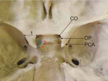
The optic strut and the anterior clinoid process represent bony structures that are closely related to anatomically and clinically significant elements such as the cavernous sinus, the internal carotid artery, the optic nerve and the pituitary gland. The objective of our study was to quantify dimensions of the optic strut and anterior clinoid process, and to determine variations in positions and forms of these structures. A descriptive anatomical study was performed on 200 dry human skulls. We analyzed dimensions and variations in position of the optic strut, dimensions of the anterior clinoid process as well as the incidence and forms of the caroticoclinoid foramen. The average thickness of the optic strut on skulls belonging to males was 3 mm and 2.8 mm on those belonging to females. The optic strut was most commonly attached to the anterior two fifths on the lower side of the anterior clinoid process. On the male skulls the average width of the anterior clinoid process was 9.4 mm (right) and 9.1 mm (left). Its length was 9.9 and 9.3 mm. On female skulls the average width of the process was 8.7 mm (right) and 8.3 mm (left), while the length measured 9.3 mm on the right and 8.9 mm on the opposite side. In our sample, a complete caroticoclinoid foramen appeared in 4.25%, a contact form in 2.75%. At last, an incomplete form of the foramen was observed in 9.75%. The anatomic variations of the investigated structures must be considered during the approaches to the cavernous sinus and neurovascular elements of the sellar region.

Downloads

Download data is not yet available.
Anatomical variations and morphometric study of the optic strut and the anterior clinoid process

Downloads

Additional Files

Published

2012-05-20

How to Cite

1.
Kapur E, Mehić A. Anatomical variations and morphometric study of the optic strut and the anterior clinoid process. Biomol Biomed [Internet]. 2012May20 [cited 2023Dec.4];12(2):88-93. Available from: https://www.bjbms.org/ojs/index.php/bjbms/article/view/2502

Issue

Section

Anatomy

INTRODUCTION

The anterior clinoid process is a part of the roof to the cavernous sinus in its frontal portion. During surgeries on tumors and aneurysms in the parasellar and suprasellar region, the anterior clinoid process and the optic strut must be moved from the lesser wing of sphenoid bone to provide a better approach to the operating field, primarily to the internal carotid artery and the optic nerve, and to minimize the need for brain retraction [1,2]. The superficial and the thin deep layer of the dura cover the upper and lower sides of the anterior clinoid process. Removal of the anterior clinoid process (anterior clinoidectomy) allows a full approach to the anterior portion of the cavernous sinus and to the vertical segment of the internal carotid artery. It’s a significant problem in neurosurgical procedures to find a massive anterior clinoid process, which by removing can cause optic nerve and internal carotid artery injury [3]. The caroticoclinoid foramen was first described by Henle (1885) as a bony foramen formed by merging of the anterior and middle clinoid processes’ tops. According to Williams [4] and Lang [5] this foramen is formed by the ossification of the caroticoclinoid ligament in early childhood. The existance of this foramen is of great importance to brain surgeons since it disables retraction or mobilization of the cavernous segment of the internal carotid artery, even after release of proximal and distal dural ring [1], as well as proper tumor extirpation in sellar region, especially meningeoma of the tuberculum sellae [6, 7]. A preoperative detection of caroticoclinoid foramen, particularly by CT imaging, has a huge clinical significance, for its appearance conditions an inappropriate retraction of the cavernous segment of the internal carotid artery which can lead to its rupture with fatal outcome [8].

MATERIALS AND METHODS

Samples

As material for osteological analysis we used 200 dry human skulls of the osteological collection of Department of Anatomy at Medical Faculty, University of Sarajevo, of both genders and different age. Out of 200 analyzed skulls, 109 or 54.5% belonged to males, and 91 or 35.5% originated from females (Table 1).

TABLE 1: Gender and age distribution of the investigated skulls

Procedure

On the osteological material we conducted following morphometric measurements (using manuel caliper) and analysis:

  1. dimensions and variations in the position of the optic strut,

  2. dimensions of the anterior clinoid process,

  3. incidence and forms of the caroticoclinoid foramen.

Statistical analysis

In the study we used the method of statistical and comparative analysis and processed the acquired data using SPSS-15.0 statistical software. The statistical analysis included calculating basic parameters of descriptive statistics like mean value, standard deviation and standard error of the mean value for single analyzed group. The results are presented in tables. We tested statistical significance of mean values by crossing individual parameters of analyzed groups. The testing was performed by Stundent’s t-test, and p<0.05 is considered statistically significant.

RESULTS

The optic strut is a bony formation located between upper side of the body and the lesser wing of sphenoid bone. This formation separates optic canal from the medial portion of superior orbital fissure. Position of the optic strut was determined based on relation between the length of anterior clinoid process and the distance measuring from the optic strut to the top of the anterior clinoid process. On examined skulls of both genders it has been found that the optic strut was attached to the anterior fifth of anterior clinoid process in 11.6% cases on the right side, and in 14.5% on the left. Its attachment to the anterior two fifths of the process was registered in 42% on the right side and the 47.8% on the left side. The least seen positions of the attachment site are entirely to the front, by the base of anterior clinoid process – in 1.4% on the right and 4.3% on the left side, as well as entirely to the back, by the top of the process - in 2.9% on the right and 0.9% on the left side (Table 2). On all the skulls we measured the width of anterior clinoid process in its base, and the length from the base’s midline to the top as shown in Figure 1. The anterior clinoid process in almost every observed male skull was triangular in shape, its base oriented to the front and top to the back and medially. In eleven skulls – in 5.5% cases, we found the process to be quadrangular in shape. In the total of 109 analyzed male skulls, the mean value of PCA width on the right side measured 9.4±1.4 mm, ranging between 5.9 and 12.1 mm. On the left side the PCA width range was between 5.8 and 14.0 mm, mean value 9.1 mm and standard deviation of 11.7mm. The length of the process in males measured 9.9±1.6 mm on the right, and 9.3±1.4 mm on the left (Table 3). On the skulls originated from females, the mean value of PCA width on the right side measured 8.7±1.5 mm, ranging from 5.7 to 11.6 mm, and 8.3±2.1 mm on the left, mini-malvalue being 4.9, maximal value 12.7 mm. The process’ mean value length in females on the right side was 9.3±1.6 mm, and 8.9±2.0 mm on the left side (Table 3). There were statistically significant differences in mean width and in mean length of the anterior clinoid process between man and woman on the both sides of the examined skulls (p<0.05).

TABLE 2: Location of the optic strut in relation to the anterior clinoid process (PCA – processus clinoideus anterior)
FIGURE 1: The measured parameters on the anterior clinoid process. 1-width in the base, 2-length from base to the top, 3-length from the top to the posterior margin of the optic strut (PCA-processus clinoideus anterior, OP-optic strut, CO-canalis opticus).
TABLE 3: Dimensions of the anterior clinoid process (width and length in milimetars) considering gender and side of the examined skulls.

The caroticoclinoid foramen (foramen caroticoclinoideum) is formed when tops of the anterior and middle clinoid processes merge on the upper side of sphenoid bone. The merge can appear to be complete and incomplete. Aside from these two forms of the foramen, we also analyzed the so called contact form when there’s a suture between the two clinoid processes (Figure 2). The complete form of caroticoclinoid foramen on the total of 109 male skulls (218 sides) was found to be bilateral in only one case (Figure 3), or 1.83%. Unilaterally, it appeared in six skulls (2.75%), in four cases on the right side, and in remaining two cases on the left. An incomplete caroticoclinoid foramen was found in larger number of male skulls. It appeared bilaterally in five skulls – 4.58% cases, and we registered this form to appear unilaterally in eleven male skulls (5.04%), of which seven were on the right and four on the left side. In six skulls we found the complete form to be on one side, there was also an incomplete form of caroticoclinoid foramen on the opposite side. The incidence of the contact form of the foramen with presence of the interclinoid suture was higher on the left side (1.37%) than on the right (0.45%) in male skulls we observed (Figure 4). The contact form of the foramen was found bilaterally in two male skulls equaling 3.66% (Figure 5). Based on previous results, we can conclude that the caroticoclinoid foramen in all three forms appeared in 37 cases, which is 16.97%. It should be kept in mind that the entire statistics was performed on a sample of 218 sides of the investigated male skulls (Table 4). The transverse diameter of the foramen measured in average 5.32±0.52 mm on the right, and 5.21±0.73 on the left. In male skulls with the incomplete form of the foramen the distance between tops of the anterior and middle clinoid processes measured 3.12±0.92 mm on the right side and 2.94±1.34 on the left. While analyzing female skulls (91 skulls - 182 sides), we found complete caroticoclinoid foramen bilaterally in two skulls (2.19%). The incomplete form on a single side was registered in five skulls - 2.74% cases. Out of those five, three were on the right and two on the left side. Similar to male skulls, incomplete caroticoclinoid foramen was found in larger number of female skulls as well: in five skulls bilaterally (5.49%) and eight skulls unilaterally (4.39%). In the group of eight skulls where incomplete foramen was found unilaterally, five were observed on the right and three on the left side. In three out of five female skulls with unilaterally presented complete foramen there was an incomplete form found on the opposite side. We haven’t found the contact form of caroticoclinoid foramen bilaterally in female skulls. Incidence of this form unilaterally however was 1.09% (two skulls) on the left side and 0.54% on the right (one skull). Of the 91 analyzed female skulls we found the three forms of caroticoclinoid foramen to appear in 30 cases, which is 16.48% (Table 5). The transverse diameter of the foramen in complete and contact form measured in average 4.97±0.63 mm on the right side, and 4.99±0,54 mm on the left. In female skulls with incomplete form of the foramen, the distance between tops of the anterior and middle clinoid processes measured 3.02±0.76 mm on the right side, and 2.84±0.97 mm on the left. The total number of different forms of connection between the anterior and the middle clinoid process on the entire osteological sample is displayed in Table 6.

FIGURE 2: llustration of three forms of caroticoclinoid foramen. PCA-processus clinoideus anterior, PCM-processus clinoideus medius A – incomplete form, B – contact form, C – complete form.
FIGURE 3: Male skull (age 58). A complete form of the caroticoclinoid foramen bilaterally (1).
FIGURE 4: Male skull (age 45, lef side). Contact form of the caroticoclinoid foramen. 1-suture between the anterior and the middle clinoid process, 2-anterior clinoid process, 3-middle clinoid process.
FIGURE 5: Male skull (age 64). Complete (on the left) and contact form of the caroticoclinoid foramen (on the right). 1- caroticoclinoid foramina, 2-interclinoid suture.
TABLE 4: The incidence of complete, incomplete and contact form of the caroticoclinoid foramen in male skulls (n=218)
TABLE 5: The incidence of complete, incomplete and contact form of caroticoclinoid foramen on female skulls (n=192)
TABLE 6: The incidence of complete, incomplete and contact form of caroticoclinoid foramen on the entire sample (n=400), considering sides of skulls.

DISCUSSION

On male skulls, average width of the base of anterior clinoid process measured 9.4 mm on the right side and 9.1 mm on the left, maximum values being 12.1 and 14 mm. Its length measured from the base to the top was 9.9 mm and 9.3 mm. In female skulls the mentioned dimensions of anterior clinoid were less by 3 to 9 mm in average. The dimension of PCA given in our study are significantly larger compared to the dimension given by other authors, that are refering to skulls of the population of Central America and East Asia [9], while compared to the skulls of European origin in the study of Hauser and Di Stefano, the dimensions are similar. These differencies could be explained by racial features. Along with a strong and massive anterior clinoid process there’s an additional problem of its possible pneumatization, which can be visualised by CT scanning, leading to sphenoid and ethmoid sinus opening during clinoidectomy [10, 11]. The optic strut represents a bony formation that connects the body of sphenoid bone and its lesser wing. Positioned like that, the optic strut separates optic canal from the medial portion of superior orbital fissure. Surgical procedures on cavernous sinus and suprasellar region demand a total removal of the optic strut as well as anterior clinoid process [2]. The optic strut itself or its parts that are not removed can lead to an optic nerve or internal carotid artery injury. The strut is placed laterocaudally to the optic nerve, so to remove it without damaging the nerve we recommend resection in the direction from anterior and medially to posterior and laterally. In addition, based on analysis we conducted on dry skulls we advise the optic strut to be removed before anterior clinoid process so the process could be completely released. Otherwise, we risk the anterior process’ bony fragments that are left behind, may cause damage of the internal carotid and the optic nerve [12]. Optic strut’s thickness on male skulls we investigated was 3 mm ± 1.2 mm in average, and 2.8 mm ± 0.9 mm on female skulls. We observed no statistically significant differencies regarding gender. The optic strut was most commonly attached to front two fifths on the lower side of anterior clinoid process (42% on the right side and 47.8% on the left). We noticed that, when the optic strut had been attached to the posterior fifth of anterior clinoid process (2.9% on the right and 0.9% on the left), it was the thickest, with maximal values of 4.5 mm in male skulls and 4 mm in female skulls. These data on size and dimensions of the optic strut should be kept in mind during surgical procedures on cavernous sinus and region of orbital apex. Knowing the precise location of the optic strut and its variations are very important while analyzing CT images, since it represents a valuable landmark in proper evaluation and differentiation of the optic canal, superior orbital fissure and anterior clinoid process on coronal and axial scans [13]. In our sample consisting of 109 male and 91 female skulls (400 sides) the complete form of caroticoclinoid foramen was discovered in 17 cases - 4.25%. The contact form of the foramen when tops of anterior and middle clinoid processes connect into the so called interclinoid suture [14] was detected in 11 cases, which is 2.75% in a sample of 400 sides. The incomplete form was present in 39 cases - 9.75%. An insight into the results show us that the complete, contact and incomplete forms of caroticoclinoid foramen were more often found in men, and were more often found on right side of the skulls regardless of gender. There are considerable racial differencies in the incidence of different forms of the foramen too. In the study by Lee et al. [9], the complete and incomplete forms of caroticoclinoid foramen were found in 17.1% cases, Maxi (1950 – quoted by Erturk) [14], registered them in 23.4 % (among population of Central America), while Keyers (1935 – quoted by Lee) [9] registered their appearance in 27.46% among American caucasions, predominantly on the left side. Azeredo et al. [15] registered caroticoclinoid foramen in the skulls of Portuguese population in 6.27%, and in the study by Reisch et al. we can see that the incidence among Germans was 14%. Erturk [14] describe three forms of caroticoclinoid foramen: a complete form (complete fusion of the anterior and the middle clinoid process), an incomplete form (if the processes don’t fuse completely) and a contact form (when the tops of the processes fuse with a suture inbetween). This author found the three forms of the foramen in 23.68% cases. The incidence of different forms of the foramen varies within the range from 6.27% [15] to 35.67% [14] (Table 7). When it’s a question of side of a skull the foramen appeares on, most studies proclaim higher incidence on the right side, with an exception of studies performed in USA [5], that indicate higher incidence of caroticoclinoid foramen on the left side, as well as more significant number of complete or incomplete foramina bilaterally.

TABLE 7: Comparison of the caroticoclinoid foramen and inter- clinoid bone bridges’ incidence by different studies.

CONCLUSION

The autors opine that anatomical knowlege of optic strut and anterior clinoid process, may be clinically important for surgeons operating in the region of cavernous sinus or the surrounding structures. Our investigations may be helpfull for pre-operative planning. Presence of any variations may result in unnecessary injury to the complicated neurovascular structures in the vicinity of cavernous sinus.

DECLARATION OF INTEREST

Authors have no conflict of interest to declare.

REFERENCES

  1. (). Direct microsurgical repair of intracavernous vascular lesions. J Neurosurg.
  2. (). Selective extradural anterior clinoidectomy for supra-and parasellar processes: Tehnical note. Journal of Neuro surgery.
  3. , , , , , (). Surgical anatomy and efficient modifications of procedures for selective extradural anterior clinoidectomy. Minim Invasiv Neurosurg.
  4. , (). Gray's Anatomy.
  5. (). Structure and postnatal organization of heretofore uninvestigated and infrequent ossification of the sella turcica region. Acta Anat.
  6. , , , , , (). The anatomy of caroticoclinoid foramen and its relation with the internal carotid artery. Surg Radiol Anat.
  7. , (). Surgical managament of pituitary tumors.
  8. (). The cavernous sinus. Neurosurg Clin N Am.
  9. (). Anterior clinoid process and optic strut in Koreans. Yonsei Med J.
  10. , , (). Surgical anatomy of the anterior clinoid process. J Clin Neurosci.
  11. , , , , , (). Remarkable anatomic variations in paranasal sinus region and their clinical importance. Eur J Radiol.
  12. , , , (). Surgical approaches to the cavernous sinus: a microsurgical study. Neurosurgery.
  13. , , , (). Computed tomography and magnetic resonanace imaging of the orbital apex. Radiol Clin North Am.
  14. , , (). Anatomy of the clionoidal region with special emphasis on the caroticoclinoid foramen and interclinoid osseous bridge in a recent Turkish population. Neurosurg Rev.
  15. , , (). Anatomical variations of the clinoid process of the human sphenoid bone. Arq Cent Estud Curso Odontol.