TREM-1 is a positive regulator of TNF-α and IL-8 production in U937 foam cells

Authors

  • Yu-Shi Wang Department of Cardiovascular, the First Hospital of Jilin University
  • Xiang-Jun Li School of Pharmaceutical Sciences, Jilin University
  • Wai-Ou Zhao Department of Cardiovascular, the First Hospital of Jilin University

DOI:

https://doi.org/10.17305/bjbms.2012.2503

Keywords:

TREM-1, foam cell, TNF-α, IL-8, atherosclerosis

Abstract

The purpose of our study was to investigate the expression levels of TREM-1 (triggering receptor expressed on myeloid cells-1) in U937 foam cells and determine whether TREM-1 regulates the production of tumor necrosis factor-alpha and interleukin-8 in these cells.

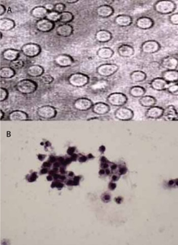
Human U937 cells were incubated with phorbol 12-myristate 13-acetate and then oxidized human low-density lipoprotein to induce foam cell formation. Oil red O staining was used to identify the foam cells. The production of IL-8 and TNF-α by U937 foam cells was assayed by enzyme-linked immunosorbent assay. The expression of TREM-1 mRNA in U937 foam cells was detected by reverse transcription-polymerase chain reaction. Moreover, U937 foam cells were transfected by small interfering RNA using Lipofectamine 2000 to knockdown TREM-1. Western blot was performed to assay protein expression of TREM-1 and ELISA was used to examine the effect of TREM-1 knockdown on IL-8 and TNF-α production.

PMA and ox-LDL induced U937 cells to form foam cells. The production of TNF-α and IL-8 was found to be significantly elevated in U937 foam cells, concomitant with a significant up-regulation of TREM-1 mRNA. TREM-1 siRNA was able to partially silence the expression of TREM-1 protein and remarkably inhibited TNF-α and IL-8 production in U937 foam cells, suggesting that TREM-1 is a positive regulator of TNF-α and IL-8 production in U937 foam cells.

Our finding that TREM-1 controls the production of IL-8 and TNF-α in U937 foam cells defines a potentially critical role of TREM-1 in the pathogenesis of atherosclerosis and implicates TREM-1 as a potential therapeutic target for the disease.

Downloads

Download data is not yet available.
TREM-1 is a positive regulator of TNF-α and IL-8 production in U937 foam cells

Downloads

Additional Files

Published

2012-05-20

How to Cite

1.
Wang Y-S, Li X-J, Zhao W-O. TREM-1 is a positive regulator of TNF-α and IL-8 production in U937 foam cells. Biomol Biomed [Internet]. 2012May20 [cited 2023Sep.26];12(2):94-101. Available from: https://www.bjbms.org/ojs/index.php/bjbms/article/view/2503

Issue

Section

Immunology

INTRODUCTION

Atherosclerosis is a complex, progressive disease that is newly recognized to have a strong inflammatory component [1, 2]. In the early stage of atherosclerosis, monocytes migrate into the arterial intima and differentiate into macrophages that can take up oxidized low-density lipoprotein (ox-LDL) to stimulate differentiation into foam cells [3, 4]. The accumulation of foam cells in the artery wall causes the formation of fatty streak(s), the earliest visible lesion of atherosclerosis [5]. The uptake of ox-LDL by macrophages results in the production of numerous proinflammatory cytokines that can amplify the inflammatory response and promote the evolution of atheroma [6]. Interleukin-8 (IL-8) and tumor necrosis factor-alpha (TNF-α) are two important proatherosclerotic cytokines secreted by ox-LDL-activated macrophages [7-10]. Both of these molecules have been found to be up-regulated at the sites of formation of atherosclerotic plaques [11, 12]. IL-8 may induce the firm adhesion of monocytes to endothelial cells and mediate the intimal accumulation of macrophages in atherosclerotic lesions [13, 14], while TNF-α can enhance macrophage foam cell formation by inhibition of intracellular lipid catabolism and, therefore, contribute to atherosclerotic development and progression [15]. Accumulating evidence indicates that infectious processes may contribute to atherosclerosis [16]. Infectious agents, such as cytomegalovirus [17], Chlamydop hila pneumoniae [18], Helicobacter pylori [19], and parvovirus [20], have been shown to augment the production of cytokines in macrophages and provide inflammatory stimuli that can accelerate atherogenesis. TREM-1 (triggering receptor expressed on myeloid cells-1) is an activating receptor that is selectively expressed on neutrophils and monocytes/macrophages and can be up-regulated by bacterial and fungal stimuli [21]. Engagement of TREM-1 on monocytes can trigger the release of large amounts of proinflammatory cytokines, including IL-8 and TNF-α, and amplify inflammatory responses [21, 22]. At present, it remains unclear whether TREM-1 is upregulated during foam cell formation and, if it is, whether TREM-1 regulates the production of proinflammatory cytokines by macrophage-derived foam cells. In the present study, we established an in vitro foam cell formation model by stimulating human myelomonocytic U937 cells with phorbol 12-myristate 13-acetate (PMA) and ox-LDL to investigate the expression of TREM-1 in macrophage-derived foam cells and its relationship with the secretion of TNF-α and IL-8. Furthermore, small interfering RNA (siRNA) was employed to knockdown TREM-1 in order to examine TREM-1 effects on the production of TNF-α and IL-8 in U937 foam cells.

MATERIALS AND METHODS

Cell culture and induction of foam cell formation

Human myelomonocytic cell line U937 was purchased from KeyGen Biotech (Nanjing, China) and was maintained in RPMI-1640 medium (Gibco, USA) containing 10% fetal bovine serum (FBS) in a 5% CO2 incubator at 37°C. U937 cells during the logarithmic growth phase (at a density of 1.0 × 10⁹ cells/L) were stimulated with 100 nmol/L of PMA (Sigma, USA) for 72 h to induce the formation of macrophage-like U937 cells. After 12 hours of culture in serum-free medium, PMA-induced U937 cells were divided into three groups and incubated with RPMI-1640 medium containing 10% FBS (PMA group), 100 mg/L of LDL (PMA+LDL group) or 100 mg/L of ox-LDL (PMA + ox-LDL group; Yuanyuan Biotechnology, Guangzhou, China). Experiments were performed in quintuplicate. After 24 h of culture, supernatants were collected to measure the contents of TNF-α and IL-8, and the cells were harvested for detection of TREM-1 mRNA expression by reverse transcription-polymerase chain reaction (RT-PCR).

Identification of U937 foam cells

U937 foam cells were identified by oil red O staining. Briefly, adherent U937 foam cells were stained with freshly prepared 0.3% oil red O solution for 20 min. Cell nuclei were then counterstained with hematoxylin solution for 5 min. After rinsing with 70% ethanol, cells were mounted onto glass slides with an aqueous mounting reagent. Stained cells were observed under an inverted light microscope (TE2000; Nikon, Japan).

RT-PCR

Total RNA was isolated from cells using the TRIzol Reagent (Gibco) according to the manufacturer’s protocol. Reverse transcription was performed using M-MLV reverse transcriptase and oligo-dT primers (Fermentas, USA) following the manufacturer’s instructions. PCR was then carried out to determine the expression levels of TREM-1 and glycer-aldehyde-3-phosphate dehydrogenase (GAPDH, control) mRNAs using the following parameters: pre-denaturation at 94°C for 5 min; 30 cycles of denaturation at 94 °C for 30 s, annealing at 55 °C for 30 s, and extension at 72 °C for 30 s; and, a final extension at 72 °C for 10 min. The sequences of the TREM-1 and GAPDH primers were as follows: TREM-1 forward, 5-TGCTGTGGATGCTCTTTGTC-3’ and reverse, 5’-CACAGTTCTGGGGCTGGTAT-3’; GAPDH forward, 5’-ACCACAGTCCATGCCATCAC-3’ and reverse, 5’-TCCACCACCCTGTTGCTGTA-3’. The PCR products were resolved by electrophoresis on a 2% agarose gel containing ethidium bromide (0.5 μg/ml). The expression level of TREM-1 mRNA relative to GAPDH transcripts was determined by densitometric scanning (Gel Image system-1600; Tanon, Shanghai, China).

Cytokine measurement

TNF-α and IL-8 in culture supernatants were assayed by enzyme-linked immunosorbent assay (ELISA) using commercial kits (Westang, Shanghai, China), according to the manufacturer’s instructions. Briefly, 100 μl culture supernatants were added into the 96-well plate. After incubation for 2 h at 37 °C, the plate was washed three times with cleaning solution [TBST: 20 mmol/L Tris-HCl, 150 mmol/L NaCl, 0.05% (V/V) Tween 20]. With the exception of the negative control, all the wells were supplemented with 100 μl of horseradish peroxidase (HRP)-antibody working solution and incubated at 37 °C for 1 h. After washing as above, a drop of substrate A and B were added sequentially. After incubation at 37 °C for 15 min, a drop of end solution was added into each well, and the absorbed density (OD) was detected at 450 nm.

siRNA transfection

The following two oligonucleotides encoding a TREM-1 siRNA were obtained from Dharmacon (Lafayette, USA): sense, 5’-CCGGAAGTGTATGTGATCAGAGTAAT TCAAGAGATTACTCTGATCACATACACTTTTTTTG-3’; anti-sense, 5’-AATTCAAAAAAAGTGTATGTGATCAGAGTAATCTCTTGAATTACTCTGATCACATACACTT-3’. Macrophage-like U937 cells induced with PMA as described above were used for siRNA transfection. In addition to the TREM-1 siRNA group (TREM-1 siRNA), an empty liposome group (empty liposome) and a random siRNA group (negative siRNA) were used as negative controls. Transfection was performed using Lipofectamine 2000 according to the manufacturer’s protocol. After 48 h of transfection, cells were treated with 100 mg/L of ox-LDL as described above and subjected to detection of TREM-1 protein expression by Western blot, and TNF-α and IL-8 production by ELISA.

Western blot

Western blot was performed using routine procedures. Briefly, total protein was harvested by cell lysis solution (20 mmol/L Tris, 150 mmol/L NaCl, 1 mM EDTA, 1% Triton X-100, 2.5 mmol/L sodium pyrophosphate, 1 mmol/L p-Glycerolphosphate, 1 μg/ml Leupetin, 1 mmol/L phenylmethylsulfonyl fluoride, pH 7.5) and the quantity was estimated by Lowry analysis [23]. Cellular proteins (50 μg) were resolved by SDS-PAGE using 15% polyacrylamide gels and transferred to polyvinylidene fluoride membranes. After incubation with rabbit anti-TREM-1 polyclonal antibody (1:100; sc-48762; Santa Cruz Biotechnology, Santa Cruz, CA, USA), rabbit anti-PKC polyclonal antibody (1:200; sc-10800; Santa Cruz Biotechnology, Santa Cruz, CA, USA) or rabbit anti-GAPDH polyclonal antibody (1:100; sc-25778; Santa Cruz Biotechnology, Santa Cruz, CA, USA), the bands were detected by using an HRP-conjugated secondary antibody (1:100; Santa Cruz Biotechnology, Santa Cruz, CA, USA). The immunoblot signals were visualized using the Immobilon Western Chemilum HRP Substrate (WBKLS0100; Millipore Corporation, Billerica, MA, USA). The protein expression level of TREM-1 relative to GAPDH was determined by densitometric scanning.

Statistical analysis

Numerical data have been expressed as mean ± standard deviation (SD). Statistical analysis was performed using the SSPS 11.0 software package. Intergroup differences were compared using one-way analysis of variance (ANOVA) and p<0.05 was considered to be significantly different.

RESULTS

PMA and ox-LDL induced the differentiation of U937 cells into foam cells

As shown in Figure 1A, the untreated U937 cells exhibited the characteristic round shape. After incubation with PMA for 72 h, the majority of U937 cells shifted to an ameboid shape and began to aggregate and adhere, indicating the differentiation from monocytes into macrophage-like cells (Figure 1B). In the presence of ox-LDL, PMA-induced macrophage-like cells showed obvious foamy changes as many oil red O-positive lipid droplets were observed in the cytoplasm. Some cells showed an enlarged size due to uptake and accumulation of an excessive amount of ox-LDL (Figure 1C). In contrast, no obvious lipid droplets were observed in PMA-induced macrophage-like cells with (data not shown) or without (Figure 1B) LDL treatment.

FIGURE 1: PMA and ox-LDL induced the differentiation of U937 cells into foam cells. (A). Inverted microscopy showed the characteristically round U937 cells prior to treatment. (B). After incubation with PMA for 72 h, the U937 cells shifted to an ameboid shape, and aggregated and adhered, indicating their differentiation from monocytes into macrophage-like cells. (C). In the presence of ox-LDL, PMA-induced macrophage-like cells showed obvious foamy changes: many oil red O-positive lipid droplets were observed in the cytoplasm; some cells showed an enlarged size due to uptake and accumulation of excessive ox-LDL (magnification ×200).

TNF-α and IL-8 levels were elevated in U937 foam cells

To determine the effects of ox-LDL treatment on the levels of TNF-α and IL-8 in PMA-induced U937 cells, ELISA was performed (Figure 2). The levels of both TNF-α and IL-8 in PMA+ox-LDL U937 cells were found to be significantly higher than those in PMA and PMA+LDL groups (all p <0.05): TNF-α, 100.38 ± 10.24 (PMA+ox-LDL) vs. 13.68±1.56 (PMA) and 15.87 ± 1.63 (PMA+LDL); IL-8, 97.88 ± 9.37 (PMA+ox-LDL) vs. 20.39 ± 2.40 (PMA) and 23.43 ± 3.59 (PMA+LDL). The levels of both TNF-α and IL-8 in PMA+LDL group were slightly higher than those in the PMA group, but there were no significant differences observed (p>0.05). These results indicated that the levels of both TNF-α and IL-8 were elevated in U937 foam cells.

FIGURE 2: TNF-α and IL-8 levels were elevated in U937 foam cells. The levels of TNF-α (A) and IL-8 (B) secreted by PMA-induced U937 cells in the presence of ox-LDL were determined by ELISA. Data have been shown as mean ± SD (n = 5). *p > 0.05 vs. PMA; **p < 0.05 vs. PMA; *** p < 0.05 vs. PMA+LDL.

TREM-1 mRNA expression was up-regulated in U937 foam cells

To examine the impact of ox-LDL treatment on the expression of TREM-1 mRNA in PMA-induced U937 cells, the level of TREM-1 mRNA relative to GAPDH was determined by RT-PCR (Figure 3). The relative level of TREM-1 mRNA in PMA+ ox-LDL U937 cells was significantly higher than those in PMA and PMA+LDL groups: 0.73 ± 0.06 (PMA+ox-LDL) vs. 0.42 ± 0.05 (PMA) and 0.51 ± 0.06 (PMA+LDL) (both p<0.05; Figure 3 A and B). However, there was no significant difference observed in the relative level of TREM-1 mRNA between PMA and PMA+LDL groups (p>0.05). These data clearly indicated that TREM-1 mRNA expression was up-regulated in U937 foam cells.

FIGURE 3: TREM-1 mRNA expression was up-regulated in U937 foam cells. The expression levels of TREM-1 mRNA in U937 foam cells were determined by RT-PCR. Data have been shown as mean ± SD (n = 5). (A). Representative image showing the expression of TREM-1 and GAPDH mRNAs in U937 foam cells; (B). Quantitative analysis of the expression level of TREM-1 mRNA relative to GAPDH mRNA. *p > 0.05 vs. PMA; **p < 0.05 vs. PMA; ***p < 0.05 vs. PMA+LDL.

TREM-1 siRNA partially silenced the expression of TREM-1 protein in PMA-induced U937 cells

To examine whether TREM-1 is involved in the regulation of TNF-α and IL-8 production in U937 foam cells, siRNA-mediated silencing of the TREM-1 gene was performed. Western blot analysis indicated that the expression of TREM-1 protein was significantly down-regulated in PMA-induced U937 cells transfected with TREM-1 siRNA, as compared to untransfected cells or cells transfected with empty liposome or negative siRNA (p<0.05, Figure 4 and Table 1).

FIGURE 4: TREM-1 siRNA partially silenced the expression of TREM-1 protein in PMA-induced U937 cells. After PMA-induced U937 cells were transfected with TREM-1 siRNA (TS), negative siRNA (NS) or empty liposome (EL), and then treated with ox-LDL, the expression levels of TREM-1 protein were measured by Western blot. Untransfected U937 cells untreated (PM) or treated with ox-LDL (PO) were used as controls. Data have been shown as mean ± SD (n = 5). (A). Representative image showing the expression of TREM-1 and GAPDH proteins in U937 cells; (B). Quantitative analysis of the expression level of TREM-1 protein relative to GAPDH protein. *p < 0.05 vs. the PMA group (PM); # p < 0.05 vs. the PMA+ox-LDL group (PO), Negative siRNA group (NS) or Empty liposome group (EL), respectively.
TABLE 1: siRNA inhibited ox-LDL-induced TREM-1 expression and TNF-α and IL-8 secretion in U937 foam cells

In contrast, no significant differences were noted in the expression levels of TREM-1 protein among untransfected U937 cells and U937 cells transfected with empty liposome or negative siRNA. Thus, TREM-1 was partially silenced by the corresponding siRNA in PMA-induced U937 cells.

TREM-1 knockdown suppressed TNF-α and IL-8 production in U937 foam cells

After PMA-induced U937 cells transfected with TREM-1 siRNA were treated with ox-LDL, the production of TNF-α and IL-8 in U937 foam cells was detected by ELISA. As shown in Figure 5 and Table 1, TREM-1 knockdown significantly inhibited ox-LDL-induced TNF-α and IL-8 secretion in U937 foam cells. In contrast, the levels of TNF-α and IL-8 in foam U937 cells transfected with empty liposome or negative siRNA were not significantly different from those detected in untransfected cells. Apparently, siRNA-mediated silencing of the TREM-1 gene was capable of significantly suppressing the production of TNF-α and IL-8 in U937 foam cells.

FIGURE 5: TREM-1 knockdown suppressed TNF-α and IL-8 production in U937 foam cells. After PMA-induced U937 cells were transfected with TREM-1 siRNA (TS), negative siRNA (NS) or empty liposome (EL), and then treated with ox-LDL, the levels of TNF-α (A) and IL-8 (B) secreted by these cells were measured by ELISA. Untransfected U937 cells untreated (PM) or treated with ox-LDL (PO) were used as controls. Data have been shown as mean ± SD (n = 5). *p < 0.05 vs. the PMA group (PM); # p < 0.05 vs. the PMA+ox-LDL group (PO), Negative siRNA group (NS) or Empty liposome group (EL), respectively.

TREM-1 knockdown suppressed ox-LDL-induced up-regulation of PKC expression in U937 foam cells

To determine whether ox-LDL treatment alters the expression of PKC protein in PMA-induced U937 cells, Western blot was performed (Figure 6). The expression level of PKC protein in the PMA+ox-LDL group was significantly higher than that in the PMA group (p<0.05). However, TREM-1 knockdown significantly inhibited ox-LDL-induced up-regulation of PKC expression in U937 foam cells. In contrast, the expression levels of PKC protein in foam U937 cells transfected with empty liposome or negative siRNA were not significantly different from that detected in untransfected cells. Collectively, these results indicated that TREM-1 knockdown suppressed ox-LDL-induced up-regulation of PKC expression in U937 foam cells.

FIGURE 6: TREM-1 knockdown suppressed ox-LDL-induced up-regulation of PKC expression in U937 foam cells. After PMA induced U937 cells were transfected with TREM-1 siRNA (TS), negative siRNA (NS) or empty liposome (EL), and then treated with ox-LDL, the expression levels of PKC in these cells were determined by Western blot. Untransfected U937 cells untreated (PM) or treated with ox-LDL (PO) were used as controls. Data have been shown as mean ± SD (n = 5). (A). Representative image showing the expression of TREM-1 and GAPDH proteins in U937 cells; (B). Quantitative analysis of the expression level of TREM-1 protein relative to GAPDH protein. *p < 0.05 vs. the PMA group (PM); # p < 0.05 vs. the PMA+ox-LDL group (PO), Negative siRNA group (NS) or Empty liposome group (EL), respectively.

DISCUSSION

Infectious agents can contribute to the pathogenesis of atherosclerosis by promoting macrophage production of inflammatory cytokines [16]. Moreover, TREM-1 plays a critical role in monocyte/macrophage-mediated inflammatory responses to microbial infection [24]. Based on these observations, we hypothesized that TREM-1 may be involved in regulating the production of proinflammatory cytokines during foam cell formation. In the present study, we demonstrated that ox-LDL treatment significantly up-regulate the expression of TREM-1 mRNA in PMA-induced macrophage-like cells, and TREM-1 knockdown remarkably inhibited ox-LDL-induced TNF-α and IL-8 secretion, suggesting that TREM-1 is a positive regulator of TNF-α and IL-8 production in U937 foam cells. Given that TNF-α and IL-8 are proinflammatory cytokines that play an important role during macrophage foam cell formation and atherosclerotic development and progression [13-15], these findings suggest a possible critical role of TREM-1 in the pathogenesis of atherosclerosis. Multiple studies have demonstrated that TNF-α and IL-8 are up-regulated in macrophage foam cells in atherosclerotic plaques and involved in the potentiation of atherosclerosis [7-15]. The activation of p38 mitogen-activated protein kinase (MAPK) and nuclear factor κB (NF-κB) may be implicated in the secretion of IL-8 in lipid-laden macrophages [8], while TNF-α production may depend on the activation of activator protein 1 (AP-1) [9], phosphoinositide 3-kinase (PI3K) and extracellular signal-regulated kinase (ERK) [6]. In the present study, we found that TREM-1 could control the release of TNF-α and IL-8 in U937 foam cells. Similarly, previous studies also showed that TREM-1-mediated TNF-α and IL-8 secretion was observed in monocytes/macrophages [21]. Although the mechanism underlying TREM-1-mediated release of IL-8 and TNF-α in U937 foam cells is still unclear, the connection between TREM-1 and the cytokine-inducing Toll-like receptor 4 (TLR4)/NF-κB pathway may provide a clue to this mechanism [25]. Yang et al. [26] found that blocking of TLR4 was able to substantially inhibit NF-κB activity and ox-LDL-induced IL-8 expression. Ornatowska et al. [27] demonstrated that TREM-1 silencing in macrophages resulted in decreased expression of key proteins in the TLR4 signaling pathway. Thus, it is reasonable that the TLR4/NF-κB signaling pathway might be involved in TREM-1-mediated TNF-α and IL-8 production in U937 foam cells. Protein kinase C (PKC) is a family of serine/threonine protein kinases that play a key role in the regulation of the inositol phosphate/diacylglycerol signalling pathway. Upon activation, PKC enzymes are translocated from the cytosol to the plasma membrane and exert diverse biological functions by phosphorylating target proteins. Feng et al. [28] found that PKC could promote the expression of scavenger receptor CD36 and lipid droplet-associated PAT-proteins in monocytes, thus contributing to foam cell formation. Oso et al. [29] demonstrated that inhibition of PKCbeta prevents LDL uptake and foam cell formation by reducing scavenger receptor an expression in human macrophages. Another study [30] indicated that PKC activation could modulate adipophilin-mediated lipid accumulation in THP-i macrophages. In the present study, we discovered that the expression pattern of PKC in U937 foam cells was similar to that of TREM-1, suggesting that PKC activation may be involved in TREM-1-mediated IL8/TNF-α production in U937 foam cells. However, the mechanisms behind the link between PKC activation and TREM-1 link remain to be further studied. Of note, the PKC family consists of at least 12 isozymes that are divided into three subfamilies. Therefore, elucidation of the roles of different PKC isoforms in foam cell formation will lead to a better understanding of the pathogenesis of atherosclerosis. At present, the mechanism underlying ox-LDL-induced TREM-1 up-regulation remains elusive. Although a natural ligand for TREM-1 has not yet been identified, previous studies have shown that TREM-1 expression can be modulated in vitro and in vivo by various TLR ligands, including the gram-negative bacterial lipopolysaccharide, the primary ligand of TLR4 [31]. This raises the possibility that the TLR signaling pathway may also be involved in ox-LDL-induced TREM-1 up-regulation. Interestingly, TREM-1 and TLR ligands have been shown to induce inflammatory responses via intersecting and mutually stimulating pathways [21]. In addition, LDL oxidation is capable of inducing the formation of a number of highly reactive substances, some of which may also directly mediate TREM-1 up-regulation. As stated above, macrophage foam cell formation is a characteristic feature of atherosclerotic plaques. Inhibition of foam cell formation and reduction of inflammatory cytokine production may be of therapeutic value in the treatment of atherosclerosis [5]. Our finding that TREM-1 controls the release of IL-8 and TNF-α in U937 foam cells defines a possible critical role of TREM-1 in the pathogenesis of atherosclerosis and implicates TREM-1 as a potential therapeutic target for the disease. In animals with LPS-induced endotoxemia, blockage of TREM-1 signaling by administration of either a soluble form of the TREM-1 molecule, small-molecule inhibitor or siRNA have been found to be able to improve animal survival [24, 32, 33]. If our results are verified in vivo, these approaches might also be applicable to the treatment of atherosclerosis.

CONCLUSION

In conclusion, the present study provides evidence that, in U937 foam cells, up-regulated expression of TREM-1 is concomitant with an increase in the levels of TNF-α and IL-8, and siRNA-mediated down-regulation of TREM-1 expression suppresses the production of TNF-α and IL-8. Apparently, TREM-1 can stimulate the release of TNF-α and IL-8 in U937 foam cells. These results suggest a critical role of TREM-1 in foam cell formation and the pathogenesis of atherosclerosis and may aid in the design and development of new strategies to combat atherosclerosis.

Acknowledgements

ACKNOWLEDGEMENTS

The authors thank Dr. Xiang-Jun Li and Medjaden Bioscience Limited for assisting in the preparation of this manuscript.

DECLARATION OF INTEREST

There was no conflict of interest in this study.

REFERENCES

  1. , , (). Inflammation and atherosclerosis. Circulation.
  2. (). Atherosclerosis-an inflammatory disease. N Engl J Med.
  3. , (). Monocytes in atherosclerosis: subsets and functions. Nat Rev Cardiol.
  4. (). Monocyte recruitment and foam cell formation in atherosclerosis. Micron.
  5. , (). The macrophage foam cell as a target for therapeutic intervention. Nat Med.
  6. , , , , , (). Toll-like receptor 4–dependent and – independent cytokine secretion induced by minimally oxidized low-density lipoprotein in macrophages. Arterioscler Thromb Vasc Biol.
  7. , , , , , (). Oxidized LDL induces monocytic cell expression of interleukin-8, a chemokine with T-lymphocyte chemotactic activity. Arterioscler Thromb.
  8. , , , (). Low-density lipoprotein modified by macrophage-derived lysosomal hydrolases induces expression and secretion of IL-8 via p38 MAPK and NF-kappaB by human monocyte-derived macrophages. Arterioscler Thromb Vasc Biol.
  9. , , , (). Human monocytes/macrophages release TNF-alpha in response to Ox-LDL. Arterioscler Thromb Vasc Biol.
  10. , (). Production of tumor necrosis factor and interleukin-1 by macrophages from human atheromatous plaques. Am J Pathol.
  11. , , , , , (). Interleukin-8 is induced by cholesterol loading of macrophages and expressed by macrophage foam cells in human atheroma. J Biol Chem.
  12. , , , , , (). Synthesis of TNF alpha and TGF beta mRNA in the different micro-environments within atheromatous plaques. Cardiovasc Res.
  13. , , , , , (). MCP-1 and IL-8 trigger firm adhesion of monocytes to vascular endothelium under flow conditions. Nature.
  14. , , , (). A leukocyte homologue of the IL-8 receptor CXCR-2 mediates the accumulation of macrophages in atherosclerotic lesions of LDL receptor-deficient mice. J Clin Invest.
  15. , , (). Interleukin-ibeta and tumour necrosis factor-alpha impede neutral lipid turnover in macrophage-derived foam cells. BMC Immunol.
  16. , , (). Roles of infectious agents in atherosclerosis and restenosis: an assessment of the evidence and need for future research. Circulation.
  17. , , , , , (). Cytomegalovirus infection stimulates expression of monocyte-associated mediator genes. J Immunol.
  18. , , , , , (). Chlamydophila pneumoniae infection leads to smooth muscle cell proliferation and thickening in the coronary artery without contributions from a host immune response. Am J Pathol.
  19. , (). Helicobacter pylori and atherosclerosis: can current data be useful for clinical practice?. Int J Cardiol.
  20. , , , , , (). Human parvovirus b19 infection in patients with coronary atherosclerosis. Arch Med Res.
  21. , , (). Cutting edge: inflammatory responses can be triggered by TREM-1, a novel receptor expressed on neutrophils and monocytes. J Immunol.
  22. , , (). The TREM receptor family and signal integration. Nat Immunol.
  23. , , , , , (). Analysis of cyclooxygenase 2 (COX-2) expression during malignant melanoma progression. Cancer Biol Ther.
  24. , , , (). TREM-1 amplifies inflammation and is a crucial mediator of septic shock. Nature.
  25. , (). Uncovering the TREM-1-TLR connection. Am J Physiol Lung Cell Mol Physiol.
  26. , , , , , (). Role of Toll-like receptor 4/NF-kappaB pathway in monocyte-endothelial adhesion induced by low shear stress and ox-LDL. Biorheology.
  27. , , , , , (). Functional genomics of silencing TREM-1 on TLR4 signaling in macrophages. Am J Physiol Lung Cell Mol Physiol.
  28. , , , , , (). Induction of CD36 expression by oxidized LDL and IL-4 by a common signaling pathway dependent on protein kinase C and PPAR-gamma. J Lipid Res.
  29. , , , , , (). Inhibition of protein kinase Cbeta prevents foam cell formation by reducing scavenger receptor A expression in human macrophages. Circulation.
  30. , , , , , (). Effects of PKC activity on lipid-accumulation mediated by Adipophilin in THP-1 Macrophage. Progress in Biochemmistry and Biophysics.
  31. , , , (). Lipopolysaccharide-induced up-regulation of triggering receptor expressed on myeloid cells-1 expression on macrophages is regulated by endogenous prostaglandin E2. J Immunol.
  32. , , , , , (). A soluble form of the triggering receptor expressed on myeloid cells-1 modulates the inflammatory response in murine sepsis. J Exp Med.
  33. , , , , , (). TREM-1 promotes survival during septic shock in mice. Eur J Immunol.