Possible involvement of calcium channels and plasma membrane receptors on Staurosporine-induced neurite outgrowth

Authors

  • Hossein Zhaleh Department of Biology, Faculty of Science, Razi University Department of Chemical Biotechnology Engneering, Science and Research Branch, Islamic Azad University
  • Mehri Azadbakht Department of Biology, Faculty of Science, Razi University
  • Ali Bidmeshki Pour Department of Biology, Faculty of Science, Razi University

DOI:

https://doi.org/10.17305/bjbms.2012.2526

Keywords:

staurosporine, neurite outgrowth, calcium channels, plasma membrane calcium receptors, PC12 cell

Abstract

Staurosporine as a protein kinases inhibitor induced cell death or neurite outgrowth in PC12 cells. We investigated the involvement of calcium channel and plasma membrane receptors on staurosporine inducing neurite outgrowth in PC12 cells. PC12 cells were preincubated with NMDA receptor inhibitors (1.8 mM ketamine and 1μM MK801, treatment 1) or L-Type Calcium channels (100 μM nifedipine and 100 μM flavoxate hydrochloride, treatment 2) or calcium-calmoduline kinasses (10 μM trifluoprazine, treatment 3) and nifedipine, MK801, flavoxate hydrochloride and ketamine (treatment 4 or without pretreatments (control). Then, the cells were cultured in RPMI culture medium containing 214nM staurosporine for induction of neurite outgrowth. The percentage of Cell cytotoxicity and apoptotic index was assessed. Total neurite length (TNL) and fraction of cell differentiation were assessed. After 24h, the percentage of cell cytotoxicity were increased in treatments 1, 2 and 4 compared with control (p<0.05). After 6h, apoptotic index was similar between all treatments. After 12h, apoptotic index were increased in treatment 4 compared with control (p<0.05). After 24h, apoptotic index were increased in treatments 1, 2 and 4 compared with control (p<0.05). TNL were decreased in treatments 1, 2 and 4 compared with control in different times of assessment (6, 12 and 24 h) (p<0.05). The fraction of cell differentiation were decreased in treatments 1, 2 and 4 compared with control (p<0.05). It can be concluded that the possible involvement of L-type calcium channel and the N-methyl D-aspartate receptor on staurosporine-induced neurite outgrowth process in PC12 cells.

Downloads

Download data is not yet available.
Possible involvement of calcium channels and plasma membrane receptors on Staurosporine-induced neurite outgrowth

Downloads

Additional Files

Published

2012-02-20

How to Cite

1.
Zhaleh H, Azadbakht M, Bidmeshki Pour A. Possible involvement of calcium channels and plasma membrane receptors on Staurosporine-induced neurite outgrowth. Biomol Biomed [Internet]. 2012Feb.20 [cited 2023Sep.22];12(1):20-5. Available from: https://www.bjbms.org/ojs/index.php/bjbms/article/view/2526

Issue

Section

Biochemistry

INTRODUCTION

Staurosporine (STS) as a protein kinase inhibitor [1, 2] has dual effects on neuronal cells; induction of cell death and cell differentiation. STS induces apoptosis in high concentrations (μM) [3, 4] and neuronal differentiation in low concentrations (nM) by neurite extension in several types of cells [2, 3, 5, 6]. Although the detailed mechanism of STS action as a neurogenic morphogen remains unclear, it seems that it is associated with the inhibition of some protein kinases which may contribute to neurite outgrowth [7]. In previous studies it has been determined that STS can inhibit PKA, Ca2+/calmodulin-dependent kinase II, cyclin dependent kinases, ion channels (Kv1.3, L-type Ca2+ channel, voltage-gated K+ channel) in myocyte [8-12]. Although, the role of STS in the inhibition of protein kinases during neurite outgrowth was clear but its function on plasma membrane calcium channels and receptors remains to be fully known [10,11]. Calcium plays an important role in the regulating a great variety of neuronal processes such as neuronal cell differentiation. In most neurons, multiple mechanisms exist whereby increases in intracellular calcium concentration may occur including for example in calcium entry through N-methyl-D-Aspartate (NMDA) glutamate receptors and various voltage-gated calcium channels such as L-type calcium channels (LTCC), as well as in the release of calcium from intracellular stores [13-15]. Calcium influx through LTCCs is particularly effective in neuronal migration, activation of transcription factors (e.g CREB), changes in gene expression that underlie plasticity and adaptive neuronal responses (e.g c-fos) [1, 16-22]. Although STS induced increasing of intracellular calcium in treated cells, its effect on plasma membrane and calcium channels and receptors located in the plasma membrane during neuronal differentiation and neurite outgrowth are not well known. In this study we aimed to determine whether plasma membrane calcium channels and receptors involves in staurosporine-induced neurite outgrowth.

MATERIALS AND METHODS

Cell culture

PC12 cells were cultured in complete culture medium containing RPMI1640 culture medium (Gibco), supplemented with 0.2 % bovine serum albumin (BSA, Gibco), 1% NEAA (Sigma), 2 mM L-glutamine (Sigma), 100 IU/ml penicillin and 100 μg/ml streptomycin (Sigma) in 10-cm tissue culture dishes. The cultures were incubated at 37°C in a humidified incubator containing 95% air and 5% CO2. Culture medium was replaced every 2 days. When cell cultures reached to 80% confluency, they were trypsinated using trypsin-EDTA 0.25% (Sigma) and the cells were subcultured at a density of 104 cells/well in 24-well culture plates.

Cell treatment

One day after plating PC12 cells, cells were washed with phosphate buffer saline (PBS), pH 7.4. For inhibition of L-Type Calcium channel, NMDA receptor and CaM kinase, cells were preincubated with, adding 1.8 mM ketamine and 1μM MK801, 15 min (treatment 1), 100 μM nifedipine and 100 μM flavoxate hydrochloride, 30 min (treatment 2) and 10 μM trifluoperazine, 30 min (treatment 3). In our experiment, we combined ketamine, MK801, flavoxate hydrochlo-ride and nifedipine for inhibition of L-Type Calcium channels and NMDA receptors (treatment 4). Then, cells were cultured in differentiation medium containing complete culture medium supplemented with 214 nM staurosporine for 24h. PC12 cells were cultured in differentiation medium without inhibitor preincubation seems as control group. The cells were placed in the incubator at 37°C with 5% CO2.

Cell cytotoxicity measurement

Cell cytotoxicity was quantified by measuring the release of lactate dehydrogenase (LDH) from damaged or destroyed cells into the medium. Cytotoxicity was measured with LDH Cytoxicity Detection Kit (Roche, Germany). This kit detects LDH release from dead cells. Therefore, increase of LDH activity in each treatment show that the treatment solution has further dead cells or cytotoxicity effects on PC12 cells. Cells were plated in 96 well culture plates with 104 cells/mL density for 12h. Then cells were pretreated in different treatments for certain time. Then, cells were cultured by differentiation medium for 24h. The percentage of cytotoxicity was measured by protocol of company; colorimetery of LDH activity measured by calculated the absorbance of samples at 490 or 492 nm using an ELISA Reader (EL800; USA). The references wavelength should be more than 600 nm. All experiments were replicated independently at least 3 times. Within each experiment, we replicated each condition 4 times.

Quantification of cell death incidence

Hoechst / PI nuclear staining was carried out as previously described [23]. Briefly, cells were plated in 24 well culture plates with 104 cells/mL density for 12h. Then cells were pre-treated in different treatment mediums for certain time. These were grown for a range of times in differentiation medium (6, 12 and 24h). Then cells were incubated for 15 min at 37°C with Hoechst 33342 dye (10 mg/ml in PBS), washed twice in PBS. PI (50 mg/ml in PBS) was added just before microscopy. Cells were visualized using an inverted-florescence microscope (Olympus IX-71, Japan). Nuclear morphology was scored as follows: 1, viable cells had blue-stained nuclei with smooth appearance; 2, viable apoptotic cells had blue-stained nuclei with multiple bright specks of condensed chromatin; 3, non-viable apoptotic cells had red-stained nuclei with either multiple bright specks of fragmented chromatin or one or more spheres of condensed chromatin (significantly more compact than normal nuclei); 4, non-viable necrotic cells had red-stained, smooth and homogeneous nuclei that were about the same size as normal (control) nuclei. The apoptotic index were calculated by the fraction of numbers of apoptotic cells on the total cell count in 100 (300 cells), respectively. All experiments were replicated independently at least 3 times. Within each experiment, we replicated each condition 4 times.

Measurement of total neurite length

Measurement of total neurite length was conducted as reported by previous study [24]. The assay is based on the measurement of total neurite length. Total neurite length (length of largest neurite on 100 cells) was assessed. Cells were plated in 24 well culture plates with 104 cells/well density for 12h. Then cells were pretreated in different treatments for certain time. These were then grown for a range of times at differentiation medium (6, 12 and 24h), fixed, and the morphology assessed by an inverted microscope (Olympus IX-71, Japan). Digital photos were taken of random fields of neurons derived from the treatments. Total neurite length was measured (Motic software; Ver.2). All experiments were replicated independently at least 3 times. Within each experiment, we replicated each condition 4 times.

The fraction of cell differentiation assessment (f (%))

Fraction of cell differentiation was carried out as previous study [25]. PC12 cells were plated at a density of 2–104 cells/well on 24 well plates. Cells were pretreatment with different treatment mediums. These were then grown for a range of times at differentiation medium (6, 12 and 24 h), fixed, and the morphology microscopically assessed (Motic software; Ver. 2). The fraction of cell differentiation was evaluated under an inverted microscope by the fraction of neurite-bearing cells were the fraction of numbers of neurite-bearing cells with at last one neurite longer than the cell body diameter on the total cell count (300 cells). All experiments were replicated independently at least 3 times. Within each experiment, we replicated each condition 4 times.

Statistical analysis

Data were expressed as Mean ± SEM. All calculations were performed by SPSS (version 16; SPSS Inc.). The differences in the percentage of cytotoxicity, incidence of apoptotic index, total neurite length and fraction of cell differentiation, in PC12 cells between treatments were analyzed using t-test at significant level (p<0.05).

RESULTS

Cell cytotoxicity

The percentage of cytotoxicity of inhibitors in PC12 cells cultured in culture medium containing 214 nM staurosporine was assessed by evaluation of the lactate dehydrogenase activity. In PC12 cells the percentage of cytotoxicity were increased in treatments 1, 2 and 4 (36% ± 2%, 32% ± 2% and 46% ± 3%, respectively) compared with control (20% ± 2%), (p<0.05). The percentage of cytotoxicity in treatment 3 (21% ± 3%) were decreased compared with treatments 1, 2 and 4 (p<0.05) and was similar to control (Figure 1).

FIGURE 1: The effects of calcium channel and receptor on cell viability in PC12. treatment1: pretreatment with ketamine; treatment2: pretreatment with nifedipine; treatment3: pretreatment with trifluoperazine; treatment4: pretreatment with ketamine and nifedipine. Control: without pretreatment. All data represented by mean ± S.E.M (p<0.05).

Effects of inhibitors on apoptosis index

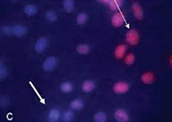
The evaluation of apoptotic index of inhibitors for PC12 cells cultured in culture medium containing 214 nM staurosporine was assessed by PI/Hoechst florescence staining. After 6h, the apoptotic index were increased in treatments 1 and 2 (18% ± 3% and 19% ± 2%); respectively and were similar in treatments (15% ± 3%) compared with control cells (16% ± 2%) but these differences were not significant. The apoptosis index in treatment 4 (26% ± 4%) was increased compared with control and treatments 1-3 (p<0.05). After 12 h, the apoptosis index were increased in treatments 1 (31% ± 2%), 2 (28% ± 2%) and 4 (43% ± 4%) compared with control (21% ± 3%) (p<0.05). The apoptotic index in treatment 3 (22% ± 4%) was decreased compared with treatments 1, 2 and 4 p<0.05) and was similar to control. After 24 h, the apoptosis index were increased in treatments 1, 2 and 4 (42% ± 4%, 38% ± 3% and 55% ± 5%, respectively) compared with control (23% ± 4%) (p<0.05). The apoptosis index in treatment 3 was increased (25% ± 3%) compared with control but this difference was not significant (Figures 2;A and B).

FIGURE 2: The effects of calcium channels and receptors on cell death in PC12. (A) Morphology of cells as examined by fluorescence microscopy a: cells without pretreated with, b: Cells pretreated with treatment1, c: Cells pretreated with treatment2, d: cells pretreated with treatment3, e: Cells pretreated with treatment4 for 24 hours. (B) Quantitative analysis of necrotic and apoptotic cells by fluorescence microscopy in various treatments. Viable cell: white and short arrow, apoptotic cell: long arrow, necrotic cell: double short arrow, Control: cells without pretreatment; treatment1: pretreatment with ketamine; treatment2: pretreatment with nifedipine; treatment3: pretreatment with trifluoperazine; treatment4: pretreatment with ketamine and nifedipine. All data represented by mean ± S.E.M (p<0.05). Magnitude is 400×.

Neurite outgrowth measurement

The average of total neurite length for PC12 cells was assessed. The total neurite length (TNL) was calculated. Af-ter inhibitors preincubation, TNL significantly were decreased compared with control cells. After sh, TNL were decreased in treatments 1, 2 and 4 (123 ± 0.67, 118 ± 0.72 and 98 ± 0.83, respectively) compared with control cells (142 ± 0.89) (p<0.05). TNL in treatment 3 (141 ± 0.64) was similar to control. After 12h, TNL were decreased in treatments 1, 2 and 4 (102 ± 0.92, 98 ±0.87 and 83 ± 0.93, respectively) compared with control cells (128 ± 0.94) (p<0.05). TNL in treatment 3 (12s ± 0.85) was similar to control. After 12h, TNL were decreased in treatments 1, 2 and 4 (76 ± 0.85, 71 ±0.88 and 6s ± 0.82, respectively) compared with control cells (108 ± 0.81) (p<0.05). TNL in treatment 3 (102 ± 0.92) was similar to control (Figures 3 and 4).

FIGURE 3: The effects of calcium channels and receptors on PC12 cells morphology. A. without pretreatment; B. pretreatment with trifluoperazine; C. pretreatment with ketamine; D. pretreatment with nifedipine; E. pretreatment with ketamine and nifedipine. All data represented by mean ± S.E.M (p<0.05). Magnitude is 200×.
FIGURE 4: The effects of calcium channels and receptors on total neurite length in PC12 cells. treatment1: pretreatment with ketamine; treatment2: pretreatment with nifedipine; treatment3: pretreatment with trifluoperazine; treatment4: pretreatment with ketamine and nifedipine. Control: without pretreatment; All data represented by mean ± S.E.M (p<0.05).

Fraction of cell differentiation assessment

The evaluation of the fraction of cell differentiation of inhibitors for PC12 cells cultured in culture medium containing 214nM staurosporine was assessed. After 6 h, f (%) were not significantly decreased in treatments 1, 2 and 4 (98% ± 1%, 98% ± 0.7% and 96% ± 1%, respectively) compared with control (100%). f (%) in treatment 3 (100%) similar to control. After 12h, The fraction of cell differentiationf (%) was decreased in treatment 4 (92% ± 1.2%) (p<0.0s). f (%) were not significantly decreased in treatments 1 and 2 (95% ± 2% and 94% ± 2%) compared with control (100%) (p<0.05). f (%) in treatment 3 (p<0.05) was similar to control cells. After 24h, f (%) were decreased in treatments 1, 2 and 4 (87% ± 3%, 78% ± 3% and 63% ± s%, respectively) compared with control cells (98 % ± 2%), (p<0.05).f (%) in treatment 3 was similar to control (Figure 5).

FIGURE 5: The effects of calcium channels and receptors on fraction of cell differentiation in PC12 cells. treatment1: pretreatment with ketamine; treatment2: pretreatment with nifedipine; treatment3: pretreatment with trifluoperazine; treatment4: pretreatment with ketamine and nifedipine. Control: without pretreatment; All data represented by mean ± S.E.M (p<0.05).

DISCUSSION

The current study investigated the involvement of calcium channel and plasma membrane receptors on staurosporine inducing neurite outgrowth in PC12 cells. In this work, we used PC12 cells as the best cell model for study of effect of materials on neurite outgrowth [25]. PC12, a neuron-like cell line, expresses voltage-dependent Ca channels appear to dihydropyridine-sensitive voltage-dependent Ca channels demonstrable by different techniques [26, 27]. Staurosporine was employed as a strong inducer of neurite outgrowth with inhibition of protein kinases in vitro model. The results obtained in this study showed that nifedipine and ketamine could effectively inhibit neurite outgrowth induced by staurosporine and increase cell death incidence in PC12 cells. We observed that when cells were preincubated with nifedipine and flavoxate hydrochloride or ketamine and MK801, they dramatically suppressed the neurite outgrowth and increased cell death and cytotoxicity in PC12 cells. Meanwhile, preincubation with ketamine and MK801 together with nifedipine and flavoxate hydrochloride result in powerful inhibition of neurite outgrowth and induce cell death in PC12 cells. It could be suggested that the possible involvement of voltage dependent calcium channels and NMDA receptors on staurosporine-calcium dependent signal transduction. Meanwhile, PC12 application of trifluoperazine does not the same effects on either of cytotoxicity or neurite outgrowth. It was shown this possible that staurosporine leads to inhibition of calmodulin in 214 nM concentrations. It is unclear that how extracellular Ca2+ causes the intracellular events that leads to the differentiation in PC12 by staurosporine. It seems staurosporine leads to regulation of neurite outgrowth process with activation of different plasma membrane calcium channels and increasing of intracellular calcium concentration. Development, neuronal survival and differentiation can be influenced by a variety of local signals or signals derived from intermediate or final target tissues [28]. Previously, it has been shown that external Ca2+ evoke the signal transduction through the Ca2+ influx via extracellular Ca2+ - sensing receptor localized to neurons and their nerve terminals [29]. It demonstrated that neurite outgrowth of PC12 is induced via the Ca2+-signal transduction pathway by the Ca2+ influxes through channels [30]. On the other hand, recent study showed that staurosporine leads to intracellular calcium overload, which induce apoptosis in PC12 cells [31]. In the recent study, showed that staurosporine caused a large increase in [Ca2+]c even after the depletion of Ca2+ from the ER, the IP3-sensitive Ca2+ store, in the absence of perfusate Ca2+. This result indicates that IP3-insensitive, non-ER compartments are responsible for the staurosporine-induced [Ca2+]c increase in rat submandibular acinar cells [32]. We reported previously that Staurosporine use extracellular calcium stores tend to increase intracellular calcium concentration [33]. In addition, previously, it is known that cytosolic Ca2+ increase caused by staurosporine that mobilize Ca2+ from different sources might cause apoptosis in astrocytes [34]. Ca2+ in DDTIMF-2 smooth muscle cells by influx but also by intracellular mobilization from thapsigargin-sensitive and -insensitive Ca2+ stores. Furthermore, the high local Ca2+ gradient just under the plasma membrane, which can be preserved over long periods of time in Ca2+- free medium despite the presence of EGTA, indicates that the efflux mechanism is also affected [35]. The stores of Ca2+ ion entry from extracellular into intracellular during staurosporine-induced neurite outgrowth is still not completely understood. Many studies in different cells showed that staurosporine result in an increase cytosolic calcium concentration and induction of apoptosis in NGF-differentiated cells [36, 37]. In another study showed that the rate of apoptotic cells is greater in differentiated cells than undifferentiated cells [28]. Different study showed that neurotrophins factors like NGF result in increase of mRNA incoding of calcium channels like voltage-dependent calcium channels and glutamate-sensitive ion channels like NMDA [38-42]. It has shown that compared with undifferentiated cells maybe activation of calcium channels and plasma membrane receptors by staurosporine lead to increase of staurosporine-induced apoptosis in differentiated cells. If true, these receptors and channels play important role in increasing intracellular calcium concentration during staurosporine-induced cell differentiation in PC12 cells. Meanwhile, We suggest it possible that staurosporine by a protein kinase-independent mechanism (PKC, PKA and CaMKs) by activation of plasma membrane Ca+2 channels lead to enhance of neurite outgrowth and increases cell viability and fraction of cell differentiation in PC12 cells.

CONCLUSION

According to the results of present study, application of staurosporine with activation of calcium channels may lead to enhance of neurite outgrowth and have effects on neuronal cell differentiation in PC12 cells. However, more key receptors and enzymes need to be investigated in these effects.

Acknowledgements

ACKNOWLEDGEMENTS

This study was supported by Faculty of Sciences from the Razi University.

DECLARATION OF INTEREST

There is no conflict of interest.

REFERENCES

  1. , , , (). Analysis of gene expression during staurosporine induced neuronal differentiation of human prostate cancer cells. Oncol Rep.
  2. , , , (). Staurosporine is a potent activator of neuronal, glial, and “CNS stem cell-like” neurosphere differentiation in murine embryonic stem cells. Mol Cell Neurosci.
  3. , , , , , (). Staurosporine-induced apoptosis in chang liver cells is associated with down-regulation of Bcl-2 and Bcl-Xl. Int J Mol Med.
  4. , , , , , (). Staurosporine induced apoptosis is independent of p and p and achieved via arrest at G2/M and at G1 in U251/MG human glioma cell line. Cancer Chemother Pharmacol.
  5. , , , (). Staurosporine induces astrocytic phenotypes and differential expression of specific PKC isoforms in C6 glial cells. J Neurochem.
  6. , , , , (). Inhibition of protein kinases in rat pheochromocytoma (PC12) cells promotes morphological differentiation and down-regulates ion channel expression. Proc Biol Sci.
  7. , , , , , (). Expression of neural markers in human umbilical cord blood. Exp Neurol.
  8. , (). Staurosporine, K-252 and UCN-01: potent but nonspecific inhibitors of protein kinases. Trends Pharmacol Sci.
  9. , , , , , (). Staurosporine: an effective inhibitor for Ca2+/Calmodulin dependent protein kinase II. J Neurochem.
  10. , , , , (). Staurosporine directly blocks Kv1.3 channels expressed in Chinese hamster ovary cells. Naunyn Schmiedebergs Arch Pharmacol.
  11. , , (). The protein kinase inhibitor, staurosporine, inhibits L-type Ca2+ current in rabbit atrial myocytes. Biochem Biophys Res Commun.
  12. , , , , , (). Staurosporine inhibits voltage-dependent K+ current through a PKC-independent mechanism in isolated coronary arterial smooth muscle cells. J Cardiovasc Pharmacol.
  13. , , , (). Calcium regulation of gene expression in neuronal cells. J Neurobiol.
  14. , , , , , (). Relative abundance of subunit mRNAs determines gating and Ca2+ permeability of AMPA receptors in principal neurons and interneurons in rat CNS. Neuron.
  15. , (). What is the role of dopamine in reward: hedonic impact, reward learning, or incentive salience?. Brain Res Brain Res Rev.
  16. , , (). Regulation of gene expression in hippocampal neurons by distinct calcium signaling pathways. Science.
  17. , , , , , (). L-type calcium channels and GSK-3 regulate the activity of NF-ATc4 in hippocampal neurons. Nature.
  18. , , (). Membrane depolarization and calcium induce c-fos transcription via phosphorylation of transcription factor CREB. Neuron.
  19. , (). Role of ion flux in the control of c-fos expression. Nature.
  20. , , , , (). Activity dependent regulation of BDNF and NGF mRNAs in the rat hippocampus is mediated by non-NMDAglutamate receptors. EMBO J.
  21. , , , (). Calcium channels: mechanisms of selectivity, permeation, and block. Annu Rev Biophys Biophys Chem.
  22. , (). Selective role of N-type calcium channels in neuronal migration. Science.
  23. , , , , , (). Hydrogen peroxide induces the activation of the phospholipase C-g1 survival pathway in PC12 cells: protective role in apoptosis. Acta Biochim Biophys Sin (Shanghai).
  24. , , , , , (). simple procedure for quantification of neurite outgrowth based on stereological principles. J Neurosci Methods.
  25. , (). Regulation of neurite outgrowth by extracellular Ca2_for neural cells PC12 and PC12D. Coll and Surf B: Biointerfaces.
  26. (). Calcium antagonists: High-affinity binding and inhibition of calcium transport in a clonal cell line. J Biol Chem.
  27. , , (). Three types of neuronal calcium channel with different calcium agonist sensitivity. Nature.
  28. , (). Axon guidance and the patterning of neuronal projections in vertebrates. Science.
  29. , , , (). Cloned and expressed rat Ca2-sensing receptor. J Biol Chem.
  30. , , , , (). Calcium influx induces neurite growth through a Src/Ras signaling cassette. Neuron.
  31. , (). Calcium overload is essential for the acceleration of staurosporine-induced cell death following neuronal differentiation in PC12 cells. Exp mol Med.
  32. , , , , , (). Staurosporine mobilizes Ca2+from Secretory granules by inhibiting protein kinase C in rat submandibular acinar cells. J Dent Res.
  33. , , (). Effects of extracellular calcium concentration on neurite outgrowth in PC12 cells by staurosporine. Neurosc Letters.
  34. , , , , , (). Apoptotic effect of ethanol is potentiated by caffeineinduced calcium release in rat astrocytes. Neurosci Lett.
  35. , , (). Staurosporine induced Ca2+increase in DDTIMF-2 smooth muscle cells. Amer phys soci.
  36. , (). NGF-differentiated and undifferentiated PC12 cells vary in induction of apoptosis by ethanol. Life Sci.
  37. , , , , (). TNF-alpha induces apoptosis through JNK/Bax-dependent pathway in differentiated, but not naive PC12 cells. Cell Cycle.
  38. , , , (). Differential expression by nerve growth factor of two types of Ca2+channels in rat phaeochromocytoma cell lines. J Physiol.
  39. , , , (). Time-dependent expression of Na and Ca channels in PC12 cells by nerve growth factor and cAMP. Neurosci Res.
  40. , , (). Enhanced expression of Ca2+channels by nerve growth factor and the v-src oncogene in rat phaeochromocytoma cells. J Physiol.
  41. , (). Modifications of intracellular Ca2+signalling during nerve growth factor-induced neuronal differentiation of rat adrenal chromaffin cells. Eur J Neurosci.
  42. , , , , , (). NMDA-dependent NGF mRNA expression by human astrocytoma cells is mediated by nitric oxide. Neuroreport.