Intrauterine blood transfusion in immune hydrops fetalis, corrects middle cerebral artery Doppler velocimetry very quickly

Authors

  • Ahmet Yalinkaya Department of Obstetrics and Gynecology, School of Medicine, Dicle University
  • Mehmet Sıddık Evsen Department of Obstetrics and Gynecology, School of Medicine, Dicle University
  • Yusuf Celik Department of Biostatistic and Medical Informatics, School of Medicine, Dicle University
  • Muhammet Erdal Sak Department of Obstetrics and Gynecology, School of Medicine, Dicle University
  • Hatice Ender Soydinc Department of Obstetrics and Gynecology, School of Medicine, Dicle University
  • Mehmet Zeki Taner Department of Obstetrics and Gynecology, School of Medicine, Dicle University

DOI:

https://doi.org/10.17305/bjbms.2012.2532

Keywords:

Rh isoimmunization, middle cerebral artery, peak systolic velocimetry, intrauterine transfusion

Abstract

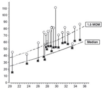
The aim of our study was to evaluate the middle cerebral artery velocimetry before and after intrauterine blood transfusion in immune hydrops fetalis. The current study was conducted in a tertiary research hospital, from February 2009 to January 2011. Nineteen intrauterine blood transfusions performed during the study period. The factors recorded were age of the mothers, gestational weeks, pre-transfusion fetal hematocrit and post-transfusion fetal hematocrit, and also middle cerebral artery peak systolic velocimetry (MCA-PSV) was detected and recorded before and after intrauterine transfusion. A control group of twenty two cases for normal MCA doppler velocimetry was also included to the study. During the study, a total of eleven rhesus isoimmunized pregnancies underwent intrauterine blood transfusions at our perinatal diagnose unit. Before transfusion seventeen severe and two moderate anemias were detected and mean MoM of MCA-PSV was 1.76±0.38 MoM. Post transfusion mean MoM of MCA-PSV in the patient group and control group were 1.08±0.22 MoM and 0.96±0.21 MoM, respectively. The mean MCA-PSV values were higher in RI fetuses than post transfusion and control group. In current study, we found that MCA-PSV is a valuable parameter in detecting fetal anemia requiring intrauterine transfusion and mean MCA-PSV values is higher than 1.5 MoM in fetuses with anemia. And also decrease in MCA-PSV just after transfusion in anemic fetuses showed the quick response of the fetus to correction of anemia.

Downloads

Download data is not yet available.
Intrauterine blood transfusion in immune hydrops fetalis, corrects middle cerebral artery Doppler velocimetry very quickly

Downloads

Additional Files

Published

2012-02-20

How to Cite

1.
Yalinkaya A, Sıddık Evsen M, Celik Y, Erdal Sak M, Ender Soydinc H, Zeki Taner M. Intrauterine blood transfusion in immune hydrops fetalis, corrects middle cerebral artery Doppler velocimetry very quickly. Biomol Biomed [Internet]. 2012Feb.20 [cited 2023Jun.6];12(1):37-40. Available from: https://www.bjbms.org/ojs/index.php/bjbms/article/view/2532

Issue

Section

Translational and Clinical Research

INTRODUCTION

Rhesus isoimmunization (RI) is a frequent cause of fetal anemia caused by the transport of maternal antibodies from the placenta and subsequently destroys fetal red blood cells. In the United States the ratio of fetuses under risk of anemia due to RI has a ratio of 35 per 100.00 live births and of these fetuses only 10 percent will require intrauterine blood transfusion because of severe anemia [1-3]. The detection of anemia in RI fetuses is the most important point in the treatment of immune hydrops fetalis. Invasive techniques such as amniocentesis (AC) and cordocentesis (CC) used to identify the fetuses with severe anemia [4-5]. Mari et al. [6] reported that MCA-PSV value, detected by Doppler ultrasonography, increases in fetuses with anemia. In a prospective multicenter trial Zimmermann et al. [7] studied the MCA-PSV in fetuses with anemia to find the need of invasive procedures and found that 90 of 125 fetuses avoided from AC or CC. In current study, we aimed to evaluate the pre- transfusion and just after transfusion MCA-PSV values of RI fetuses with anemia and compare with healthy fetuses.

MATERIALS AND METHODS

Subjects

This prospective case control study was performed at perinatal diagnose unit of Dicle University, School of Medicine from February 2009 to January 2011. This is a tertiary and reference hospital in the southeast region of Turkey. Most of the patients referred from the outside health centers. The health services are mostly free of charge and supported by the government. The study was approved by the Ethics Committee of the instuition, and written informed consent was received by all of the patients.

Procedures

A total nineteen intrauterine blood transfusion to eleven pregnancies with RI fetuses were included to this study. In five patient one, in four patients two and in two patients three transfusions at different gestational ages were determined. The factors recorded were age of the mothers, gestational weeks, pre-transfusion fetal hematocrit and post-transfusion fetal hematocrit, and middle cerebral artery Doppler velocimetry. The patient group had Rh-negative blood group, positive indirect coombs test, and a obstetric history of fetal anemia requiring intrauterine transfusion and/or intrauterine or postpartum fetal demise due to fetal anemia. All pregnancies were accurately dated by the last menstrual period and /or by first-trimester ultrasonographic investigation. The MCA-PSV was detected by using Doppler ultrasonography pre transfusion and post transfusion (just after procedure, approximately in five minute). All of the transfusions were performed by experienced perinatologist with ultrasound (Volusun 730 PRO 4D ultrasound device). Fetal hydrops was defined as the presence of generalized skin thickening of greater than 5 mm and at least two of the following conditions: ascites, pleural effusion, pericardial effusion or placental enlargement. The transfusions were performed by CC. The amount of blood transfused to the fetuses was calculated according to the formula advised by Mandelbrot et al. [8]. The severity of fetal anemia was determined according to the reference values reported previously [6].

Statistical analysis

Mean and standard deviation (SD) were calculated for continuous variables. Chi-Square and Independent Sample t test were used to evaluate associations between the categorical and continuous variables. Two-sided p values were considered statistically significant at p<0.05. Statistical analyses were carried out by using the statistical packages for SPSS 15.0 for Windows (SPSS Inc., Chicago, IL, USA).

RESULTS

The demographic and clinical characteristics of the cases are shown in Table 1. The mean age and gestational age of the pregnant women in patient and control group were 32.8±4.3 years old (range, 27 to 41 years) and 29.7±6.5 years old (range, 21 to 42 years), and 29.1±3.4 weeks (rage, 20.4 to 35.1 weeks) and 28.6±3.5 weeks (range, 23 to 34.2 weeks), respectively. There were no significant differences in maternal and gestational age and gestational weeks between groups (p> 0.05) The fetal hemoglobin levels of the fetuses before the transfusions were shown in Figure 1. Of these nineteen transfusions, seventeen fetuses had severe and two fetuses had moderate anemia. In RI fetuses, mean pre-transfusion MCA-PSV values were 1.76±0.38 MoM, seventeen of these fetuses had a MoM of MCA-PSV greater than 1.5 MoM and two lower than 1.5 MoM. These two fetuses had previous transfusion before the evaluation. Just after the transfusion the mean MoM of MCA-PSV values were 1.08±0.22 MoM and all were lower than 1.5 MoM. There was a statistically significant decrease between the groups before and after the transfusions (p<0.001) (Figure 2). In control group, mean MCA-PSV were 0.96±0.21 MoM, there was statistically significant difference between control group and pre-tranfusion MCA-PSV MoM (p<0.001), whereas control group measurements were similar with post-transfusion MCA-PSV MoM (p=0.08).

TABLE 1: The clinical and demographic characteristics of the pregnant women
FIGURE 1: The mean hemoglobin level of the fetuses before the transfusions.
FIGURE 2: The mean MCA-PSV values before and after transfusions

DISCUSSION

Detecting the fetal anemia in RI fetuses by non-invasive procedures such as Doppler ultrasonography has been used by investigators previously [6,9]. Previous reports reported higher MCA-PSV in anemic fetuses due to the decreased viscosity of the blood, increased cardiac output, vasodilatation and brain sparing effects [10,11]. Zimmermann et al. [7] reported a positive correlation of 98 % between fetal anemia and MCA-PSV. In current study, we conducted a prospective case control study to evaluate the MCA-PSV values of RI fetuses before and just after the intra uterine blood transfusions. Eleven RI fetuses underwent a total of nineteen transfusions during the study period. The MCA-PSV values were determined before and just after the transfusions. Before transfusion seventeen severe and two moderate anemias were detected and mean MoM of MCA-PSV was 1.76±0.38 MoM. The pre-transfusion seventeen MCA-PSV values were higher, and the other two were lower than 1.5 MoM according to the same gestational age. Post transfusion mean MoM of MCA-PSV in the patient group and control group were 1.08±0.22 MoM and 0.96±0.21 MoM, respectively and both were under 1.5 MoM. The mean MCA-PSV values were higher in RI fetuses than post transfusion and control group. Previous studies reported that, MCA-PSV levels of > 1.5 MoM was found to be a good predictor of anemia in RI fetuses with no intrauterine blood transfusions[6,12,13]. Vyas et al.[14] also found similar findings to the literature and reported that MCA-PSV values were higher in fetuses with anemia than in normal fetuses. Similar to these studies, we found that MCA-PSV levels were higher in anemic RI fetuses when compared with the normal fetuses. Previously the gold standards in detecting the anemia in RI fetuses were AC and CC. AC has been used for last 40 years and CC 16 years. Both of these procedures are invasive and related with risks for fetus. Mari et al. [6] found that 70 % of fetuses underwent CC were either non-anemic or mildly anemic. Therefore, they recommended avoiding or delaying this procedure. Detti et al. [15] performed a study to determine the need of the transfusion of the RI fetuses and studied on sixty four fetuses that had one previous transfusion. With traditional criteria, they found that forty-six fetuses (72%) were not or mildly anemic; 7 fetuses (11%) were moderately anemic, and 11 fetuses (17%) were severely anemic. Middle cerebral artery peak systolic velocity for the prediction of severe, moderate, and mild anemia at a sensitivity of 100% showed false-positive rates of 6%, 37%, and 70%, respectively. As a result they reported that in fetuses previously transfused once, timing of the second transfusion can be determined noninvasively by Doppler ultrasonography on the basis of an increase in the peak velocity of systolic blood flow in the middle cerebral artery. We also found that MCA-PSV is a valuable parameter in detecting the fetal anemia requiring intrauterine transfusion and mean MCA-PSV values in higher than 1.5 MoM in fetuses with anemia. Also the decrease in MCA-PSV just after the transfusion in anemic fetuses showed the quick response of the fetus to correction of anemia.

CONCLUSION

Middle cerebral artery-PSV values seem to be an effective method in detecting the need of transfusion of the RI fetuses. It also helps to determine to predict the timing of the following transfusions. Another important finding is by the correction of the fetal anemia, the MCA-PSV values quickly straighten out to normal ranges.

DECLARATION OF INTEREST

We declare no conflict of interest.

REFERENCES

  1. (). Hemolytic disease of the fetus and newborn. Creasy & Resnik's Maternal-Fetal Medicine.
  2. (). Fetal hemolytic disease. High Risk Pregnancy Management Options.
  3. , , , , , (). Births: Final data for 2003. Natl Vital Stat Rep.
  4. , (). Rh disease: Intrauterine intravascular fetal blood transfusion by cordocentesis. Turkish J Pediatr.
  5. , , , , , (). Accurate prediction of fetal hemoglobin by Doppler ultrasonography. Obstet Gynecol.
  6. , , , , , (). Non invasive diagnosis by Doppler ultrasonography of fetal anemia due to maternal red-cell alloimmunization. N Engl J Med.
  7. , , , (). Longitudinal measurement of peak systolic velocity in the fetal middle cerebral artery for monitoring pregnancies complicated by red cell alloimmunisation: a prospective multicentre trial with intention-to-treat. BJOG.
  8. , , , , (). Assessment of fetal blood volume for computer-assisted management of in utero transfusion. Fetal Ther.
  9. , , , (). Failure of ultrasonographic parameters to predict the severity of fetal anemia in rhesus isoimmunization. Am J Obstet Gynecol.
  10. , , , (). Effects of hematocrit variations on regional hemodynamics and oxygen transport in the dog. Am J Physiol.
  11. , (). Cerebral blood flow velocity in infants with polycythemia and hyperviscosity: effects of partial exchange transfusion with Plasmanate. J Pediatr.
  12. , , , , , (). Increasing the noninvasive management of rhesus isoimmunization. Int J Gynaecol Obstet.
  13. , , , , , (). Doppler ultrasonography versus amniocentesis to predict fetal anemia. N Engl J Med.
  14. , , (). Doppler examination of the middle cerebral artery in anemic fetuses. Am J Obstet Gynecol.
  15. , , , , , (). Doppler ultrasound velocimetry for timing the second intrauterine transfusion in fetuses with anemia from red cell alloimmunization. Am J Obstet Gynecol.