A novel and simple approach to distinguish chronic prostatitis/ chronic pelvic pain syndrome IIIb from IIIa using virtual touch tissue quantification

Authors

  • Xiaozhi Zheng Department of Ultrasound, The Fourth Affiliated Hospital of Nantong University (The First People’s Hospital of Yancheng)
  • Ping Ji Department of Ultrasound, The Fourth Affiliated Hospital of Nantong University (The First People’s Hospital of Yancheng)
  • Hongwei Mao Department of Ultrasound, The Fourth Affiliated Hospital of Nantong University (The First People’s Hospital of Yancheng)
  • Jing Wu Department of Ultrasound, The Fourth Affiliated Hospital of Nantong University (The First People’s Hospital of Yancheng)

DOI:

https://doi.org/10.17305/bjbms.2011.2546

Keywords:

chronic prostatitis/chronic pelvic pain syndrome, virtual touch tissue quantification, shear wave velocity

Abstract

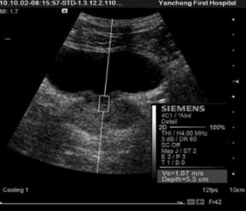
Virtual touch tissue quantification (VTTQ) is a new, promising technique for detecting the stiffness of tissues. To evaluate the performance of VTTQ in discrimination between chronic prostatitis/chronic pelvic pain syndrome(CP/CPPS) IIIa and IIIb, VTTQ was performed in 147 patients with clinical definite CP/CPPS. The shear wave velocity (SWV) at inner gland and outer gland was quantified by implementing an acoustic radiation force impulse. The performance of different ratios of SWV at outer gland and inner gland in discrimination between CP/CPPS IIIa and IIIb was compared. CP/CPPS IIIb and IIIa was detected in 69 and 78 patients, respectively. The SWV values of outer gland in the patients with CP/CPPS IIIa were significantly greater than that of inner gland, while there were no significant difference between outer gland and inner gland in the patients with CP/CPPS IIIb. The area under the receiver operating characteristic curve for the ratio one (<1.5) of SWV at outer gland and inner gland to distinguish CP/CPPS IIIb from IIIa was 0.72, while it was 0.88 for the ratio two (<1.1). The diagnostic sensitivity, specificity and accuracy for CP/CPPS IIIb were 100%, 69.2%, 83.7%, respectively for the ratio one and 100%, 84.6%, 91.8%, respectively for the ratio two. These data suggested that CP/CPPS IIIa and IIIb have different SWV values in inner gland and outer gland, and VTTQ can effectively distinguish CP/CPPS IIIb from CP/CPPS IIIa using the ratio of SWV at outer gland and inner gland.


Downloads

Download data is not yet available.
A novel and simple approach to distinguish chronic prostatitis/ chronic pelvic pain syndrome IIIb from IIIa using virtual touch tissue quantification

Downloads

Additional Files

Published

2011-11-20

How to Cite

1.
Zheng X, Ji P, Mao H, Wu J. A novel and simple approach to distinguish chronic prostatitis/ chronic pelvic pain syndrome IIIb from IIIa using virtual touch tissue quantification. Biomol Biomed [Internet]. 2011Nov.20 [cited 2023Apr.1];11(4):205-8. Available from: https://www.bjbms.org/ojs/index.php/bjbms/article/view/2546

Issue

Section

Translational and Clinical Research

INTRODUCTION

Chronic prostatitis/chronic pelvic pain syndrome (CP/CPPS, NIH category III), is the most prevalent form of prostatitis, which is characterized by chronic pelvic pain without an infection of the urinary tract [1-5]. To date, the pathogenesis of CP/CPPS is also unclear. Five different causes, including infection, detrusor-sphincter dysfunction, immunological dysfunction, interstitial cystitis and neuropathic pain, have been discussed [3, 4]. According to the presence or absence of inflammatory cells in the expressed prostatic secretions (EPS), CP/CPPS is split into inflammatory CP/CPPS (category IIIa) and non-inflammatory CP/CPPS (IIIb). However, white blood cell (WBC) counts have not been shown to correlate with symptoms or with the presence or absence of infection [4, 6]. Moreover, the relevance of abnormal WBC counts in the EPS is questionable. In addition, prostatic massage often leads to rectal discomfort, whose acceptance rate is low. Better methods for discrimination between CP/CPPS IIIa and IIIb are needed. Virtual touch tissue quantification (VTTQ) is a new, promising implementation of the ultrasound acoustic radiation force impulse (ARFI) imaging, which can effectively and objectively detect the tissue stiffness by measuring the shear wave velocity (SWV) values [7-9]. Our previous studies demonstrated that VTTQ not only can easily detect the age-related changes in prostate stiffness [10] and but also can discriminate between prostate cancer and benign prostatic hyperplasia. For CP/CPPS, it has not been possible to histologically confirm a correlation between prostatitic inflammation and symptoms at present, but its subtypes, IIIa and IIIb, maybe have different stiffness in the prostate. In the present study, we quantified the stiffness of inner gland and outer gland of prostate in CP/CPPS IIIa and IIIb, and evaluated the performance of VTTQ in discrimination between CP/CPPS IIIa and IIIb in order to explore a better strategy for CP/CPPS diagnosis.

MATERIALS AND METHODS

Study Population

The study was approved by the local human research ethics committee and free informed consent was obtained from all the subjects. One hundred and forty seven patients (mean age: 39.7±12.3 years, range: 26-53 years) with clinical definite CP/CPPS based on abnormal or normal findings on expressed prostatic secretion and post prostatic massage urine in the 4-glass or 2-glass test were enrolled in this study. The control group consisted of 65 healthy volunteers (mean age: 37.8±13.7 years, range: 25-51 years). The inclusion criteria were: (a) absence of any history of focal or diffuse disease at any of the examined organs, assessed by subject’s history, clinical symptoms, laboratory data, radiology and computer tomography; (b) normal visualization of the prostate on transrectal ultrasound.

Examination protocol

VTTQ was performed in all the subjects, using a Siemens ACUSON S2000 US system (Siemens, Germany), with convex probes (4C1), tissue harmonic imaging (THI; 4 MHz) and mechanical index of 1.7. To begin with, a target region of interest (ROI) (box with fixed dimension of 1×0.5 cm) was identified on a conventional ultrasound image. Then, an acoustic push pulse was transmitted immediately on the right side of the ROI, where the SWVs were calculated and expressed with a numerical value (meter/second, m/s), as a result of multiple measurements made for the same spatial location [6]. The subjects were placed in the recumbent position. The operators performed three measurements at inner gland and outer gland through the abdomen and perineum, respectively, after the subjects properly emptied their bladders (Figure 1).

FIGURE 1: The measurement of shear wave velocity (SWV) of the prostate at inner gland from the abdominal view (A) and outer gland from the transperineal view (B) with virtual touch tissue quantification.

Statistical analyses

Data were expressed as the mean±SD. Differences between the mean values of the two groups were analyzed by unpaired t tests. A McNemar test was used to compare the sensitivity, specificity, and accuracy for different diagnostic criteria. A receiver operating characteristic curve (ROC) analysis was used to evaluate and compare the performance of the two ratios of SWV at outer gland and inner gland (<1.1 and <1.5) in discrimination between CP/CPPS IIIa and IIIb. Differences were considered significant at p<0.05. All statistical analysis was performed with SPSS version 13 software for Windows (SPSS Inc, Chicago, IL).

RESULTS

CP/CPPS IIIa was detected in 69 patients (46.9%) in all patients, and CP/CPPS IIIb was detected in the remaining 78 patients(53.1%). As shown in Figure 2, the SWV values of outer gland in the patients with CP/CPPS IIIa were significantly greater than that of inner gland, while there were no significant difference between inner gland and outer gland in the patients with CP/CPPS IIIb. The SWV values at out gland of CP/CPPS IIIa were significantly greater than that of normal prostate (1.83±0.61 vs. 1.37±0.39), and were slightly greater than that of CP/CPPS IIIb (1.83±0.61 vs. 1.63±0.58), however, the SWVs at inner gland were significantly lower than that of normal prostate and CP/CPPS IIIb (1.07±0.43 vs. 1.36±0.45, 1.67±0.64). While the SWV values at inner gland and outer gland of CP/CPPS IIIb were all greater than that of normal prostate. As all known, the area under the ROC (AUC) is positively correlated with the discrimination performance. The performance of two ratios of SWV at outer gland and inner gland (<1.1 and <1.5) in discrimination between CP/CPPS IIIa and IIIb are shown in Figure 3. The AUC for the ratio, <1.5, to distinguish CP/CPPS IIIb from IIIa was 0.72, while it was 0.88 for the ratio, <1.1. The diagnostic sensitivity, specificity and accuracy of the two ratios of SWV at outer gland and inner gland (<1.1 and <1.5) for CP/CPPS IIIb are shown in Table 1. The overall specificities and accuracies of the ratio, <1.1, were significantly higher than those of the ratio, <1.5 (84.6% vs. 69.2%, 91.8% vs. 83.7%), although there were no significant differences in the sensitivities between the two ratios (100% vs. 100%).

FIGURE 2: The comparison of shear wave velocity (SWV) values between inner gland and outer gland in the normal prostate(NP), chronic prostatitis/chronic pelvic pain syndrome (CP/CPPS) IIIa and IIIb (*p<0.05, **p<0.01).
FIGURE 3: Receiver operating characteristic curve showing the performance of the different ratios of shear wave velocity at outer gland and inner gland in discrimination between chronic prostatitis/chronic pelvic pain syndrome (CP/CPPS) IIIa and IIIb.
TABLE 1: The sensitivity, specificity and accuracy of the different ratios of shear wave velocity at outer gland and inner gland for chronic prostatitis/chronic pelvic pain syndrome (CP/CPPS) IIIb.

DISCUSSION

The results presented here indicate that VTTQ can effectively detect the stiffness of prostate in patients with CP/CPPS, and CP/CPPS IIIa and IIIb have different SWV values at outer gland and inner gland. VTTQ can effectively distinguish CP/CPPS IIIb from IIIa using the ratio of SWV at outer gland and inner gland. ARFI imaging is a new ultrasound imaging modality to evaluate the stiffness of deep tissues by short-duration acoustic radiation forces that produce localized displacements in a “pushed” ROI [11, 12]. Due to the non-invasive and easily accessible nature of VTTQ, this technology makes it possible to conduct a thoroughly evaluation of prostate stiffness at an optional site without any discomfort and special preparation. In our study, we can easily detect the stiffness of prostate by measuring SWV values via the abdomen or perineum in all subjects with a satisfactory reproducibility. Normal prostate is assumed as a linear, isotropic, elastic body. Theoretically, the stiffer the prostate, the faster the shear wave will be propagated. In our study, CP/CPPS IIIa has greater SWV values at outer gland than at inner gland and those of normal prostate, which possibly correlates with its pathogenesis. Maybe, the inflammation often occurs at outer gland of the prostate. Interestingly, CP/CPPS IIIb has greater SWV values both at outer gland and inner gland than those of normal prostate. Just as the pathophysiology of CP/CPPS remains an enigma, the cause of this phenomenon can not be clarified now. Another phenomenon we can see from the results is that the ratio of SWV at outer gland and inner gland of CP/CPPS IIIb is nearly equal to one, while it is always far greater than one in CP/CPPS IIIa. So the difference of the ratios of SWV at outer gland and inner gland just provide a new method for the diagnosis of CP/CPPS. In our study, several ratios of SWV at outer gland and inner gland for the diagnosis of CP/CPPS IIIb were chosen according the AUC under the ROC. Two ratios (<1.1 and <1.5) are finally determined. Compared to the ratio, <1.5, the ratio,<1.1 not only have a 100% sensitivity, but also have higher specificities and accuracies, which can be a novel and simple approach to distinguish CP/CPPS IIIb from CP/CPPS IIIa. In the present study, the specimen of subjects is limited, which needs to be collected in the future. The pathogenesis for causality between the stiffness of prostate and the category of CP/CPPS requires further study. There are also some problems with the use of VTTQ for the quantitation of prostate stiffness. The limited detected depth (maximum 5.5 cm), the fixed box dimension (1×0.5cm) of the target ROI, and the sensitivity to movement artifacts maybe become obstacles to the extensive application of this new technology.

CONCLUSION

VTTQ can simply and effectively discriminate between CP/CPPS IIIa and CP/CPPS IIIb with much higher sensitivity, specificity and accuracy. Although several limitations mentioned above, this method still holds considerable clinical promise for the diagnosis of CP/CPPS.

Acknowledgements

ACKNOWLEDGMENTS

The authors gratefully acknowledge the technical assistance and helpful discussion of Lin L, Qiao XL at the department of ultrasound, The First People’s Hospital of Yancheng, Jiangsu Province, P. R. China.

DECLARATION OF INTEREST

The authors have no conflict of interest to declare.

REFERENCES

  1. , , , , , (). Management of chronic prostatitis/chronic pelvic pain syndrome: a systematic review and network meta-analysis. JAMA.
  2. , , (). Prostatitis: diagnosis and treatment. Am Fam Physician.
  3. , , , , (). Prostatitis and male pelvic pain syndrome: diagnosis and treatment. Dtsch Arztebl Int.
  4. , (). Prostatitis: Infection, neuromuscular disorder, or pain syndrome? Proper patient classification is key. Cleve Clin J Med.
  5. , (). Management of prostatitis. Prostate Cancer Prostatic Dis.
  6. , , (). Chronic prostatitis: epidemiology and role of infection. Urology. discussion 13
  7. , , (). Acoustic Radiation Force Impulse (ARFI) technique in ultrasound with Virtual Touch tissue quantification of the upper abdomen. Radiol med.
  8. , , (). Tissue quantification with acoustic radiation force impulse imaging: Measurement repeatability and normal values in the healthy liver. Am J Roentgenol.
  9. , , , , , (). Shear wave velocity is a useful marker for managing nonalcoholic steatohepatitis. World J Gastroenterol.
  10. , , , , , (). A novel approach to assessing the changes in prostatic stiffness with age using virtual touch tissue quantification. J Ultrasound Med.
  11. , , , (). Acoustic radiation force impulse imaging: in vivo demonstration of clinical feasibility. Ultrasound Med Biol.
  12. , , , (). Solid appearance of pancreatic serous cystadenoma diagnosed as cystic at ultrasound acoustic radiation force impulse imaging. JOP.