Radiographic estimation in seropositive and seronegative rheumatoid arthritis

Authors

  • Vjollca Sahatçiu-Meka Physical Medicine Department, Clinic for Orthopaedics, Faculty of Medicine, University of Prishtina
  • Sylejman Rexhepi Rheumatology Department, Clinic for Internal Medicine, Faculty of Medicine, University of Prishtina
  • Suzana Manxhuka-Kërliu Institute for Pathology, Faculty of Medicine, University of Prishtina
  • Mjellma Rexhepi Rheumatology Department, Clinic for Internal Medicine, Faculty of Medicine, University of Prishtina

DOI:

https://doi.org/10.17305/bjbms.2011.2571

Keywords:

rheumatoid arthritis, seropositive, seronegative, radiography estimation

Abstract

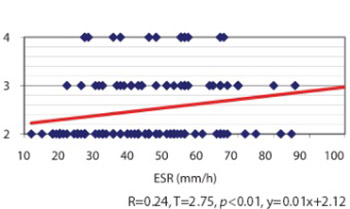
Long since it have been suggested that a subpopulation of patients with rheumatoid arthritis, diagnosed with negative rheumatoid factor tests, represents a clinical entity quite distinct from that of seropositive rheumatoid arthritis (RA). Our aim was to establish a scientific comparative analysis between seronegative and seropositive rheumatoid arthritis, regarding some radiological and clinical parameters, applied for the first time on patients from Kosovo. Two hundred fifty patients with rheumatoid arthritis according to the American College of Rheumatology criteria were retrospectively studied by analysis the radiographic damage and clinical parameters of the disease, using a data base. All examinees were between 25-60 years of age (Xb=49.96, SD=10.37) with disease duration between 1-27 years (Xb = 6.41, SD=6.47). All patients underwent a standardised evaluation radiographs. Baseline standardised poster anterior radiographs of hands and feet and radiographs of other joints, depending on indications, were assessed. Erythrocyte sedimentation rate values correlated with the radiological damages and statistical difference was found for seronegative subset (r=0.24, p<0.01). Longer duration of the disease resulted in the increase of radiological changes in both subsets (r=0.66, p<0.01) seronegative, (r=0.49, p<0.01) seropositive. Anatomic changes of IInd and IIIrd level were nearly equally distributed in both subsets, 76 (60.8%) seronegative, 75 (60%) seropositive. Radiological damages are nearly equal in both subsets, elevate in relation to the duration of the disease and correlate with ESR values. Regarding the sero-status, differences within sex, with some exceptions, are not relevant. Although there are some definite quantitative and qualitative differences regarding sero-status, obviously there is a great deal of overlap between the two groups.

Downloads

Download data is not yet available.
Radiographic estimation in seropositive and seronegative rheumatoid arthritis

Downloads

Additional Files

Published

2011-08-20

How to Cite

1.
Sahatçiu-Meka V, Rexhepi S, Manxhuka-Kërliu S, Rexhepi M. Radiographic estimation in seropositive and seronegative rheumatoid arthritis. Biomol Biomed [Internet]. 2011Aug.20 [cited 2023Nov.30];11(3):180-4. Available from: https://www.bjbms.org/ojs/index.php/bjbms/article/view/2571

Issue

Section

Translational and Clinical Research

INTRODUCTION

Rheumatoid arthritis (RA) is an auto-immune, chronic inflammatory disease characterised by synovitis and bone destruction [1]. Although the etiopathogenesis of RA is unknown, the majority of scientists support the immunology based theory on discovery of rheumatoid factor (RF) [2]. A positive test for rheumatoid factor is by no means pathogomonic of rheumatoid arthritis, but is present in 70 to 90% of patients with the disease, as well as in 5-8% in healthy population. Patients with a high titer of IgM-RF are more likely to have erosive joint disease, extra-articular manifestations, and greater functional disability. In contrast, patients with negative rheumatoid factor in general exhibit a milder disease course. Recently, various test methods based on the principle of agglutination (Waaler-Rose and Latex tests) are being applied, by which only the presence of IgM-RF is proven. Rheumatoid factor could be found in different immunoglobuline classes (G, A, D and E) defined by ELISA [3]. The inflammation in RA causes a shift in the bone metabolism towards increased osteoclast - mediated bone turn-over [4]. This dysregulation causes reduced bone mass, which is known to be an early feature in RA patients, visualised as juxta-articular bone demineralisation on radiographs [5]. One of the 7 diagnostic criteria for the diagnoses of RA, established by the American College of Rheumatology (ACR) in 1987, is the presence of bone erosion on radiograph [6]. Genetic information is necessary for prediction of radiographical changes in patients with RA. Severe radiological changes are associated with allele HLA-DRB1*04. Within 2 years of disease onset, approximately 70% of all patients develop erosive disease, and show a light progress from the ninth year onwards. The patients with erosion, particularly on feet, in the early phase of disease are associated with a destructive course of RA [7]. The same problem appears in patients with arthritis of large joints at first presentation, in particular the knee [8]. Radiographic progression in rheumatoid arthritis has in several studies been shown to be predicted by serological markers widely used in daily clinical practice [9, 10]. Quantification of localised bone loss has been proposed as an outcome measure in early RA [11]. Plain X-ray offers high specificity in the differential diagnoses of rheumatic diseases [12]. There are other useful tools like Magnetic Resonance Imaging (MRI), Computed Tomography (CT), Doppler Sonography, Bone Scintigraphy, Ultrasonography, etc., which are suitable for evaluating the intensity of synovitis, for early diagnosis of synovitis, and for the assessment of joints and periarticular structures in all rheumatological disorders respectively [13,14]. In response to the continuing debate as to whether seronegative and seropositive rheumatoid arthritis are part of the same disease spectrum, or are distinct disorders, we aimed to perform a comparative analysis regarding some clinical and radiological features.

MATERIALS AND METHODS

Patients

Using the data base, 250 patients with rheumatoid arthritis, diagnosed according to the American College of Rheumatology ACR (1987) revised diagnostic criteria, were retrospectively studied by analysis the radiographic damage and clinical parameters of the disease, using the data base. The studied group consisted of 125 (93 female, 32 male) seronegative patients with titers lower than 1/64 as defined by Rose-Waaler test, whereas the control group consisted of 125 (93 female, 32 male) seropositive patients with titers of 1/64 or higher. Patients who belonged to 2nd and 3rd functional class (ARA) are taken into consideration. Their age ranged from 25 to 60 years (Xb=49.96) (seronegative Xb=46.63, SD=10.31, seropositive Xb=47.30, SD=10.47). Disease duration was between 1-27 years (Xb=6.41) (seronegative Xb=6.45, SD=5.99, seropositive Xb=6.36, SD=6.94). At baseline, all patients underwent a standardized evaluation including laboratory tests and radiographs. Conventional hand radiographs were used as a “test subject”. Patients belonged to II-IV anatomic stage (ARA). Erythrocyte sedimentation rate (ESR) was measured by the Westergren method, ranging from 0 to 140 mm/h. The correlation between different clinical parameters, laboratory and anatomic stages were investigated. For the presentation of the results the structure, prevalence, arithmetic average (Xb), standard deviation (SD), variation coefficient (CV%) and variation interval (Rmax-Rmin) were used. Probability level was expressed by p<0.01 and p<0.05. The correlation between the duration of RA and anatomical stages (ARA), ESR and anatomical stages (ARA), regarding sero-status was measured by Point-biserial correlation.

RESULTS

The largest number of patients (Table 1) has met IInd anatomic class (ARA) [76 (60.8%) seronegative, 75 (60%) seropositive]. Differences between sero-groups, regarding anatomical classes were not significant (χ2=0.15, p>0.05). Apart from this, no significant statistical difference regarding sex was found. Patients who belonged to IInd anatomic class (Table 2) had shorter disease duration, while the cohort of over 10 years of duration was dominated by patients from the IIIrd anatomic class, valid for both sero-groups. Seropositive patients with duration of the disease up to 10 years have passed faster in the third and fourth anatomical stage, while others of over 10 years, had approximately equal distribution through sero-groups. Correlation coefficient for both sero-groups was high and positive [seronegative (r= 0.66, p<0.01), seropositive (r=0.49, p<0.01)], which means that extended duration of disease increases the number of radiological changes with high reliability (Figure 1, 2). Low erythrocyte sedimentation rate values, up to 30mm/h (Table 3) were more frequent in IInd anatomic class 49 (32.5%) and showed reduction in IIIrd anatomic class 12 (17.4%) and IVrth anatomic class 5(16.7%). Erythrocyte sedimentation rate average values, 30-70mm/h, have shown a tendency to increase by stages (II, III, IV: 56.3%, 71%, 76.7%). For maximum ERS values, 71-110 mm/h, the changes were too small. Accelerated ESR values correlated with anatomical stages (r=0.14, p<0.05). Greater correlation and positive (r=0.24, p<0.01) was found among seronegative patients, while seropositive group correlation was smaller and of no statistical significance (r=0.06, p>0.05) (Figure 3, 4).

TABLE 1: Radiological changes (ARA) regarding to sero-status and sex.
TABLE 2: Correlation between the duration of AR and anatomical stages (ARA) according to sero-status
FIGURE 1: Correlation and regression. Anatomic stage-Duration (years)-SPRA
FIGURE 2: Correlation and regression. Anatomic stage-Duration (years)-SPRA
TABLE 3: Correlation between accelerated values of ESR and anatomical stages (ARA) according to sero-status
FIGURE 3: Correlation and regression. ERS - Anatomic stage - SNRA
FIGURE 4: Correlation and regression. ERS - Anatomic stage - SNRA

DISCUSSION

Patients with clinical features of rheumatoid arthritis, but negative rheumatoid factor present a diagnostic challenge. It has recently been suggested that a subpopulation of patients with RA, diagnosed on clinical, radiologic and pragmatic grounds, but with negative rheumatoid factor tests, represents a clinical entity quite distinct from that of seropositive RA [15]. The nature of the destructive process, as defined by radiological examination, may be different in patients with seropositive rheumatoid arthritis from that seen in individuals with so-called 'seronegative rheumatoid arthritis' [16]. Presence of anti-cyclic citrullinated peptide antibodies is correlated to disease activity and to bone erosions development [17]. Vittecoq et al. [18] have concluded that the antibody anti-CCP, compared with rheumatoid factor, is of insufficient value to predict an early erosive and progressive RA, while Vencovsky [19] considers that combined analysis of above parameters increase this possibility. This study was undertaken to determine whether the two populations differ radiologically. It was found that the differences between sero-groups in relation to anatomical stages did not show significant statistical difference, which is valid to both sexes. Opinions of some authors [20] who confirm that seropositivity did not correlate with bone erosion are close to these findings. Unlike our data, some authors claim that seropositivity results in more severe anatomical changes in joints [21, 22, 23] and those patients with high titer of IgM-RF have significantly higher progressive radiological index [24]. Related data provided by el-Khoury et al. [25] confirmed that radiograms of seronegative patients differ significantly from radiograms of seropositive patients concerning the lower rate of juxta-articular osteoporosis, relative lack of subchondral erosion, predominance of changes across the carpal part, greater number of contractures and the asymmetry of the attacked joint. To this perception contributes the study of Krahe et al. [26] as well, confirming that the extent of periarticular destruction was significantly greater amongst seropositive than amongst seronegative patients, both at the beginning and the end of the study, but there was no significant difference in the rate at which this progressed. HLA-DR alleles such as HLA-DR4 and HLA-DR1 are associated with the risk to develop RA [27]. Listing et al. [28] have noticed no significant differences in frequency of DR4, in the patients with and without erosion, while Vehe et al. [29] and Reneses et al. [30] have found that DR4 is prevalent in the patients with erosive RA, regardless of the status of FR. Furthermore, in patients with RA, Mattey et al. [31] have found that DRB1 are not predictive for erosive damages to early seropositive patients, but are predictive for seronegative patients. Anatomic damages are proportional to the intensity and the duration of the inflammatory process, deteriorate rapidly during the first 2 years of the disease, show a weak progress from the 9th year of duration and onward [32]. Much of these findings were confirmed in our study, where seropositive patients pass earlier in anatomic stages III-IV and that the duration of the disease increases radiological changes, with no statistically significant difference according to sero-status. Currently available biomarkers of more severe disease include elevated ESR or C-reactive protein levels (CRP) and IgM-RF or the antibody anti-CCP positivity [1,33]. Erythrocyte sedimentation rate is more closely related to the progression of joint damage than C-reactive protein or hemoglobin [30]. Findings in some studies add to the understanding of the antibody anti-CCP and ESR as important predictors of bone involvement in RA [9]. Our findings provide evidence that accelerated ESR values correlate with anatomical stages (r=0.14, p<0.05). Greater correlation and positive (r=0.24, p<0.01) was found among seronegative patients, whereas seropositive group correlation was smaller and not of any statistical significance (r=0.06, p>0.05). Our collected data are comparable to those of van Leeuwen et al. [33] who confirm that acute phase proteins are in correlation with radiological damages. Some authors consider that erosive damages at 1st year in patients with recent-onset RA are significantly influenced by SE homozygosity and the presence of baseline erosions, but not by RF status, anti-CCP status, or -308 TNF-alpha genotype [30] the others claim that female gender, DRB1*04 alleles (rather than the SE), and the presence of anti-CCP antibodies at baseline (independently of the titer) are the most important predictors of progression [34].

CONCLUSION

Radiological damages are nearly equal in both subsets, elevate in relation to the duration of the disease and correlate with ESR values. Regarding the sero-status, differences within sex, with some exceptions, are not relevant. Although there are some definite quantitative and qualitative differences regarding sero-status, obviously there is a great deal of overlap between the two groups.

DECLARATION OF INTEREST

Authors declare no conflict of interest.

REFERENCES

  1. , , (). Genetic influences modulating the radiological severity of rheumatoid arthritis. Ann Rheum Dis.
  2. (). Arthritis and Allied conditions.
  3. , , (). Genetic basis for rheumatoid arthritis. Arch Immunol Ther Exp.
  4. (). [An update on diagnostic and prognostic biomarkers of early rheumatoid arthritis] Cas Lek Cesk.
  5. (). Pathogenesis of bone and cartilage destruction in rheumatoid arthritis. Rheumatology (Oxford).
  6. , , , , (). The 5-yr HAQ-disability is related to the first year's changes in the narrowing, rather than erosion score in patients with recent-onset rheumatoid arthritis. Rheumatology (Oxford).
  7. , , , , , (). Clinical usefulness of genetic information for predicting radiographic damage in rheumatoid arthritis. J Rheumatol.
  8. , , , (). Arthritis of the large joints, in particular the knee, at first presentation is predictive for a high level of radiological destruction of the small joints in rheumatoid arthritis. Ann Rheum Dis.
  9. , , , , , (). Antibodies to cyclic citrullinated protein and erythrocyte sedimentation rate predict hand bone loss in patients with rheumatoid arthritis of short duration: a longitudinal study. Arthritis Res Ther.
  10. , (). How to predict prognosis in early rheumatoid arthritis. Best Pract Res Clin Rheumatol.
  11. , , (). Hand bone loss as an outcome measure in established rheumatoid arthritis: 2-year observational study comparing cortical and total bone loss. Arthritis Res Ther.
  12. , , , , , (). [Bone marrow edema: definition, diagnostic value and prognostic value]. J Radiol.
  13. , , , , , (). High-resolution MRI of the wrist and finger joints in patients with rheumatoid arthritis: comparison of 1.5Tesla and 3.0 Tesla. Eur Radiol.
  14. , , , (). Power Doppler sonography and pulse-inversion harmonic imaging in evaluation of rheumatoid arthritis synovitis. AJR Am J Roentgenol.
  15. , (). [Anti-CCP antibody test: diagnostic and prognostic values in rheumatoid arthritis]. Rev Med Brux.
  16. , (). The hand radiograph as a diagnostic discriminant between seropositive and seronegative ’ rheumatoid arthritis’: a controlled study. Ann Rheum Dis.
  17. , , , (). Disability and health-related quality of life among patients with rheumatoid arthritis: association with radiographic joint damage, disease activity, pain, and depressive symptoms. Scand J Rheumatol.
  18. , , , , , (). Rheumatoid factor is the strongest predictor of radiological progression of rheumatoid arthritis in a three-year prospective study in community-recruited patients. Rheumatology (Oxford).
  19. , , , , , (). Autoantibodies can be prognostic markers of an erosive disease in early rheumatoid arthritis. Ann Rheum Dis.
  20. , , , , , (). Disease activity and joint damage progression in early rheumatoid arthritis: relation to IgG, IgA and IgM rheumatoid factor. Ann Rheum Dis.
  21. , , , , , (). Raised IgA rheumatoid factor (RF) but not IgM RF or IgG RF is associated with extra-articular manifestations in rheumatoid arthritis. Scand J Rheumatol.
  22. , , , , , (). Prognostic markers of radiographic progression in early rheumatoid arthritis. Arthritis Rheum.
  23. , , , (). The effect of HLA-DRB1 genes, rheumatoid factor, and tretment on radiographic disease progression in rheumatoid arthritis over 6 years. J Rheumatol.
  24. , , , , , (). Rheumatoid factor measured by fluoroimmunoassay: a responsive measure of rheumatoid arthritis disease activity that is associated with joint damage. Ann Rheum Dis.
  25. , , , (). Seronegative and seropositive rheumatoid arthritis: radiographic differences. Radiology.
  26. , , , (). [Follow-up study in chronic polyarthritis as represented in roentgen image]. Rofo.
  27. , , , (). Extraarticular manifestation of seronegative and seropositive reuma-thoid arthritis. Bosn. J Basic Med Sci.
  28. , , , , , (). HLA-DRB1 genes, rheumatoid factor and elevated C-reactive protein: independent risk factors of radiographic progression in early rheumatoid arthritis. Berlin Collaborating Rheumatological Study Group. J Rheumatol.
  29. , , , , , (). Erosive rheumatoid factor negative and positive rheumatoid arthritis are immunogenetically similar. J Rheumatol.
  30. , , , , , (). The value of HLA-DRB1 shared epitope, -308 tumor necrosis factor-alpha gene promoter polymorphism, rheumatoid factor, anti-citrullinated peptide antibodies, and early erosions for predicting radiological outcome in recent-onset rheumatoid arthritis. J Rheumatol.
  31. , , , , , (). Independent association of rheumatoid factor and HLA-DRB1 shared epitope with radiographic outcome in rheumatoid arthritis. Arthritis Rheum.
  32. , , , , (). Measurement and prediction of radiological progression in early rheumatoid arthritis. J Rheumatol.
  33. , , , , , (). The acute-phase response in relation to radiographic progression in early rheumatoid arthritis: a prospective study during the first three years of treatments. Br J Rheumatol.
  34. , , , (). The effect of HLA-DRB1 genes, rheumatoid factor, and tretment on radiographic disease progression in rheumatoid arthritis over 6 years. J Rheumatol.