Choledochal cyst: presentation of the disease with a case report.
DOI:
https://doi.org/10.17305/bjbms.2011.2574Keywords:
common bile duct, choledochal cyst, cholangiographyAbstract
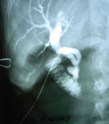
Choledochal cyst is a rare disease of the biliary tract. There are five main types of choledochal cysts with a few recognized sub-types. The etiology of choledochal cysts still is unclear. The incidence of biliary tract cancer in patients with choledochal cysts increases with age. In the past, choledochal cysts were often treated using drainage procedures; however, the optimal treatment used today is likely to involve the complete excision of the extrahepatic duct, cholecystectomy, and Roux-en-Y hepaticojejunostomy. Endoscopic treatment of type III choledochocele should be limited to the management of smaller lesions. We report a case of 75 years old patient with distal choledochal diverticuly, Todani’s type III- choledochocele. Delay in the diagnosis increases the frequency of associated biliary pathology, malignant alternation and suboptimal surgical therapy. Often, intraoperative finding of choledochal cyst is the first contact with this rear entity, so awareness of possible presence of this uncommon disease is very important for surgeon.
Citations
Downloads
Downloads
Additional Files
Published
How to Cite
Accepted 2017-10-17
Published 2011-08-20


