Intra-amniotic administration of exogenous pulmonary surfactant for improving in lung maturity of fetal rabbits with intrauterine infection caused by premature rupture of membranes

Authors

  • Jing Liu Department of Neonatology & NICU, Bayi Children’s Hospital Affiliated With General Hospital of Beijing Military Command
  • Jing Wu Department of Neonatology & NICU, Bayi Children’s Hospital Affiliated With General Hospital of Beijing Military Command Department of Neonatology, He-Xian Memorial Hospital Affiliated to Southern Medical University
  • Na Yang Department of Neonatology & NICU, Bayi Children’s Hospital Affiliated With General Hospital of Beijing Military Command Anhui Medical University
  • ZhiChun Feng Department of Neonatology & NICU, Bayi Children’s Hospital Affiliated With General Hospital of Beijing Military Command

DOI:

https://doi.org/10.17305/bjbms.2011.2590

Keywords:

premature rupture of membranes, intrauterine infection, lung ultrastructure, pulmonary surfactant, fetal rabbits

Abstract

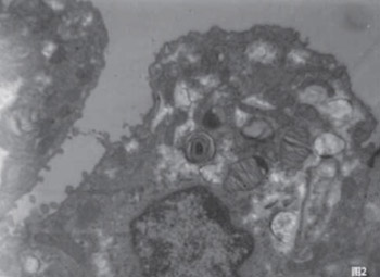
This study was to investigate the effect of intra-amniotic administration of pulmonary surfactant (PS) on lung maturation in conditions of experimentally induced premature rupture of membranes (PROM) and intrauterine infection of rabbits. To establish animal (rabbit) models of intrauterine infection caused by PROM, E. coli was intrauterinely injected in 24-and 26-day pregnant animals. Twenty healthy pregnancy adult Japanese white rabbits were divided into three groups: the infection group (8 rabbits), infection group with intra-amniotic PS administration (8 rabbits) and healthy controls (4 rabbits). Ultrastructure changes in the lung were observed under an electron microscope 19.5 hours after intervention. The results showed that the lung infection levels of fetal rabbits in the infection group and the infection group with PS had no significant difference. Bacillus bodies and infiltrated inflammatory cells can be occasionally seen in the alveoli, bronchial lumen, and cytoplasm. The type II alveolar epithelial cells (AT-II) were decreased in the infection group than that in control group. Lamellar bodies showed vacuolation changes and different levels of apoptosis. In the infection group with PS, the count of AT-II did not show an obvious decrease. Lamellar bodies increased, and different secretion forms appeared. Also, there was little apoptosis and no obvious collagen fiber hyperplasia in antenatal administration of PS group. We believe that once PROM has happened, intrauterine infection and inflammation stimulated a cascade reaction on the fetal lung, leading to abnormal changes in the alveolar ultrastructure. Intra-amniotic administration of PS can improve the fetal lung ultrastructure and its maturity.

Downloads

Download data is not yet available.
Intra-amniotic administration of exogenous pulmonary surfactant for improving in lung maturity of fetal rabbits with intrauterine infection caused by premature rupture of membranes

Downloads

Additional Files

Published

2011-05-20

How to Cite

1.
Liu J, Wu J, Yang N, Feng Z. Intra-amniotic administration of exogenous pulmonary surfactant for improving in lung maturity of fetal rabbits with intrauterine infection caused by premature rupture of membranes. Biomol Biomed [Internet]. 2011May20 [cited 2023Sep.23];11(2):103-7. Available from: https://www.bjbms.org/ojs/index.php/bjbms/article/view/2590

Issue

Section

Short Communication

INTRODUCTION

Premature rupture of membranes (PROM) is a common clinical pathological phenomenon with an incidence rate of 2.7 to 21.4%. Prolonged PROM can adversely affect the health of a fetus or a neonate because of the significant reduction in amniotic fluid, intrauterine infection, and other risks [1-8]. Infection and inflammatory reaction after PROM are also important reasons for respiratory distress syndrome (RDS), bronchopulmonary dysplasia (BPD), or pulmonary hypoplasia [9, 10]. These pulmonary diseases are major factors that lead to fetuses or neonates deaths. Therefore, the mortality of fetuses and neonates with PROM can be significantly reduced if antenatal measures are taken suitably. Antenatal supplementary pulmonary surfactant (PS) for the prevention of RDS has been attempted in both animal and human studies [11, 12], and some of them illustrated this method has a significant role in decreasing the incidence and severity of premature RDS [12-16], but the mechanism still remains unknown. The changes in lung ultrastructure related to PROM animal model and the effect of antenatal PS on lung ultrastructure have not been studied or clarified. Therefore, the purpose of this study was to investigate the effects of intrauterine infection on the pulmonary ultrastructure of fetal rabbits and to determine whether or not intra-amniotic administration of PS could promote lung maturity of fetal rabbits.

MATERIALS AND METHODS

Preparation of animal models

Preparation of the bacterium liquid: Standard E. coli strain (ATCC 25922 strain, offered by the National Institute for Pharmaceutical and Biological Products) was inoculated in blood agar and cultivated at 37 °C for 16 to 18h to acquire purified E. coli. This was first diluted in physiological saline, and then to 104/mL with Maxwell turbidimetry. Experimental model: the intrauterine infection model was made according to Guen et al. [17] and modified properly. Twenty healthy adult female Japanese white rabbits of reproductive age were selected (purchased from Beijing Baierkangnate Experimental Rabbit Breeding Biotechnology Development Co. Ltd). After successful pregnancy, they were divided into three groups: the intrauterine infection groups (8 rabbits), intrauterine infection with intra-amniotic PS administration groups (8 rabbits) and healthy controls (4 rabbits). Each group was further classified into 2 subgroups of 24 days (equivalent to 30 gestational weeks of humanity) (4 rabbits) and 26 days (equivalent to 32 gestational weeks of humanity) (4 rabbits), respectively. After they were anesthetized with 25 mg of ketamine/kg, a 2-cm vertical incision was performed along the median line below the gravid uterus:

  1. Intrauterine infection groups: a standard of E. coli strain 103 (0.1 mL) was in administrated into the bilateral uterine horns of the pregnant rabbits;

  2. Intrauterine infection+PS groups: besides intrauterine inoculation of the bacteria, 200 mg/kg PS (Curosurf: Chiesi Farmaceutici S.p.A., Italy) was administered through a needle into the amniotic cavity of each pregnant rabbit.

  3. Healthy controls: neither E. coli nor PS was intrauterine inoculated.

The incision was then closed up level by level, and the animals were returned to their cages. Exactly 19.5 hours (equivalent to 1 gestational week of humanity) after the establishment of the model, the fetal rabbits (including healthy controls) were taken out by hysterotomy for further observations. The preparation for transmission electron microscopy (TEM): 10 fetal rabbits were randomly selected for the following observations. The lung tissue specimen was prepared according to literature [18,19] and modified properly.

RESULTS

Lung tissue ultrastructure of normal fetal rabbits. The pulmonary alveolus cavity, alveolar type II cells (AT II), and alveolar type I cells (AT I) were examined under an electron microscope. AT I cells had a big nucleus, a small cytoplasm, and a few cell organs in the cytoplasm. On the free surface of AT II cells were some microvilli in different lengths and thicknesses, an obvious nucleus, and a plentiful cytoplasm. There were also plentiful mitochondria, rough endoplasmic reticulum, Golgi complex, lysosome, and other cellular organs. A number of lamellar bodies arranged in concentric circles or are parallel, as well as sedimentary glycogen ponds were visible (Figure 1).

FIGURE 1: Lung tissue ultrastructure of normal fetal rabbits (×10000) of gestational age of 24 days (A) and 26 days (B). Showing AT-II & I cells, nucleus, cytoplasm, mitochnodria, and many other cellular organs.

Changes in fetal rabbit lung ultrastructure in the infection group. The thalli of E. coli were occasionally seen in the bronchial tube lumen, alveolar space, and epithelial cell cytoplasm of the infection group. The thallus karyotheca was thick, and the chromatin was engrained. Under low power lens, infiltrated neutrophilic granulocyte, macrophage, and phagocytosis grains were visible, and perinuclear space can be seen between the nucleus and the cytoplasm (Figure 2). “Lung ultrastructure of the fetal rabbits in the 24-day infection group. Compared with the normal controls, AT II cells in the lung decreased in this group. Only a few microvilli on free cell surfaces were seen. The nucleus structure was loose and of edema. There were fewer cell organs in the cytoplasm, such as the mitochondria, endoplasmic reticulum, and golgiosome, and cytomembrane invagination was evident. Occasional lamellar bodies and sedimentary glycogen ponds in the cell were present (Figure 3A). Lung ultrastructure of the fetal rabbits in the 26-day infection group. Compared with the 24-day infection group, this group had a larger count of AT II cells. But they were less than controls. A number of lamellar bodies were seen. The mitochondria in the cytoplasm swelled, and the mitochondrial cristas broken. Besides, apoptosis phenomenon such as nuclear chromatin condensation occurred in more AT II cells (Figure 3B). Changes in fetal rabbit lung ultrastructure in infection with PS group. Fetal rabbit lung ultrastructure in 24-day infection with PS group. Compared with the infection group, the 24-day infection group with PS had more AT II cells and clearer microvilli on the free cell surfaces. The mitochondria, endoplasmic reticulum, golgiosome, and other cell organs in the cytoplasm were better developed, although there were still various degrees of dropsy. Lamellar bodies increased, and changes in different shapes such as concentric circle and laminiplantation occurred (Figure 4). Fetal rabbit lung ultrastructure in 26-day infection with PS group. Although the mitochondria in the cell still swelled, the count of AT II cells and that of AT II cells with lamellar bodies increased obviously. Moreover, the distribution of different shapes appeared, and one part scattered into alveolar space, showing changes in secreting PS status or vacuolation (Figure 5).

FIGURE 2: Fetal rabbit lung ultrastructure in intrauterine infection groups (×10000). The thalli of E. coli in alveolar space (A) and phagocytic vacuole and phagocytic grains (B). Showing the thick thallus karyotheca and engrained chromatin.
FIGURE 3: Changes in lung ultrastructure in intrauterine infection group (×10000). Showing the decreased AT- II cells, few microvilli, edema and loose nucleus at gestational age of 24 days (A). The decreased AT-II cells, swelled mitochondria and apoptosis bodies at gestational age of 26 days are shown on (B).
FIGURE 4: Changes in fetal rabbit lung tissue ultrastructure in 24-day infection with PS group (×10000). Showing clearer microvilli on the surface of AT-II cells and well developed cellular organs in cytoplasm.
FIGURE 5: Changes in fetal rabbit lung tissue ultrastructure in the 26-day infection group with PS (×10000). Showing the increased AT-II cells and more lamellar bodies.

DISCUSSION

Many factors can lead to PROM, but data on epidemiology, clinic, histology, microbiology, and molecular biology indicate that ascending infection or inflammation plays a major role. With infection, the proteolytic enzyme produced by the microorganism in the uterine neck and the vagina vault hydrolyzes the extracellular matrix of the fetal membrane. This reduces the tensile strength of tissues, so that collagenous fiber II is lessened, and membrane fragility increases. The endotoxin of the infection microorganism can also induce the production of prostaglandin and cause uterine contraction that results in PROM. When the fetus is exposed in the inflammatory environment for a prolonged time, the level of inflammatory factor increases and regulates fetal lung growth in many aspects. This hinders the alveolarization process and the growth of lung capillaries, which increases the incidence of lung diseases such as RDS, BPD, and CLD [10,20-23]. In our present study, intra-amniotic administration of PS was implemented in animal models with intrauterine infection, and the results showed that the lung ultrastructure of fetal rabbits with intrauterine infection can be improved significantly, which can be used for decreasing the degree and incidence rate of neonatal lung diseases such as RDS. Alveolar epithelial cells are a layer of epithelial cells covering the pulmonary alveolus surface, categorized into type I and type II pulmonary alveolus cells. The lamellar body is one kind of globular lamellar structure with concentric circles or parallel arrangement in alveolar epithelial type II cells, mainly composed of phosphatide, protein, and osamine polysaccharide. It is not only the storage area of PS in type II cells but is also the secretory granule of PS. The lamellar body secretes PS in exocytosis manner. Along with fetal lung maturity, lamellar bodies increase gradually. Therefore, the count of lamellar bodies in type II cells can reflect the maturity of the fetal lung. Galan et al. [13] once has studied the effect of intra-amniotic administration of exogenous PS (Exosurf) in preterm rabbit fetuses, and their results showed those rabbit pups receiving Exosurf had significantly better pressure-volume relationships and lower opening pressures than the rabbit pups with no treatment. Together with ours results of this study, we believe that intrauterine administration of exogenous PS may provide an additional means of RDS prophylaxis in the antepartum period. The following are the main findings of this study:

  1. The count of lamellar bodies in the pulmonary alveolus II cells of fetal rabbits in the infection group and that of type II cells with lamellar bodies were less than that of the normal group. Obviously, the evacuation of lamellar bodies showed the phenomenon of vacuole and apoptosis. The possible mechanism was that the products, such as phospholipase, released after the activation of neutrophilic granulocytes and macrophages can disturb and destroy PS and deactivate it in the infection environment. The excessive apoptosis of type II epithelial cells caused the cell count to reduce, decreasing the PS. The inflammatory factor after inflammation stimulation can directly damage the pulmonary alveolus and the blood capillary basilar membrane, and many serum proteins’ exosmosis deactivated the PS system. It is certainly that all of these hypothesis need to be confirmed in the following study.

  2. Compared with the infection group and control groups, the pulmonary alveolus cavities of the fetal rabbits in administration of PS group were larger and more regular. The count of pulmonary alveolus type II cells and lamellar bodies increased obviously. This indicated that intrauterine infection could hinder fetal lung development, whereas intra-amniotic administration of PS could improve the process of lung maturity of fetal rabbits. As a qualitative observation by electron microscope, no quantitative data obtained from present preliminary study, so it is necessary for us to make some quantitative evaluation in the future studies to confirm the results of this study.

CONCLUSION

In brief, the results of this study showed that intra-amniotic administration of PS after the occurrence of PROM with intrauterine infection could improve the lung maturity of fetal rabbits, and this is the first findings to provide an experimental basis for the use of antenatal PS for prevention of RDS in premature infants.

Acknowledgements

ACKNOWLEDGEMENTS

This work was supported by China Postdoctoral Science Foundation (20080431405 & 200801041).

DECLARATION OF INTEREST

Authors do not have any commercial affiliations, or potential conflicts of interest associated with this work submitted for publication.

REFERENCES

  1. , , (). Intrauterine infection and preterm delivery. N Engl J Med.
  2. , , (). Preterm prelabour repture of membranes: effect of chorioamnionitis on overall neonatal outcome. J Obstetr Gynecol.
  3. , , (). Preterm premature rupture of membrane management-inpatient versus outpatient: A retrospective review. Am J Perinatol.
  4. , , (). Maternal and fetal outcomes of spontaneous preterm premature rupture of membranes. JAOA.
  5. , , (). Prevalence, management, and outcomes of preterm prelabour rupture of the membranes of women in Canada. J Obstet Gynaecol Can.
  6. , (). Preterm premature rupture of membranes: diagnosis and management. Am Fam Physician.
  7. , , (). Early mortality in twin pregnancies complicated by premature rupture of membranes in the United States. J Obstet Gynaecol.
  8. , , (). The incidence rate of premature rupture of membranes and its influence on fetal – neonatal health: A Report from Mainland China. J Tropi Pediatr.
  9. , , (). Chorioamnionitis and inflammation of the fetal lung. Am J Obstet Gynecol.
  10. , , (). Antenatal infection in the rabbit impairs postnatal growth and lung alveolarisation. Eur Respir J.
  11. , , (). Antenatal modes of surfactant administration for RDS prevention: a review. J Natl Med Assoc.
  12. , , (). The effects of antenatal corticosteroids and surfactant replacement on neonatal respiratory distress syndrome. Bosn J Basic Med Sci.
  13. , (). Effect of intra-amniotic administration of Exosurf in preterm rabbit fetuses. Obstet Gynecol.
  14. , , (). Evidence of fetal pulmonary aspiration of intra-amniotic administered surfactant in animal experiment. J Perinat Med.
  15. , , (). Intraamniotic surfactant supply as RDS prevention. Med Wieku Rozwoj.
  16. , , (). Prophylaxis of neonatal respiratory distress syndrome by intra-amniotic administration of pulmonary surfactant. Chin Med J.
  17. , , (). Persistent bacteremia in rabbit fetuses despite maternal antibiotic therapy in a novel intrauterine-infection model. Antimicrob Agents Chemother.
  18. , , (). Ultrastructure of canine type II pneumocytes during hypothermic ischemia of the lung: a study by means of conventional and energy filtering transmission electron microscopy and stereology. Anat Rec.
  19. , , (). Human SP-A and a pharmacy-grade porcine lung surfactant extract can be reconstituted into tubular myelin-a comparative structural study of alveolar surfactants using cryo-transmission electron microscopy. Clin Physiol Funct Imaging.
  20. , , (). Chorioamnionitis and early lung inflammation in infants in whom bronchopulmonary dysplasia develops. Pediatrics.
  21. (). Antenatal inflammation and lung injury: prenatal origin of neonatal disease. J Perinatol.
  22. , (). Antenatal infection/inflammation and postnatal lung maturation and injury. Respir Res.
  23. , , (). The association between early tracheal colonization and bronchopulmonary dysplasia. J Perinatol.