Skin and kidney histological changes in graft-versus-host disease (GVHD) after kidney transplantation

Authors

  • Tadeja Pintar Abdominal Surgery Department, University Medical Centre of Ljubljana
  • Mario Alessiani Department of Surgery and IRCCS Polikliniko San Matteo, University of Pavia
  • Alojz Pleskovič Abdominal Surgery Department, University Medical Centre of Ljubljana
  • Aleš Pleskovič Department of Internal Medicine, University Medical Centre of Ljubljana
  • Ruda Zorc-Pleskovič Institute of Histology and Embryology, University Ljubljana, Faculty of Medicine
  • Aleksandra Milutinović Institute of Histology and Embryology, University Ljubljana, Faculty of Medicine

DOI:

https://doi.org/10.17305/bjbms.2011.2594

Keywords:

experimental model, graft versus host disease, kidney transplantation skin biopsies

Abstract

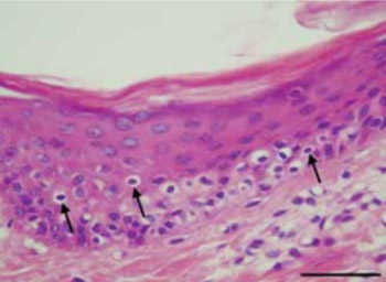
Kidney transplantation (Ktx) is generally performed during end stage renal disease due to a loss of the kidneys’ ability to filter wastes from the circulatory system. Acute graft-versus-host disease (GVHD) after Ktx is a life-threatening complication that progresses to organ failure, systemic complications, and death. The current study evaluated the significance of histologic findings of GVHD as obtained from skin biopsies following Ktx in swine. A swine model of Ktx with tacrolimus-based immunosuppression was used to assess possible correlations between acute-graft-cellular rejection and skin histological findings for prediction of GVHD. Animals were divided into a Ktx treatment group or a control group with no Ktx and skin and kidney biopsies were histologically assessed at postoperative days 0, 15, 30, 45 and 60. Skin samples were analyzed and classified from grade 1 to 4 of skin GVHD and the major histopathological changes of kidney acute cellular rejection were described using Banff’s score system. We observed a significant linear correlation between the histological grading values of skin biopsy changes and the histological grading values of kidney biopsies (Kendall’s tau_b=0.993) in the Ktx experimental group. No histological changes were observed in controls. Our findings demonstrate the diagnostic value of staging skin GVHD after Ktx and suggest it’s future utility for monitoring long term Ktx-induced changes.

Downloads

Download data is not yet available.
Skin and kidney histological changes in graft-versus-host disease (GVHD) after kidney transplantation

Downloads

Additional Files

Published

2011-05-20

How to Cite

1.
Pintar T, Alessiani M, Pleskovič A, Pleskovič A, Zorc-Pleskovič R, Milutinović A. Skin and kidney histological changes in graft-versus-host disease (GVHD) after kidney transplantation. Biomol Biomed [Internet]. 2011May20 [cited 2023Jun.4];11(2):119-23. Available from: https://www.bjbms.org/ojs/index.php/bjbms/article/view/2594

Issue

Section

Pathology

INTRODUCTION

Kidney transplantation is often performed to treat patients with end stage renal disease, characterized by permanent loss of the kidneys’ ability to filter wastes from the circulatory system. The most common causes of kidney failure are diabetic nephropathy, systemic arterial hypertension, glomerulonephritis, chronic pyelonephritis and polycystic kidney disease [1]. One-year graft survival, graft half-life and patient survival after kidney transplantation has improved and the rate of graft loss has decreased over the last few decades [2]. However, additional treatment advances are warranted, as a number of complications can limit the success of kidney transplantation. The major contributing factors to acute kidney allograft rejection are immunosuppression (due to failure to optimize the immunosuppressive regimen or failure of the patient to comply with the prescribed regimen) and infection, which can trigger rejection. Graft-versus-host disease (GVHD) can also occur after solid organ transplantation. GVHD occurs when donor immunocompetent T cells recognize immuno-incompetent recipient tissues as foreign and attempt to destroy them, and is usually associated with reduced levels of immunosuppression. The skin (dermatitis), liver (hepatitis), and gastrointestinal tract (enteritis) comprise the main target organs of GVHD [3]. One of the most dangerous causes of kidney graft failure is GVHD that occurs via acute cellular rejection (ACR). Among other reasons for graft failure are thrombosis and noncompliance [4]. If not treated early, it can rapidly increase in severity and cause graft failure and death [2, 5]. Even aggressive immunosuppressive therapy might not be enough to prevent progression of diagnosed severe ACR to graft loss. Early and effective treatment after appropriate diagnosis of ACR is critical for patient care and graft survival. New immunologic strategies have been developed for protective conditioning against GVHD and graft rejection after combined organ and hematopoietic cell transplantation [6, 7] and treatment protocols based on the pathognomonic mechanisms involved in GVHD [8]. Skin rashes are frequently observed in transplanted patients, also after solid organ transplantation [3, 9]. Skin biopsies can be used to exclude other pathological conditions such as infection, drug toxicity, myeloid infiltrations and later posttransplant lymphoproliferative disease (PTLD). Further studies for induction donor chimerism have recommended skin biopsies for the detection of donor cell engraftment [10,11]. Close assessment of clinical and pathological findings is needed for the proper diagnosis of ACR. Clinical symptoms of ACR include fever, nausea, vomiting, abdominal pain, decreased urinary output and electrolyte disturbances with at least a 25% increase in serum creatinine level [10, 12]. The differential diagnosis includes drug nephrotoxicity, dehydration, urinary tract infection, change in muscle mass, exercise pattern, and any other factors that influence creatinine clearance. Skin rashes are frequently associated [5, 10]. A final diagnosis of ACR is confirmed with histological analysis of guided kidney biopsies. The major histopathological changes of kidney ACR can be described using the Banff’s score system [13], which has been used to show that grades indicating more severe acute rejection episodes were associated with a greater probability of unfavorable clinical outcomes, i.e. graft failure [2, 14]. It would of great clinical utility to demonstrate a correlation of skin biopsy results with kidney biopsies for purposes of diagnosis. Thus, the current study determined the relationship between histological skin features and kidney ACR and/or GVHD followed kidney transplantation (Ktx) in a pig model.

MATERIALS AND METHODS

Animals

Large white, unrelated pigs of average weight (mean of 25 kg) were divided into two groups: group one (group I, n=20) with Ktx, and group two (group II, n=10), with no Ktx, comprised the control group.

Procedure

Mixed lymphocyte cultures were obtained for MHC mismatching. All animals received tacrolimus-based immunosuppression from days 0 until 30 postoperatively. Animals in the control group also received tacrolimus (5 - 10 ng/ml) and no renal toxicity or dysfunction was observed (data not shown). The study period lasted 60 days and all animals received veterinary care as prescribed by National Society for Medical Research [15]. Details of graft procurement, surgical procedures, tacrolimus-based immunosuppressive therapy and animal care have been previously described [16-19]. Bilateral nephrectomy of native recipient kidney was conducted and donor kidney was heterotopically transplanted into the recipient abdomen. Anastomosis was performed as follows: termino-lateral anastomosis between renal artery and aorta; termino-lateral anastomosis between the renal vein and aorta; and Gregoire-Lich anastomosis between the ureter and the bladder [20, 21]. Kidney and skin biopsies were regularly obtained and compared to assess the correlation between skin [22, 23] and kidney histological changes. Kidney biopsies in the control group were obtained by ultrasound guided needle biopsy. Skin biopsies were always obtained from the dorsal midscapular region of the back. We obtained and analyzed 150 skin biopsies and 150 kidney biopsies from the two groups. The initial skin biopsy from day 0 (before transplantation) was obtained to exclude any pre-existing skin pathology and also for differential diagnostic procedures. Histological analyses were performed on skin and kidney samples on postoperative days 15, 30, 45 and 60. Biopsies were stained with haematoxylin-eosin (HE) and with the terminal deoxynucleotidyl transferase mediated-deoxyuridine triphosphate nick-end labelling (TUNEL) method for detection of apoptosis (Apo Taq plus Peroxidase Kit ONCOR, Gaithersburg, MD, USA) as described previously [24]. Rat thymus sections were used as positive controls [25]. The extent of the skin and kidney injury was evaluated under the light microscope at a magnification of 100x. Histological criteria for grading of kidney allograft acute rejection utilized the Banff’s score system (Table 1) [13]. Skin histopathology (Table 2) was evaluated by the previously described grading system for acute skin GVHD [9, 10, 26-28]. We divided each skin and kidney rejection category into ten subgroups according to the extent of alterations: grade I = 1.0-1.9, grade II = 2.0-2.9, grade III = 3.0-3.9, and grade IV = 3.0-4.0. The average values of grading in group I at 15, 30, 45 and 60 days after transplantation were calculated. Table 2. Histological criteria for grading of acute GVHD in skin. All histological analyses were performed by a trained pathologist unaware of the treatment protocols.

TABLE 1: Histological criteria for grading of acute kidney graft rejection according to Banff classification.
TABLE 2: Histological criteria for grading of acute GVHD in skin.

Statistical analysis

The computed values of Correlation Coefficient Kendall’s tau_b were used for determining the correlation.

RESULTS

The histological examination of the skin and kidney biopsies in all animals showed normal structure at day 0. In group I (Ktx), skin changes were evident beginning at day 15 (grade 1.535 ± 0.423) (Figures 1, 2, 3) and consisted of extensive epidermal and dermal desquamation, focal hydropic degeneration of basal cells, keratinocytes necrosis with loss of epidermis, dermal oedema and mononuclear infiltrates (Figure 1), rare apoptotic bodies, satellite cells necrosis, cytoid bodies and subepidermal clefts in the skin. Changes increased progressively from day 15 (1.533 ± 0.423) to day 30 (1.805 ± 0.591) and day 45 (2.220 ± 0.335), and were most extensively expressed at day 60 (2.500 ± 0.560) after Ktx (Figure 2). No histological changes were observed in the control group. In biopsy specimens with high-grade histological changes, subepidermal clefts were more expressed and small vessels were also affected. Endothelial swelling and intimal infiltration to complete vessel obstruction were found. Dermal collagen fiber proliferation was present and the number of melanocytes was reduced. A reduced number of melanocytes was present in biopsy specimens with low-grade histological changes, whereas complete loss of melanocytes was present in biopsy specimens with high-grade histological changes. Kidney biopsies in group I showed significant mononuclear infiltration and foci of severe tubulitis (Figure 4) or mild to moderate intimal arteritis. Changes increased from day 15 (1.667 ± 0.686) to day 30 (2.000 ± 0.655), and were extensively expressed at 45 days (2.286 ± 0.726) and 60 days (2.571 ± 0.646) (Figure 2). We found only rare apoptotic cells in the kidney in both experimental and control groups (8 in all animals of group I and 2 in group II); however, the difference was not statistically significant. We also found rare apoptotic cells in the skin in both groups (6 in group I and 2 in group II); however, the difference was not statistically significant. Finally, we found a highly significant linear correlation (computed Kendall’s tau_b = 0.993) between the histologic grading values of skin biopsy changes and the histologic grading values of kidney biopsy changes (Figure 3).

FIGURE 1: Grade I of skin, graft versus host disease in the Ktx group at day 15 of post kidney transplantation. Vacuolar degeneration (↑) of cells in stratum basale and stratum spinosum. Stratum granulosum is focally absent. (HE, magnification 400×, bar = 50 μm).
FIGURE 2: Histological skin and kidney grading at days 15, 30, 45 and 60 postoperatively, Ktx group. Data shown are the means ± SD.
FIGURE 3: Correlation between histological skin and kidney changes in the Ktx group, computed by Kendall’s tau_b = 0.993.
FIGURE 4: Grade I of acute kidney graft rejection in the Ktx group at day 15 of post kidney transplantation. Epithelial cells of proximal convoluted tubuli desquamated toward the lumen (▴), moderate tubulitis (↑) and some lymphocytes infiltrations (⇈) were observed in interstitial tissue, (HE, magnification 100×, bar = 200 μm).

DISCUSSION

The current results have demonstrated a strong linear correlation between the histologic grading values of skin and kidney biopsy changes after kidney (Ktx) transplantation. The analyses of the skin biopsies thus enable us to predict the stage of acute GVHD after Ktx. This clear correlation demonstrated between skin and transplanted organ histopathology after solid organ (kidney) transplantation concurs with a previous report of a correlation between skin GVHD after small intestinal transplantation [29, 30]. Moreover, our findings are also in accordance with the findings of Cahn and co-workers [9]. The exact mechanism responsible for simultaneous and similar histopathological changes in skin and in solid transplanted organs is not clear; however, several possible reasons include ischemia-reperfusion injury, lymphatic disruption and lymphatic tissue activation [31], immunosuppressive drug activity [32] and toxicity, and the role of chronic antigen–antibody activity and stimulation [33]. Low-grade histological changes of skin biopsies are associated with mild clinical manifestations, both skin and kidney, whereas high-grade histological changes of skin biopsies have been associated with severe clinical manifestations such as typical skin rashes, uremia, loss of weight, diarrhea, sepsis, emaciation and death [26, 28]. We found only rare apoptotic cells in both groups; however, the difference was not statistically significant. We speculate that cell destruction was via the necrotic, and not apoptotic process. The results of our study strongly support our hypothesis that a routine use of skin biopsy may be a highly useful method for the assessment of stages of acute GVHD after Ktx. The use of a large, healthy, and long-surviving animal model, such as that in the current study, allows for a controlled histopathological comparison of specific clinical situations [27]. Our model was optimal, since all the animals survived the whole experimental period. However, confirmation in humans with Ktx will be an important next step, as will future comparison with other transplanted organs.

CONCLUSION

In conclusion, we report on a close correlation between skin and kidney histological changes after Ktx. Moreover, we propose that grading of skin biopsies will serve as a highly useful diagnostic tool for acute GVHD after Ktx and long term graft survival.

Acknowledgements

ACKNOWLEDGEMENTS

This work was supported by a grant from the Italian Ministero della Sanità, Progetto Finalizzato B1370017, and from Consiglio Nazionale della Ricerche (95.04414.CT04).

DECLARATION OF INTEREST

There is no conflict of interest.

REFERENCES

  1. , , , , (). Our experiences in kidney transplantation and monitoring of kidney graft outcomes. Bosn J Basic Med Sci.
  2. , , , (). Kidney transplantation. Available at: http://www.medscape.com/viewarticle/443490
  3. , , , , , (). Graft versus host disease in intestinal transplantation. Am J Transplant.
  4. , , , , , (). Five preventable causes of kidney graft loss in the 1990s: a single-center analysis. Kidney Int.
  5. , , , , , (). Ischemia/reperfusion injury induces chronic changes in the small bowel. Transplant Proc.
  6. (). Protective conditioning against GVHD and graft rejection after combined organ and hematopoietic cell transplantation. Blood Cells Mol Dis.
  7. (). Alloreactivity as therapeutic principle in the treatment of hematologic malignancies. Studies of clinical and immunologic aspects of allogeneic hematopoietic cell transplantation with non-myeloablative conditioning. Dan Med Bull.
  8. , , , , , (). Graft-versus-host disease after liver transplantation: documentation by fluorescent in situ hybridisation and human leucocyte antigen typing. Clin Transplant.
  9. , , , , , (). Societe Francaise de Greffe de Moelle et Therapie Cellulaire, Dana Farber Cancer Institute, International Bone Marrow Transplant Registry, Prospective evaluation of 2 acute graft-versus-host (GVHD) grading systems: a joint Societe Francaise de Greffe de Moelle et Therapie Cellulaire (SFGM-TC), Dana Farber Cancer Institute (DFCI), and International Bone Marrow Transplant Registry (IB-MTR) prospective study. Blood.
  10. , , , , (). Graft-vs-Host Disease after Solid Organ Transplant. Am J Cl Pathol.
  11. , , , , , (). Graft vs host disease following kidney transplantation using an ‘o HLA antigen mismatched’ donor. Nephrol Dial Transplant.
  12. , , , , , (). Influence of donor age on renal graft function in first seven post transplant days. Bosn J Basic Med Sci.
  13. (). Transplant Pathology Internet Service 1996-2009. Available at: http://tpis.upmc.com/TPIShome
  14. (). Call for revolution: a new approach to describing allograft deterioration. Am J Transplant.
  15. (). National Society for Medical Research (NIH) Publication, revised.
  16. (). Handbook of Kidney Transplantation.
  17. , (). Kidney transplantation. Tissue and Organ Transplantation.
  18. , , (). Combined immunosuppressive therapy with tacrolimus and mycophenolate mofetil for small bowel transplantation in pigs. Transplant.
  19. , , (). Organ procurement, ACS Surgery Principles and Practice.
  20. , , , , , (). Trapianto di rene sperimentale nel suino. Modelli a confronto. Minerva Chirur.
  21. , , , , , (). Uretero-neocystostomy in a swine model of kidney transplantation: a new technique. J Surg Res.
  22. , (). Cellular Interactions and the Skin: The Epidermis as an Immune Organ. Triangle.
  23. (). Urinary system. Histology and Cell Biology: Examination and Board Review (Paperback).
  24. , , , , (). Apoptosis of myocytes and proliferation markers as prognostic factors in end-stage dilated cardiomyopathy. Cardiovasc Pathol.
  25. (). The involvement of macrophages and lymphocytes in the apoptosis of enterocytes. Arch Histol Cytol.
  26. (). Atlas of Functional Histology.
  27. , , , , , (). The influence of surgery, immunosuppressive drugs and rejection on graft function after small bowel transplantation: a large-animal study. Transpl Int.
  28. , , (). Pathology of the skin.
  29. , , , , , (). Skin biopsies predict acute graft-versus-host disease after small bowel transplantation in pigs. Acta Vet Hung.
  30. , , , , (). Prognostic value of skin histology in GVHD after intestinal transplantation. Eur J Pediatr Surg.
  31. , , , , (). Reconstruction of the intestinal lymphatic drainage after small bowel transplantation. Transplantation.
  32. , , , , , (). Micophenolat Mofetil versus Azathioprine: effects on renal graft function in early posttransplant period. Bosn J Basic Med Sci.
  33. (). How the Immune System Works.