Collet Sicard syndrome as atypical presentation of neck fibrosarcoma: a case report

Authors

  • Slađana Petrović Center for Radiology, University of Nis Faculty of Medicine
  • Danijela Grozdanović Department of Radiology, General Hospital
  • Predrag Kovačević Clinic for Plastic and Reconstructive Surgery, University of Niš Faculty of Medicine
  • Milan Višnjić Clinic for Plastic and Reconstructive Surgery, University of Niš Faculty of Medicine
  • Dragan Petrović Clinic for Maxillofacial Surgery, University of Niš Faculty of Medicine

DOI:

https://doi.org/10.17305/bjbms.2011.2600

Keywords:

fibrosarcoma, neck, Collet Sicard syndrome

Abstract

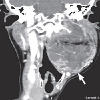
We report a 57 years old female patient with neck fibrosarcoma. Her main complaints consisted of hoarseness, difficulty swallowing, pain in the left side of her neck and left shoulder region, which all indicated the Collet Sicard syndrome, so the working diagnosis was glomus tumor Diagnostic MSCT was used, and the characteristics of the radiologic finding did not indicate any of the paraganglioma types, although the tumor was localized in the area of the carotid bifurcation, demonstrating the signs of extension into the jugular foramen. The patient has been treated surgically in general anesthesia and pathologic diagnosis was fibrosarcoma.

Downloads

Download data is not yet available.
Collet Sicard syndrome as atypical presentation of neck fibrosarcoma: a case report

Downloads

Additional Files

Published

2011-05-20

How to Cite

1.
Petrović S, Grozdanović D, Kovačević P, Višnjić M, Petrović D. Collet Sicard syndrome as atypical presentation of neck fibrosarcoma: a case report. Biomol Biomed [Internet]. 2011May20 [cited 2023Jun.2];11(2):137-40. Available from: https://www.bjbms.org/ojs/index.php/bjbms/article/view/2600

Issue

Section

Case Reports

INTRODUCTION

Fibrosarcoma has been defined as a malignant tumor of the fibroblasts that shows no other evidence of cellular differentiation and is capable of recurrence and metastasis. Fibrosarcomas are rare but may occur anywhere in the body, most commonly in the retroperitoneum, thigh, knee and distal extremities. Of all the fibrosarcomas occurring in humans, only 0.05% occur in the head and neck region [1].

CASE REPORT

A 57 years old woman was admitted for surgical treatment of her tumor in the left infratemporal region and left side of the neck. The patient had noticed the change two years earlier in the form of a growing knot in the neck, producing hoarseness, difficulty in swallowing, pain in the neck and left shoulder region. At admission, a change was found at examination in the left side of the neck, 10 cm in size, round, without evidence of local skin reaction and without palpable, enlarged lymph nodes in the neck. The lateral left wall of the pharynx was displaced medially, markedly deforming the oropharynx, and floor of the mouth was dislodged frontally and upwards. There was a moderate to severe trismus.

Preoperative MSCT detected the presence of a tumor mass sized 75×82×100 mm in the left infratemporal region and left side of the neck extending from the skull base level to the thyroid cartilage. The change was found at the bifurcation of the common carotid artery with displacement and compression of the ICA sin medially and ECA sin laterally, without any signs of their being infiltrated. The tumor inhomogenously took up the contrast, with the presence of larger areas of necrosis and with significant taking up within the septa and capsule (Figure 1). The tumor extended cranially through the jugular foramen, in which the presence of the jugular internal vein could not be detected due to infiltration. Caudal border of the tumor was the thyroid cartilage, and medial border was delineated by the parapharyngeal and carotid space. The change in the parapharyngeal space dislodged and compressed the airways (Figure 1). Due to infiltrated cranial nerves at the level of the jugular foramen, there was left vocal cord paresis as the consequence of X cranial nerve infiltration and paresis of the soft palate and pharyngeal wall due to IX cranial nerve infiltration. There were signs of left m. trapezius and m. sternocleidomastoideus atrophy due to XI cranial nerve infiltration. In addition, jugular foramen nerve involvement (IX-XI), the signs of muscle denervation, were detected at the level of the left half of the tongue, with atrophy and fat tissue replacement, indicating XII cranial nerve infiltration (Figure 2). Neck lymph nodes were not enlarged. The patient was surgically treated in general endotracheal anesthesia; tumor extirpation was performed. Histopathology of the extirpated material indicated the diagnosis of fibrosarcoma.

FIGURE 1: A) Axial MDCT scan with bone algorithm: bone destruction of the left jugular foramen (arrow) B) coronal postcontrast MDCT scan: inhomogenous tumor mass with extension in the left jugular foramen, abscence of left IJV and airway compression C) sagittal MDCT scan: inhomogenous tumor mass with extension in the left jugular foramen D) coronal MIP reconstruction: a tumor mass at carotid bifurcation with displacement of ICA and ECA
FIGURE 2: Postcontrast axial MDCT images showed denervation muscular atrophy because of low cranial nerve involvement: A) an inhomogenous tumoral mass with airway compresion and fatty infiltration of the left hemitongue - XII cranial nerve involvement (arrows) B) deplacement of arytenoid cartilage with fatty infiltration of left vocal cord - X cranial nerve involvement (arrows) C) athrophy of left m. sternocleidomastoideus (arrow) and D) atrophy of the left m. trapezius - XI cranial nerve involvement (arrows)

DISCUSSION

Head and neck soft tissue sarcomas are rare entities in adults, accounting for only about 10% of all soft tissue sarcomas and approximately 1% of all head and neck tumors [2, 3]. Fibrosarcoma is a malignant tumor arising from the fibroblasts (the cells that produce connective tissue). This is a type of sarcoma that is predominantly found in the area around the bones or in soft tissue. Of all the fibrosarcomas occurring in humans, only 0.05% occur in the head and neck region [1]. The etiology of fibrosarcoma remains obscure. Although radiation exposure has been thought to be the most important etiological factor, followed by trauma and underlying bone conditions such as the Paget’s disease, fibrous dysplasia, or chronic osteomyelitis. Our patient did not have any of the above etiological factors. Most patients with fibrosarcoma of the head and neck present in their fourth or fifth decade of life with a firm, painless mass of the face, scalp, or neck [4-6]. Most sarcomas of the head and neck region present with nonspecific signs and symptoms. In 80% of cases, these tumors manifest as a painless mass [7]. However, a variety of symptoms can occur, depending on the affected region of the head and neck. Imaging studies augment the physical examination offerring a more accurate assessment of the size and location of sarcomas in the head and neck. In addition, the information regarding bony involvement, intracranial extension, and regional nodal disease are much more precise. Computed tomography (CT) and magnetic resonance imaging (MRI) are the studies of choice in nearly all the cases. None of the imaging characteristics can be considered diagnostic, and heterogeneity in appearance is to be expected [8]. The tumor in our patient was atypical and nonspecific of fibrosarcoma by its localization and propagation. Regarding its localization in the carotid space, expanding the CCA bifurcation and displacing the ICA and ECA, the tumor would correspond first of all to glomus vagale paraganglioma, as well as to paraganglioma of the carotid body. Regarding its propagation through the jugular foramen and clinical symptoms of involvement of the low cranial nerves, glomus jugulare paraganglioma would be a correct diagnosis. Cranial nerve deficits in jugular paragangliomas usually present with the Vernet i.e. jugular foramen syndrome or Collet Sicard syndrome. The Vernet syndrome involves the paralysis of the cranial nerves IX-XI [9]. This syndrome involves hoarseness due to vocal cord paralysis (X), difficult swallowing (IX) and weakness and atrophy of the trapezius and sternocleidomastoid muscles (XI) [10, 11]. The Collet Sicard syndrome involves the Vernet syndrome with the involvement of the XII cranial nerve, causing ipsilateral atrophy of the tongue [10, 12]. Extension of fibrosarcoma into the jugular foramen and carotid space and infiltration of low cranial nerves in our patient caused the Collet Sicard syndrome as an atypical clinical presentation of fibrosarcoma. The tumor in our patient, though localized in the CCA bifurcation and extending into jugular foramen, did not show homogenous and intense postcontrast density augmentation, characteristic of paragangliomas [13], but instead the presence of large fields of necrosis with the septa and presence of the capsule. In view of differential diagnosis, due to involvement of internal jugular vein and dislocation of ICA and ECA, neurofibroma could be considered, and because of its propagation into the parapharyngeal space, a schwannoma in the carotid space was another possibility. Involvement of cranial nerves IX-XI suggested a neurinoma of the jugular foramen, but also a meningeoma of the jugular foramen [14]. Meningiomas present with a “dural tail”, while neurinomas are well-delimited lesions with smoothly marginated enlargement and non- invaded marrow space of the jugular foramen on CT scans. Some cases may present with cystic degeneration. Metastatic tumors of the neck with propagation into the jugular foramen were also one of the options in differential diagnosis. In our patient, the tumor indicated malignancy by its CT features, presence of necrotic portions with the septa and capsule, infiltration of jugular foramen and its structures, dislocation and compression of ICA and ECA sin, subcutaneous involvement, and moreover, histopathology indicated a fibrosarcoma. Radical surgery is the primary mode of treatment in patients with neck fibrosarcomas. Patients with the worst prognosis are those with unresectable disease who are treated with radiotherapy alone or radiotherapy combined with chemotherapy. Since local recurrence is the major form of treatment failure and eventual fatal outcome, negative surgical margins are of critical importance [4]. Adjuvant radiation therapy or chemotherapy may play a role in cases of high-grade or large tumors and in patients with positive margins [4, 5]. Survival in patients with fibrosarcoma is relatively favorable compared with other types of head and neck sarcomas. Five-year overall survival rates range from 62 to 82%, with 5-year disease-free survival ranging from 32% to 57% [4, 5, 7, 15-18]. High-grade malignancies portend a poor prognosis regardless of surgical margin status, and multimodality therapy should certainly be considered for these tumors [5]. Since histopathology confirmed a fibrosarcoma in our patient, and the described CT features and clinical presentation of the tumoral change did not indicate fibrosarcoma, nor any of the other tumors characteristic of the region, we thought the case was a good selection for reporting. In conclusion, fibrosarcoma has to be considered in cases with atypical clinical and imaging presentations of neck tumoral lesions.

DECLARATION OF INTEREST

None to declare

REFERENCES

  1. , , (). Fibrosarcoma of the oral cavity. Indian J Dent Res.
  2. , , , , , (). Adult soft tissue sarcomas of the head and neck treated by radiation and surgery or radiation alone: patterns of failure and prognostic factors. Review. Int J Radiat Oncol Biol Phys.
  3. , , , , , (). Report of a symposium on diagnosis and treatment of adult soft tissue sarcomas in the head and neck. Eur J Surg Oncol.
  4. , , , , (). Fibrosarcoma of the head and neck: the UCLA experience. Arch Otolaryngol Head Neck Surg.
  5. , , , (). Fibrosarcoma of the head and neck. Laryngoscope.
  6. , , (). Dermatofibrosarcoma protuberans of the upper lip: an overview and a case report. Eur J Surg Oncol.
  7. , , , , , (). Soft tissue sarcomas of the head and neck in adolescents and adults. Am J Surg.
  8. , , (). Sarcomas of the head and neck. Top Magn Reson Imaging.
  9. , , (). Neurologic manifestation of glomus tumors in the head and neck. Arch Neurol.
  10. (). Imaging and management of head and neck paragangliomas. Eur Radiol.
  11. , (). Glomus Tumors Medscape. eMedicine (Internet). Available from: http://emedicine.medscape.com/article/251009-overview
  12. , , , (). Neural infiltration of glomus temporale tumors. Am J Otol.
  13. , , (). Paragangliomas of the head and neck: radiologic – pathologic correlation. Radiographics.
  14. , , , , , (). Differentiating imaging findings in primary and secondary tumors of the jugular foramen. Neurosurg Rev.
  15. , , , , , (). Head and neck sarcoma: report of the Head and Neck Sarcoma Registry. Society of Head and Neck Surgeons Committee on Research. Head Neck.
  16. , , , (). Soft tissue sarcomas of the head and neck in adults. Am J Surg.
  17. , , (). Soft-tissue sarcomas of the head and neck. Am J Surg.
  18. , , , (). Fibrosarcoma of the head and neck. Am J Surg.