Plasma nitric oxide and left ventricular function in rabbits after cardiac lymphatic obstruction

Authors

  • Ying-Li Wang Liaocheng People’s Hospital and Liaocheng Clinical School of Taishan Medical University, Department of Cardiology
  • Xiao-Hua Wang Liaocheng People’s Hospital and Liaocheng Clinical School of Taishan Medical University, Department of Cardiology
  • Ye Jin Liaocheng People’s Hospital and Liaocheng Clinical School of Taishan Medical University, Department of Cardiology
  • De-Gui Kong Liaocheng People’s Hospital and Liaocheng Clinical School of Taishan Medical University, Department of Cardiology
  • Le-Xin Wang Charles Sturt University, School of Biomedical Sciences

DOI:

https://doi.org/10.17305/bjbms.2011.2617

Keywords:

cardiac lymphatic vessels, endothelium, nitric oxide, left ventricular function, rabbits

Abstract

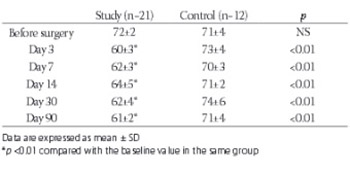
This study was designed to investigate the effect of cardiac lymphatic obstruction on plasma nitric oxide (NO) and left ventricular function. The plasma NO was measured in study group (n=21) and control group rabbits (n=12) before, and 3, 7, 14, 30 and 90 days after the obstruction of cardiac lymphatic vessels. Left ventricular ejection fraction was measured with echocardiography. There was a significant reduction in the left ventricular ejection fraction following the lymphatic obstruction (0.72±0.02 vs. 0.61±0.02, p<0.01). Plasma NO in the control group remained unchanged during the observation period (54.2±4.4 vs. 52.0±4.2 μmol/L, p>0.05). In the study group, there was a small but significant increase in the plasma NO on day 3, 7 and 14 following the lymphatic obstruction (52.3±4.1 vs. 73.4±5.9 μmol/L, p<0.01). The plasma NO returned to the baseline levels on day 30 but reduced to 44.9±3.6 pmol/L on 90 days after the lymphatic obstruction (p<0.05). In conclusion, cardiac lymphatic obstruction was associated with a significant reduction in left ventricular function. It was also associated with an increase in the plasma NO in the first 2 weeks but there was a significant reduction in the NO levels three months after the lymphatic obstruction.

Downloads

Download data is not yet available.
Plasma nitric oxide and left ventricular function in rabbits after cardiac lymphatic obstruction

Downloads

Additional Files

Published

2011-02-20

How to Cite

1.
Wang Y-L, Wang X-H, Jin Y, Kong D-G, Wang L-X. Plasma nitric oxide and left ventricular function in rabbits after cardiac lymphatic obstruction. Biomol Biomed [Internet]. 2011Feb.20 [cited 2023Nov.30];11(1):17-9. Available from: https://www.bjbms.org/ojs/index.php/bjbms/article/view/2617

Issue

Section

Biochemistry

INTRODUCTION

Cardiac transplantation is a life-saving treatment for patients with end-stage heart failure. However, the outcomes of this treatment are limited by acute allograft rejection and chronic allograft coronary artery disease or allograft vasculopathy [1, 2]. Endothelium-derived nitric oxide (NO) is a potent endogenous vasodilator, inducing vasodilation by stimulating soluble guanylate cyclase to produce cGMP [3]. Nitric oxide has been found to play a critical role in the acute allograft rejection, as well as in the development of allograft vasculopathy. The expression of the inducible isoform of nitric oxide synthase (NOS-2) is upregulated during cardiac allograft rejection in endothelial cells, in vascular smooth muscle cells, and in cardiac myocytes [4, 5]. Increased synthesis of nitric oxide by inducible nitric oxide synthase soon after the heart transplantation may contribute acute allograft rejection by inducing myocyte necrosis and ventricular failure [5, 6] After heart transplantation, the impairment of nitric oxide synthesis from the endothelium and myocytes is considered a major contributing factor for allograft vasculopathy [2].

Lymphatic vessels are an important source of nitric oxide biosynthesis [7-9]. Nitric oxide is released from lymphatic smooth muscle cells and the vascular endothelium after interacting with various vasoactive substances [7, 8]. Cardiac lymphatic flow plays a pivotal role in maintaining the homeostasis by draining the myocardial interstitial fluid and proteins back to the circulation. However, the lymphatic flow is often interrupted after heart transplantation, because the lymphatic vessels of the donor heart are not routinely connected with the recipient’s during the surgical procedure. To date, there is little knowledge about the impact of the lymphatic flow interruption on the biosynthesis of nitric oxide, which may have significant effect on the prevalence or severity of acute or chronic allograft rejection following heart transplantation. The primary purpose of this study was to investigate the plasma level of nitric oxide in a rabbit model before and after the lymphatic flow obstruction.

MATERIALS AND METHODS

Surgical preparation of the animal model

The study was approved by the Institutional Review Board. Thirty-three New Zealand white rabbits of both sexes (body weight 2.5-3.5 kg, aged 12-18 months) were assigned to the study (lymphatic obstruction) group (n=21, 12 males) and control group (n=12, 7 males). Animals in the control group underwent the same open-chest surgery as the lymphatic obstruction group, but without the ligation of the lymphatic vessels. Under general anesthesia, left thoractomy was performed in the fourth intercostal space and the heart was suspended in a pericardial cradle. In the study group, 0.5 ml of 10% methylene blue was injected into the wall of the left and right ventricular apex, clearly marking the lymphatic vessels in the epicardium and the adjacent lymph nodes. Animals in the control group underwent the same open-chest surgery as the lymphatic obstruction group, but without the ligation of the lymphatic vessels. The main epicardial lymphatic vessels, as well as the large lymph nodes between the aortic root and the pulmonary artery, and between the posterior aorta and the right superior pulmonary artery were destroyed [10]. The chest was then closed by layers and the animals were returned to the animal house for standard care by the investigators and the curators. Penicillin (average 0.8 million units) was administered intramuscularly daily for a week after the surgery to prevent wound infection.

Measurement of plasma nitric oxide

Venous blood was obtained before, and at 3, 7, 14, 30 and 90 days following the surgery. The plasma was collected from each sample by centrifuge at 3000 rpm for 5min at 4° C and stored at -25°C until detection. Thawed samples (100 μL), were diluted fourfold with deionized water and deproteinated by zinc sulfate. They were centrifuged at 3000 rpm for 5 min at room temperature and 5 μL supernatant was injected into a chemiluminescence machine (Sievers 280 NO Analyzer, Sievers Instruments, Inc., Boulder, CO), for the measurement of plasma nitric oxide.

Assessment of left ventricular function

Left ventricular ejection fraction (EF) was measured by echocardiography (HP SONOC-5500, Agilent Technologies, MA, USA) before and after operation.

Statistical analysis

Data was expressed as mean ± standard deviation. Numerical data were analyzed by one-way ANOVA. Categorical data were examined by Chi-square test. p <0.05 was considered statistically significant.

RESULTS

All animals survived the initial operation and completed the 90-day study. Pericardial fluid of 1-9 ml (between the left ventricular posterior wall and the pericardium) was detected by echocardiography in 52.4% (11/21) of the study and 8.3% (1/12) of the control group animals three days following the surgery (p <0.05). There was a gradual reduction in prevalence of pericardial fluid and at the end of week 4, none of the control or study group animals had pericardial fluid. Table 1 shows the average values of the left ventricular ejection fraction before and after operation. There was no significant difference in the ejection fraction between the study and control groups before the surgery (p >0.05). In the control group, average ejection fraction remained unchanged following operation. Following operation, there was a significant reduction in the left ventricular ejection fraction in the study group between day 3 and 90.

TABLE 1: Left ventricular ejection fraction (%) following the surgery.

As shown in Table 2, the average value of nitric oxide was similar between the control and the study group before the surgery. In the control group the average nitric oxide values remained unchanged following the sham surgery (p >0.05).

TABLE 2: Changes in plasma concentration of nitric oxide following cardiac lymphatic obstruction.

In the study group, the average plasma nitric oxide on day 3, 7 and 14 were higher than the baseline value (p<0.05). The average nitric oxide at these time points were also higher than in the control group (p <0.05). However, in the study group, the average nitric oxide on day 30 was similar to the baseline value, or the average value of the control group (p >0.05). However, the plasma nitric oxide on day 90 was lower than the baseline value (p<0.05) and the nitric oxide in the control group (p<0.05).

DISCUSSION

Previous studies have found that ligation of large cardiac lymphatic vessels, together with the destruction of the large cardiac lymph nodes, caused significant reduction or occlusion in the cardiac lymph flow [10, 11]. The lymph flow obstruction impairs the drainage of interstitial fluid and proteins in the myocardium, resulting in significant myocardial edema and fibrosis within the first four weeks of operation [11]. Cardiac lymphatic obstruction is also associated with a reduction in the left ventricular ejection fraction, compromising ventricular muscle contractility [11]. It has been proposed that the interruption of the cardiac lymphatic flow may play an important role in the pathogenesis of the heart failure following heart transplantation [12]. In the present study, pericardial effusions were detected in approximately 50% of the animals 3 days after lymphatic obstruction. Pericardial effusions were likely caused by myocardial edema, as our previous tissue examination on the same animal model showed a significant degree of edema in the ventricular myocardium [11]. The present study also demonstrated that cardiac lymphatic flow obstruction was associated with a significant elevation in plasma nitric oxide levels in the first 2 weeks following the obstruction. These results are consistent with the previous studies where elevated nitric oxide levels were identified following acute allograft rejection [5, 6]. The pathways by which cardiac lymphatic obstruction increased the plasma nitric oxide are not entirely clear. The obstruction of lymphatic vessels and the adjacent lymph nodes may cause accumulation of lymph and increase in intravascular pressure in the blocked vessels. The rises of the intravascular pressure may stimulate biosynthesis, or the release of nitric oxide from the lymphatic vessels cells or the endothelium. In addition, acute obstruction of lymphatic flow leads to hypoxia and ischemia-like changes in the coronary arteries due to the reduced drainage of interstitial fluid [13]. Hypoxia and ischemia are potent stimulators for the production of nitric oxide by the coronary endothelium [2]. Nitric oxide is an important factor in the regulation of blood flow and fluid homeostasis [14]. After heart transplantation, the expression and activity of endothelial nitric oxide synthase in the donor heart can be impaired [3]. The reduced nitric oxide activity plays a key role in the development of allograft vasculopathy [3]. The proposed factors causing the endothelial dysfunction include preexisting arteriosclerotic disease in the graft, graft ischemia before transplantation, immunosuppressive agents such as cyclosporin A, and other classic risk factors such as hyperlipidemia, hypertension, diabetes, or hyperhomocysteinemia [3]. In the present study, 4 weeks after the lymphatic obstruction, the plasma nitric oxide levels declined to a level that is below the baseline value, suggesting that chronic lymphatic obstruction may be detrimental to nitric oxide production which in turn, participating the pathogenesis of graft vasculopathy.

CONCLUSION

In conclusion, cardiac lymphatic obstruction was associated with a reduction in left ventricular function. The plasma levels of nitric oxide were increased in the first 2 weeks of lymphatic obstruction. After 4 weeks, the plasma nitric oxide levels tend to decrease to below the baseline value. Further studies are required to clarify the clinical significance of the nitric oxide changes following the lymphatic obstruction.

DECLARATION OF INTEREST

None to declare.

REFERENCES

  1. , , , , (). Role of humoral immunity in acute cardiac allograft dysfunction. J Heart Lung Transplant.
  2. , (). Cardiac allograft vasculopathy and dysregulation of the NO synthase pathway. Arterioscler Thromb Vasc Biol.
  3. , (). The obligatory role of endothelial cells in the relaxation of arterial smooth muscle by acetylcholine. Nature 1980.
  4. , , , , , (). Selective inhibition of nitric oxide production during cardiac allograft rejection causes a small increase in graft survival. Transplant.
  5. , , , , , (). Acute cardiac allograft rejection in nitric oxide synthase-2(-/-) and nitric oxide synthase-2(+/+) mice: effects of cellular chimeras on myocardial inflammation and cardiomyocyte damage and apoptosis. Circulation.
  6. , , , , , (). Induction of myocardial nitric oxide synthase by cardiac allograft rejection. J Clin Invest.
  7. , , (). Expression of inducible nitric oxide synthase in cultured smooth muscle cells from rat mesenteric lymphatic vessels. Microcirculation.
  8. , , , (). Nitric oxide production by lymphatic endothelial cells in vitro. Biochem Biophy Res Commun.
  9. , , , , , (). Characterization and microarray analysis of genes in human lymphatic endothelial cells from patients with breast cancer. Lymphatic Res Biol.
  10. (). The study of the lymphatics of the heart: an overview. Microcirc Endothelium Lymphatics.
  11. , , (). Effect of cardiac lymph flow obstruction on cardiac collagen synthesis and interstitial fibrosis. Physiol Res.
  12. , , (). Cardiac lymphatic interruption is a major cause for allograft failure after cardiac transplantation. Lymphatic Res Biol.
  13. , (). Cardiac lymphatic obstruction: ultrastructure of acute-phase myocardial injury in dogs. Mayo Clinic Proc.
  14. , , , (). Angiotensin converting enzyme activity and nitric oxide level in serum patients with dehydration. Bosn J Basic Med Sci.