HLA Association in SLE patients from Lahore-Pakistan

Authors

  • Nageen Hussain Quaid-e-Azam University of the Punjab, Department of Microbiology and Molecular Genetics
  • Ghazala Jaffery Services Institute of Medical Sciences, Department of Pathology
  • Anjum Nasim Sabri Quaid-e-Azam University of the Punjab, Department of Microbiology and Molecular Genetics
  • Shahida Hasnain Quaid-e-Azam University of the Punjab, Department of Microbiology and Molecular Genetics

DOI:

https://doi.org/10.17305/bjbms.2011.2618

Keywords:

systemic lupus erythematosus, major histocompatibility complex, Human Leukocyte Antigen, Polymerase chain reaction, rheumatology, DNA-based HLA typing

Abstract

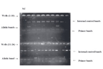
The first genetic factors to be identified as important in the pathogenesis of Systemic lupus erythematosus (SLE) were those of the major histocompatibility complex (MHC) on chromosome 6. It is now widely accepted that MHC genes constitute a part of the genetic susceptibility to SLE. The study population comprised 61 SLE patients fulfilling at least four of the American college of Rheumatology criteria for SLE and 61 healthy blood donors as controls. SLE female versus male ratio was approximately 9:1. Mean age at diagnosis was 30.35 ± 1.687 (12-68 years). DNA-based HLA Typing for HLA-A, HLA-B, and HLA-DRB1 was carried out by Polymerase chain reaction with sequence specific primers using genomic DNA obtained from blood samples. A total of 22 alleles have been studied at locus A, 37 alleles at locus B and 17 DRB1 alleles. The allelic frequencies of HLA-A, HLA-B, and HLA-DRB1 antigens in SLE patients from Pakistan were compared with the controls. A significant increase was observed in the frequency of HLA-A*01, A*03, A*11, A*23, A*26 A*69, HLA-B*27, B*40, B*49, B*51, B*52, B*53, B*54, B*95, HLA-DRBI*01, DRBI*03, DRBI*11, DRBI*14 among SLE patients indicating a positive association of these alleles with SLE. HLA-A*24, A*29, A*31, A*34, A*68, A*92, HLA-B*18, HLA-DRB1*12, were found to be decreased in the patient group as compared to controls indicating a negative association of these alleles with SLE. Thus from this study we can conclude that SLE is associated with certain MHC alleles in Pakistani population.

Downloads

Download data is not yet available.
HLA Association in SLE patients from Lahore-Pakistan

Downloads

Additional Files

Published

2011-02-20

How to Cite

1.
Hussain N, Jaffery G, Nasim Sabri A, Hasnain S. HLA Association in SLE patients from Lahore-Pakistan. Biomol Biomed [Internet]. 2011Feb.20 [cited 2023Nov.30];11(1):20-6. Available from: https://www.bjbms.org/ojs/index.php/bjbms/article/view/2618

Issue

Section

Molecular Biology

INTRODUCTION

The first genetic factor to be identified as important in the pathogenesis of SLE was the Major Histocompatibility Complex (MHC). This complex which resides on chromosome 6 comprises at least 128 genes as well as 96 pseudogenes with many of these genes playing an important role in immune responses [1]. The MHC shows a high degree of polymorphism and these polymorphisms are associated with a wide range of diseases. Defects in certain MHC genes lead to autoimmune disorders as it is a failure of self tolerance, recognition of an antigen that is self and reacts to it; one of the examples is that of Systemic Lupus Erythematosus (SLE). It is now widely accepted that MHC genes constitute a part of the genetic susceptibility to SLE. MHC is divided in to Class I, Class II, Class III regions, each containing group of genes with related functions. For example, within the MHC, there are 6 genes that encode Class I molecules, HLA-A, HLA-B, HLA-C, HLA-E, HLA-F, HLA-G and among these HLA-A, B, C are the most polymorphic and probably the most important. Furthermore, five loci encode Class II molecules, among them HLA-DP, DQ and DR are most important and polymorphic. Association of SLE with DRB1 alleles has been studied and DRB1*03 is found to be positively associated with SLE in most of the populations [2]. Number of genes resides in the MHC Class III region such as early components of the complement cascade, heat shock 70 gene complexes, tumor necrosis factor complexes and many other genes. One of the mechanism by which all these alleles may contribute to pathogenic outcomes in SLE is the alterations in the nature of antigen presentation by HLA molecules to T-Helper cells that lead to abnormal T-cell response [3]. We conducted this study to analyze the contribution of HLA-A, HLA-B, and of DRB1 alleles to the susceptibility to SLE among Pakistani SLE patients and controls.

MATERIALS AND METHODS

The human studies reported in this manuscript were approved by the Ethical Committee of the School of Biological Sciences, University of the Punjab, Lahore, Pakistan. A total of 61 SLE patients fulfilling American College of Rheumatology (ACR) criteria and 61 controls were enrolled in this study. Sample number is low because SLE is a quite rare disease in Pakistan. Rheumatology and Nephrology department of different hospitals of Lahore were chosen because most of the patients were referred to these departments for the diagnosis of lupus. Of the 61, fifty-five (90.16%) were females and six (9.83%) were males. The female versus male ratio was 9.16:1 indicating that SLE is quite common in females. Mean age at diagnosis was 30.35 ± 1.687 years and the range was 12-68 years. Mucocutaneous involvement was found in lupus patients such as malar rash 6 (9.84%), discoid rash 11 (18.03%), photosensitivity 11 (18.03%). Various syndromes were overlapping SLE and the most common one was Rheumatoid arthritis (19 patients: 31.14%) but cases of Sjogren’s syndrome (10 patients: 16.39%), Scleroderma (2 patients: 3.27%), Secondary Antiphospholipid syndrome (2 patients: 3.27%), and of Budd-Chiari Syndrome (1 patients: 1.63%) were also found along with SLE. Renal involvement was found in 20 patients: 32.78%) patients characterized by proteinuria and red cell cast. Blood samples drawn by venepuncture from each patient and control were collected in an ethylene diamine tetra acetic acid (EDTA) vial for DNA-based HLA Typing. An informed written consent was taken from all subjects recruited in the study as well as from the matched controls. Genomic DNA was purified by using Genomic DNA purification kit (Fermentas, Cat # K0512-USA) [4]. HLA-A, HLA-B, and HLA-DRB1 typing was carried out by Polymerase chain reaction with sequence specific primers (One Lambda, Lot # 006- USA) using genomic DNA from SLE patients. The concentration of DNA was estimated by taking A260/A280 ratio [5]. According to One Lambda HLA Class I Class I HLA typing kit protocol, the Polymerase Chain Reaction-Single Stranded Polymorphism (PCR-SSP) methodology was based on the principle that by using recombinant Taq polymerase, completely matched oligonucleotide primers are more efficiently used in amplifying target sequence. All kit reagents and DNA samples were brought at room temperature and then vortexed. Primer set tray was placed in a PCR tray microtube storage rack and the tray label was removed. 1 μl of deionized water was added to the negative control reaction tube on the primer set tray. Then 5.6 μl of recombinant Taq polymerase (5 units/μl) was added to the Micro SSP™ D-mix tube and vortexed. The Micro SSP™ D-mix tube was pulse spinned in a microcentrifuge to bring all liquid down from sides of the tube. Micro SSP™ D-mix (9 μl) was added to the negative control reaction tube. 111 μl of the DNA sample was then added to the Micro SSP™ D-mix tube, vortexed and again spinned. Sample-reaction mixture (10 μl) from the Micro SSP™ D-mix tube was added in to each reaction tube of the Micro SSP™ primer set tray except the negative control reaction tube. The reaction tubes were then sealed with the tray seal. The Micro SSP™ primer set tray was placed in a thermocycler and PCR program was started. Primer pairs were designed to have perfect matches only with a single allele or group of alleles. Under strictly controlled PCR conditions (Table 1), perfectly matched primer pairs resulted in the amplification of target sequences. The primer set tray was removed from the thermocycler and was unsealed gently without splashing the samples. 10 μl of the PCR reaction was transferred to a 2.5% agarose gel and the samples were electrophoresed at 150 volts for 20 minutes, bands were finally visualized by UV illuminator (Figure 1). The control primer pair amplifies a conserved region of the Human β-globin gene, which is present in all human DNA samples and is used to verify the integrity of the PCR reaction. The amplified DNA fragments of the specific HLA primer pairs were smaller than the product of the internal control primer pair, but larger than the diffuse, unincorporated primer band. The typing results were interpreted by using the worksheet provided with the trays.

TABLE 1: One Lambda PCR Program

Statistical analysis

The allelic frequencies of HLA-A, HLA-B, and HLA-DRB1 antigens in Pakistani SLE patients were compared with the controls by using online MedCalc software for calculating Odds Ratios [6]. Fisher’s exact test was calculated by using only online available Fisher’s exact test calculator described by Agresti in 1992.

FIGURE 1: A positive reaction for a specific HLA allele or allele group was visualized on the gel as in the wells 2, 8, and 15, an amplified DNA fragment can be observed between the internal control product and the unincorporated primer band. NC: Negative control in the well 1, representing the primer band.

RESULTS

The typing results were interpreted by using the worksheet provided with the trays (Appendix-VI). The allelic frequencies of HLA-A, HLA-B, and HLA-DRB1 alleles in Pakistani SLE patients were compared with the controls by using online MedCalc software for calculating Odds Ratio [7]. It is conventional to create confidence interval (CI) at 95% level as it provides a range about the observed effect size. Nomenclature of HLA alleles included in this chapter was listed in the January 2008 update of the IMGT database (A sequence database for the human major histocompatibility complex) [8].

HLA-A Alleles

A total of 22 alleles were studied at locus A, 37 alleles at locus B and 17 DRB1 alleles. Frequency of HLA-A alleles in control as well as in case groups is mentioned in Figure 2.

FIGURE 2: Frequency of HLA-A alleles in control and case groups

An increase was observed in the frequency of HLA-A*01, -A*03, -A*11, -A*23, -A*26, -A*69 alleles among total SLE patients. Odds Ratio for HLA-A*01, -A*03, -A*11, -A*23, -A*26, -A*69 alleles was more than 1 and the lower, upper bound of confidence interval didn’t include 1 that’s why these alleles can act as a significant risk to SLE. For example, Odds ratio for HLA-A*01 allele was 4.009, it means that the odds of HLA-A*01 allele was four times higher in cases than controls. There was a negative association of HLA-A*02, A*33 alleles with SLE as the OR was less than 1 and the confidence interval did not include 1. HLA-A*32 had no affect because OR was equal to 1, means that this was not significant at 5% level for 95% confidence intervals. HLA-A*25, -A*36, -A*43, -A*66, -A*74, -A*80 alleles were neither found in cases nor in controls. Association of HLA-A*29, -A*30, -A*31, -A*34, -A*68, -A*92 alleles with SLE was not proved by this study at 5% significance level (Table 2).

TABLE 2: HLA Alleles at Locus A

HLA-B Alleles

There was an increase in the frequency of HLA-B*27, -B*40, -B*49, -B*51, -B*52, -B*53, -B*54, -B*95 among total SLE patients indicating a positive association of these alleles with SLE. Frequency of HLA-B alleles in control as well as in case groups is mentioned in Figure 3. Odds Ratio for HLA-B*15, -B*46, -B*18 and -B*35 was less than 1 indicating a negative association between these alleles and SLE. Similarly, Odds ratio for HLA-B*08, -B*13, -B*37, -B*39 alleles was equal to 1 indicating no association between these alleles and SLE. HLA-B*14, -B*38, -B*42, - B*44, -B*45, -B*47, -B*48, -B*50, -B*55, -B*56, -B*59, -B*73, -B*78, -B*81, -B*82, -B*83 alleles were absent in both cases as well as in controls. Association of HLA-B*07, -B*18, -B*35, -B*41, -B*57 alleles with SLE was not proved by this study as the 95% confidence interval did include 1 (Table 3).

FIGURE 3: Frequency of HLA-B alleles in control and case groups

HLA-DRB1 Alleles

Frequency of HLA-DRB1 alleles in control as well as in case groups is mentioned in Figure 4.

TABLE 3: HLA Alleles at Locus B
FIGURE 4: Frequency of HLA-DRB1 alleles in control and case groups

The frequency of HLA-DRBI*01, -DRBI*03, -DRBI*11, -DRBI*14, -DRBI*16 was increased among total SLE patients, thus showed positive association between the alleles and SLE disease at the 95% confidence level as the Odds of these alleles was greater in cases than in controls. HLA-DRB1*04, -DRB1*07, -DRB1*08, -DRB1*15 alleles were the protective factors in the causation of SLE disease or in other words showing a negative association with SLE at the 5% significance level because the Odds of these alleles was smaller in cases than in controls. It was observed that HLA-DRB1*13 had no affect representing no relationship between these alleles and SLE. HLA-DRBI*09, -DRBI*10, -DRB3*01, -DRB3*02, -DRB4*01,- DRB5*01 were not found in both cases and in controls. Further more, association of HLA-DRB1*12 allele with SLE was not proved by this study at 5% significance level (Table 4).

TABLE 4: HLA Alleles at Locus DRB1

Association of HLA alleles with SLE

In order to prove the significant association of HLA alleles with SLE, Fischer’s exact test was applied and it was found that HLA-A*01, HLA-A*31, HLA-A*34, HLA-A*69, HLA-DRB1*01 were significantly associated with SLE (Table 5).

TABLE 5: Association of HLA alleles with SLE by Fischer Exact test

DISCUSSION

SLE is a genetically complex disease and the major contribution is from MHC genes. A lot of work has been done to find out the role of genetic factors both in murine lupus models and in human SLE. Genetic analysis of SLE in humans and in murine lupus models, suggest that SLE is a polygenic disorder. The actual genes identified in human and murine SLE may not be identical but immunological pathways can be similar [9]. The precise genetic cause of the association has been difficult to define because of the high degree of polymorphism within the genes of HLA. In the present study, those MHC genes were identified which were involved in the development of SLE in a relatively defined population in Lahore, Pakistan. The relationships between HLA and disease are of considerable importance as they provide new tools for studying the inheritance, classification, diagnosis and pathogenesis of the disease in question. It is universally accepted that genetic factors play an important role in the susceptibility to develop SLE and varies among populations. Particular attentions have been focused on the association of the disease with genetic markers located within the MHC region. Several studies have illustrated the fact that SLE is associated with certain MHC alleles in different populations. Vargas-Alcaron et al. [10] studied heat shock 70 gene polymorphism in Mexican patients with autoimmune disease. Sirikong et al. [11] investigated the association of DRB1 and DQB1 with Thai patients having SLE and found significant increase in the frequency of DRB1*1502 and DQB1*0501. Ramal et al. [12] reported that HLA associations between HLA-DRB1-DQB1 and SLE were different for Gypsy people as he found DRB1*1303, DQB1*0301 and DR5 to be significantly associated with SLE. Endreffy et al. [13] studied DRB1, DQA1, and DQB1 allele polymorphisms along with their clinical features in Hungarian patients having SLE while Azizah et al. [14] reported them in Malaysian-Chinese population, the difference is that the later study showed strong association of DQA1*0102 with anti-Ro/La while DQA1*0301 was found to be associated with dsDNA in Malaysian Chinese population. In this study, HLA-DRB1, HLA-DRB3, HLA-DRB4, HLA-DRB5 alleles were studied but few HLA-DRB1 alleles were found to be associated with SLE while HLA-DRB3, HLA-DRB4, HLA-DRB5 alleles were absent in Pakistani SLE patients as well as in the healthy controls. Pradhan et al. [15] studied HLA associations in SLE patients from Mumbai and showed a significant two-fold increase in the odds-ratio for HLA A3, A28, and B27 alleles with increase Ro, La, Sm and dsDNA autoantibodies while there is significant two-fold decrease in the odds-ratio for HLA A19, A11, B7 B15 alleles with decreased Ro, La, Sm and dsDNA autoantibodies when compared to normal controls. In this study, HLA-A*01, -A*03, -A*11, -A*23, -A*26, -A*69, HLA-B*27, -B*40, -B*49, -B*51, -B*52, -B*53, -B*54, -B*95, HLA-DRBI*01, -DRBI*03, -DRBI*11, -DRBI*14, -DRBI*16 were significantly increased in SLE patients as compared to the controls. HLA-A*02, -A*33 HLA-B*15, -B*46, -B*18, -B*35, HLA-DRB1*04, -DRB1*07, -DRB1*08, -DRB1*15 provided protection from the disease while HLA-A*32 HLA-B*08, -B*13, -B*37, -B*39, HLA-DRB1*13 had no affect. HLA-DR is expressed on a significantly high percentage of both CD4 and CD8 circulating T cells in active cutaneous lupus erythematosus [16]. HLA DRB1*02 and the allelic variant DRB1*15 have been associated with SLE in Blacks and Asians and HLA-DRBI*03, 08 and 15 have been associated with lupus in Caucasians but clear associations with HLA and SLE could not be demonstrated in racially mixed population [17]. In the present study, HLA-DRBI*01, -DRBI*03, -DRBI*11, -DRBI*14, -DRBI*16 were significantly increased in SLE patients as compared to controls. HLA-DRB1*04, -DRB1*07, -DRB1*08, -DRB1*15 provided protection from the disease while HLA-DRB1*13 had no affect on the disease. Morimoto et al. [18] studied the relation of Class II with Cutaneous manifestations in Japanese population and found a positive malar rash associations with DRB1*09 and skin ulcers with DRB1*04. In addition most studies showed an association between HLA DRBI*02, HLA DRB1*15 and lupus nephritis, although HLA alleles have rarely been associated with SLE renal histopathology. In the work presented here, HLA-DRBI*01, -DRBI*03, -DRBI*11, -DRBI*14 were found to be significantly increased in SLE patients. DRB1*01 showed a strongest association (OR: 3.086; 95% CI: 8.206-8.579) with SLE patients. Castano-Rodriguez et al. [19] found HLA-DRB1 gene as a major factor for the development of SLE in Latin Americans and showed the strongest association of HLA-DRB1*03 (OR: 2.14; 95% CI: 1.28-3.56) with SLE. Our results, HLA-DRB1*03 (OR: 2.63; 95% CI: 6.42-7.89) also showed a positive association with SLE but DRB1*01 disclosed the strongest association (OR: 3.08; 95% CI: 8.20-8.57). Wong and Tsao [20] suggested that HLA Class II containing DRB1 and DQB1 alleles are strong risk factors for SLE. Furthermore, Farjadian et al. [21] reported that the most frequent Class I and Class II alleles in Baloch population with SLE of Iran and Pakistan were A*02 (20.2%), B*40 (11.1%), DQA1*01 (42.5%), DQB1*02 (32%), and DRB1*03 (29%). In the work being discussed here, HLAB*40 (OR: 1.31; 95% CI: 3.38-3.75) and HLA-DRB1*03 (OR: 2.63; 95% CI: 6.42-7.89) were also found to be significantly increased in SLE patients but variation lies in HLA-A*02 allele which was found to provide protection from the disease in the studied SLE patients. Thus differences also lie within the Asian population but all of them showed HLA association with SLE. Thus this case-control study highlights the importance of HLA polymorphism in lupus susceptibility.

CONCLUSION

This study further confirmed and extended the association of SLE with certain MHC alleles in Pakistani population like HLA-A*01, -A*31, -A*34, -A*69, -DRB1*01 were significantly associated with SLE. Identification of genes involved in the development of SLE will provide new insight into the development of disease in susceptible individuals by HLA typing. As new therapies are developed our studies will open up new opportunities to improve diagnosis and treatment of SLE patients in this population.

Acknowledgements

ACKNOWLEDGMENT

We highly appreciate the contribution and help of all the hospital staff for the identification of Lupus patients. We wish to thank the lupus patients for their cooperation and help that made these studies possible. Encouragement from Brigadier Dr. Tahir Aziz Ahmed (Head of Immunology, Armed Forces Institute of Pathology, Rawalpindi) during these studies is sincerely acknowledged.

DECLARATION OF INTEREST

None to declare

REFERENCES

  1. , , , , , (). Nomenclature for factors of the HLA system. Tissue Antig.
  2. , (). An update on the genetics of systemic lupus erythematosus. Isr Med Assoc J.
  3. , , , (). T-cell tolerance and autoimmunity. J Autrev.
  4. , , , (). Comparison of DNA extraction methods for multiplex PCR. Anal Biochem.
  5. , , , , , (). Nomenclature for factors of the HLA system. Hum Immunol.
  6. , (). The Odds Ratios. British Med J.
  7. (). Genetics of systemic lupus erythematosus. Lupus.
  8. , , , , , , (). The IMGT/HLA database. Tissue Antigen.
  9. , (). The MHC quarterly report. Immunogen.
  10. , , , , , (). Class II allele and haplotype frequencies in Mexican systemic lupus erythematosus patients. Hum Immunol.
  11. , , , , , (). Association of HLA-DRB1*1502-DQB1*0501 haplotype with susceptibility to systemic lupus erythematosus in Thais. Tissue Antig.
  12. , , , , , (). Systemic lupus erythematosus in southern Spain: a comparative clinical and genetic study between Caucasian and Gypsy patients. Lupus.
  13. , , , (). HLA class II allele polymorphism in Hungarian patients with systemic lupus erythematosus. Ann Rheum Dis.
  14. , , , , , (). The association of the HLA class II antigens with clinical and autoantibody expression in Malaysian Chinese patients with systemic lupus erythematosus. Asian Pac J Allergy Immunol.
  15. , , , (). Autoimmune Studies and HLA Associations in SLE Patients from Mumbai. Int J Hum Genet.
  16. , , , , (). The expression of human leukocyte antigen-DR and CD25 on circulating T cells in cutaneous lupus erythematosus and correlation with disease activity. Exp Dermatol.
  17. , , , (). Molecular analysis of major histocompatibility complex alleles associated with the lupus anticoagulant. J Clin Invest.
  18. , , , , , (). Mutlicenter cooperative study of HLA class II alleles in Japanese patients with systemic lupus. Iran J Allergy Asthma Immunol.
  19. , , , , (). Meta-analysis of HLA-DRM1 and HLA DQBI Polymorphism in Latin-American patients with SLE. Autoimmun Rev.
  20. , (). Current topics in human SLE genetics. Springer Semin Immunophatol.
  21. , , , , , (). Molecular analysis of HLA allele frequencies and haplotype associations in Baloch of Iran compared with the related population of Pakistan. Tissue Antig.