Cerebral aneurysm associated with cardiac myxoma: Case Report

Authors

  • Branislava A. Ivanović University of Belgrade, Faculty of Medicine, Department of Internal Medicine Clinical Center of Serbia, Clinic for Cardiology
  • Marijana Tadić Clinical Center of Serbia, Clinic for Cardiology
  • Mile Vraneš Clinical Center of Serbia, Clinic for Cardiac Surgery
  • Bojana Orbović Clinical Center of Serbia, Clinic for Cardiology

DOI:

https://doi.org/10.17305/bjbms.2011.2629

Keywords:

atrial myxoma, left atrium, cerebral aneurysm

Abstract

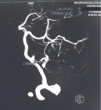
Left atrial myxomas are a rare but well known cause of cerebrovascular accidents in young people. Cerebral embolism is the most common cause of cerebral ischemic stroke. The intracranial aneurysm is rarely associated with myxoma. We report the case of a patient who had an operation of PICA aneurysm due to subarachnoid hemorrhage ten months before the discovery of the large left atrial myxoma. Fortunately, the untimely diagnosis of the myxoma did not have other consequences. In order to prevent possible complications of we should keep in mind that these two apparently different entities could be associated.

Downloads

Download data is not yet available.
Cerebral aneurysm associated with cardiac myxoma: Case Report

Downloads

Additional Files

Published

2011-02-20

How to Cite

1.
A. Ivanović B, Tadić M, Vraneš M, Orbović B. Cerebral aneurysm associated with cardiac myxoma: Case Report. Biomol Biomed [Internet]. 2011Feb.20 [cited 2023Sep.23];11(1):65-8. Available from: https://www.bjbms.org/ojs/index.php/bjbms/article/view/2629

Issue

Section

Case Reports

INTRODUCTION

Atrial myxomas represent approximately 50% of all cardiac tumors. About 75% of them are located in the left atrium [1]. They are often a source of systemic embolism (central and peripheral). To be more precise, the embolization of tumor particles or thrombotic material mixed with tumor cells occurs in 30–40% of patients with myxoma [1]. There were reports of the embolisms of cerebral vessels, coronary and renal arteries, spleen, mesenteric arteries, and lower limbs arteries [1]. In at least half of the cases cerebral arteries were affected, leading to the embolic ischemic strokes [2,3]. In addition to these neurological manifestations of myxomas, it is extremely rare to find myxoma associated with intracranial aneurysms or parenchymal brain metastases [3,4]. We report the case of a patient who had the operation of a. posterior inferior cerebelli (PICA) aneurysm due to subarachnoid hemorrhage ten months before the discovery of the large left atrial myxoma.

CASE REPORT

A 44-year-old woman was admitted to our hospital with dyspnea at rest and palpitation. Ten months earlier the patient was hospitalized at the Clinic for neurosurgery due to subarachnoid hemorrhage. The 4-vessels cerebral angiography immediately was done. It revealed small saccular aneurysm diameter 3 mm and diameter of neck 2 mm arising from the origin of left posterior inferior cerebelli artery directed backwards, upwards and lateral (Figure 1). The patient was operated and clipping of the saccular aneurysm neck was done. A month after the operation, fatigue, myalgia, and palpitations appeared and objective examination for the first time revealed a heart murmur. The patient was indicated for echocardiographic examination which she refused. In the meantime, she lost 10 kg of body weight. A month before hospitalization dyspnea on minimal exertion, provoked by changed body position, appeared. A few days before admission to hospital she had dyspnea even during conversation. There were no data about conventional cardiovascular risk factors, familial medical history of stroke or taking contraceptives. On examination patient was pale, hypotensive with normal body temperature. Her arterial pressure in the sitting position was 90/60 mmHg and 100/65 mmHg in standing position, her heart rate was 90/min. There was no jugular venous distension. The lungs were clear, auscultation revealed regular heart action, accented and split first sound with diastolic murmor on the apex. The liver and spleen were not palpated under the costal margin. Peripheral edema was not noticed. Mucocutaneous lesions or any element of excessive neuro-endocrine activity were not manifested in our patient. Routine blood tests were normal including the erythrocyte sedimentation rate (10 mm/for the first hour), the level of CRP (5 mg/l) and globulin level (23 g/l). An electrocardiogram registered sinus tachycardia. Chest roentgenogram was normal. Two-dimesional transthoracic echocardiographic (TTE) examination revealed a large polypoid tumor mass with irregular surface, diameter 7 × 4 × 3 cm, which originated from interatrial septum and prolapsed into the left ventricle through mitral valve during diastole (Figure 2). Mild mitral regurgitation was found. Apical four-chamber section detected mild dilatation of the right atrium and Doppler echocardiographic examination registered the pulmonary arterial pressure up to 65mmHg. Since our patient had a low estimated risk of coronary arteries disease multislice computed tomography (MSCT) was done. With the results of MSCT we excluded hemodynamically significant changes in the coronary arteries. The large tumor mass was also well visualized in the left atrium on both the axial slices and also with three-dimensional imaging (Figure 3). Subsequently, the patient’s left atrial mass was excised (Figure 4) without complication and the mass was histopathologically confirmed as a typical myxoma.

FIGURE 1: Conventional cerebral angiography showed saccular aneurysm arising from the origin of left posterior inferior cerebelli artery (2.4 × 3.1 mm).
FIGURE 2: TTE revealed a large polypoid tumor mass (7 × 4 × 3 cm) prolapsed into the left ventricle through mitral valve during diastole.
FIGURE 3: MSCT confirmed the presence of left atrial myxoma.
FIGURE 4: Intraoperative view of the myxoma originating from atrial septum.

DISCUSSION

Cardiac myxomas are the neoplasms which have endocardial origin and are mostly found in the left atrium [1]. About 10% of all patients with the myxoma of this localization stay completely asymptomatic. The remaining patients can develop symptoms of mitral valve obstruction (67%), systemic embolization (29%) and constitutional symptoms (34%) [2]. In our patient mitral valve obstruction symptoms dominated (dyspnea), but also existed and constitutional symptoms (fatigue, myalgia and loss of body weight). Echocardiographic examination revealed a large tumor that almost completely obliterated mitral valve with subsequent mild mitral insufficiency and secondary pulmonary hypertension. Its presence was confirmed by MSCT. At the admission to hospital due to previously mentioned symptoms our patient did not have any symptoms of central or peripheral embolization. However, there was information about subarachnoid hemorrhage and operation of left PICA aneurysm being done ten months before. Intracranial aneurysms are rarely associated with cardiac myxoma. At the end of the nineteenth century Marchand was the first author who described this phenomenon [5]. Since then about forty cases of intracranial aneurysms associated with myxoma have been reported in literature. Sabolek et al. [6] in the meta-analysis of 34 reported cases of cerebral aneurysm associated with myxoma found that patients were 6 to 68 years old, average about 35 years. In the analyzed group cerebral aneurysms were usually multiple, and were located in distal branches of both sides of middle cerebral artery. They were mostly fusiform-shaped (91% cases), rarely saccular (9%), with the diameter 3 to 10 mm [6]. The data concerning the time of aneurysm diagnosis in relation to the detection of myxoma are different. The diagnosis of aneurysms varies from 3 years before the diagnosis of tumors up to 19 years after diagnosis [7]. In majority of patients aneurysm was discovered after cardiac surgery in the period from two months up to 19 years [6,8]. The relationship between intracranial aneurysm and cardiac myxoma is still unknown. However, there are some hypotheses about the mechanism of their development [9]. According to the one of the theories, the origin of myxomatous aneurysm formation is embolization of the myxomatous cell to the intracranial vasculature with subsequent scarring, hemodynamic changes and pseudoaneurysm formation [10]. Another possible mechanism is the direct transendothelial penetration and infiltration of myxomatous tumor cells into the arterial wall which cause dilatation of arterial lumen by subendothelial growth. This theory is supported by some pathohistological studies which detect myxoma cells in the wall of aneurysms with interruption of the internal elastic lamina by invading tumor cells [9]. Alternatively, tumor cells may infiltrate cerebral vessels via vasa vasorum, thereby destroying the architecture of arterial walls. This hypothesis is almost unacceptable because vasa vasorum mostly does not exist in intracranial arteries of experimental animals and humans [11]. Time is common factor for all previously mentioned mechanisms of aneurysm formation. That could be the reason for postponed aneurysm occurrence, even many years after the operation of myxoma. In presented case the intracranial aneurysm was detected before myxoma. This is not a rarity. Sabolek et al. [6] found that in even 56% of patients intracranial aneurysm was detected before cardiosurgical resection of myxoma. Namely, it can be assumed that in the moment of appearance of neurological complications in our patient asymptomatic myxoma had already existed. Far more interesting is the finding of isolated intracranial aneurysm. However, even this rare finding with isolated aneurysm is not unique [6]. Furthermore, our patient had a saccular aneurysm while the most authors reported fusiform aneurysm. Besides the findings of Sabolek et al. [6] about the relationship between the saccular aneurysm and myxoma other authors have also find these forms in combination with fusiform in patients with myxoma [12, 13]. Specificity is the localization at the PICA. As there are other aneurysms of rare and unexpected localization such as the basilar artery and the fact that the localization of the aneurysm at the PICA is not pathognomonic for atherosclerotic and congenital aneurysm [14] we assume that there is a relation with the myxoma. In regard of the specificity in our patient arises the question whether these are two independent entities or are they related? In the absence of histopathological confirmation and other possible causes for peripheral aneurysms which include infection, tumor, choriocarcinoma embolization, trauma or Moya-Moya disease supports our assumption about the relationship between aneurysm and myxoma in this patient [15]. The natural history of intracranial aneurysms development in patients with cardiac myxoma is still unknown because coexistence of these entities is extremely rare. Roeltgen et al. [16] have suggested that the clinical course of a myxomatous aneurysm has three possible forms: resolution after surgical removal of myxoma, potential progressive enlargement with possible hemorrhage, and spontaneous resolution or stabilization. That hypothesis was confirmed in literature [9,10,17]. The poor experience in treating a small number of patients with this complication is the reason why treatment of intracranial aneurysms associated with cardiac myxoma is not precisely defined. As for one thing, appropriate and complete resection of myxoma is the method of choice because it minimizes the risk of tumor cell embolization but it does not eliminate the risk of delayed aneurysm formation regardless the mechanism of development - embolization or the metastatic way. There are no evidence-based guidelines that defined when cerebral angiography or aneurysm resection should be conducted in patients with cerebrovascular manifestations of atrial myxoma. Although fusiform aneurysms cannot be clipped because lack of a neck, successful surgical excision of these aneurysms has been reported [16]. The findings which confirm that the dividing tumor cells are responsible for the formation of these aneurysms establish the hypothesis that chemotherapy could prevent the aneurysm growth. However, results of doxorubicin therapy were equivocal [17]. In the case of our patient cardiac myxoma was not even considered as a possible reason for intracranial aneurysm development, since early diagnosis of myxoma was missed. Fortunately, there were no other complications in the meantime. In conclusion we must emphasize that intracranial aneurysm could be related with cardiac myxoma and could be the alert for prompt diagnosis of myxoma and tumor excision which consequently can prevent the other complications.

DECLARATION OF INTEREST

There is no conflict of interest.

REFERENCES

  1. (). Cardiac myxomas. N Engl J Med.
  2. , , (). Clinical presentation of left atrial cardiac myxoma. A series of 112 consecutive cases. Medicine.
  3. , , , (). Neurologic manifestations of atrial myxoma. A 12-year experience and review. Stroke.
  4. , , (). Central nervous system manifestations of cardiac myxoma. Arch Neurol.
  5. (). Zur kenntnis der embolie und thrombose der gerhirnarterien, zugleich ein beitrag zur casuistik der primaren herztumoren und der gekreuzten embolie. Klin Wochenschr.
  6. , , , , (). Multiple cerebral aneurysms as delayed complication of left cardiac myxoma: a case report and review. Acta Neurol Scand.
  7. , , (). Multiple cerebral aneurysms associated with cardiac myxoma in a patient with chronic renal failure: how can we resolve multiple cerebral aneurysms?. Thorac Cardiov Surg.
  8. , (). Giant fusiform oncotic aneurysm: MR and angiographic findings. Am J Roentgenol.
  9. , , , , , (). Histologically verified cerebral aneurysm formation secondary to embolism from cardiac myxoma. Case report. J Neurosurg.
  10. , , (). Radiologic observations in cerebral embolization from left heart myxomas. Radiology.
  11. (). Do human intracranial arteries lack vasa vasorum? A comparative immunohistochemical study of intracranial and systemic arteries. Acta Neuropathol.
  12. , , , , , (). Repeated embolism and multiple aneurysms: central nervous system manifestations of cardiac myxoma. Eur J Neurol.
  13. , , (). Multiple intracranial aneurysms as delayed complications of an atrial myxoma: case report. Neurosurgery.
  14. , , , (). Cerebral aneurysms associated with cardiac myxoma. Clinical Radiology Extra.
  15. , , (). Cerebrovascular complications of atrial myxoma. Hospital Physician.
  16. , , (). Delayed neurologic complications of left atrial myxoma. Neurology.
  17. , , (). Left atrial myxoma with cerebral emboli. Neurosurgery.