Analysis of pathohistological characteristics of pterygium
DOI:
https://doi.org/10.17305/bjbms.2010.2677Keywords:
pterygium, recurrence, pathohistological characteristicsAbstract
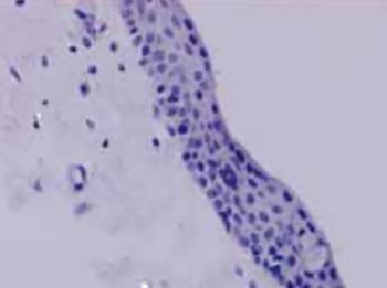
Pterygium internum (external eye layer) shows great recurrence tendency after surgical removal. Its etiology is still unclear and represents a significant problem. The main goal of our study was to explore the interrelationships of pathohistological characteristics of pterygium, namely presence of inflammation, vascularisation degree and fibrinoid changes and on the basis of their analysis to test the possibility of predicting its evolution and recurrence. The analysis was performed on the material taken from 55 patients surgically treated by the technique of Arlt. The specimens were stained using the classical histochemical methods: hematoxylin-eosin (HE), Masson’s trichrome, Gomori’s reticulin stain and PAS technique. Pterygium is mostly covered by conjunctival epithelium, while in the cap region shows morphology of modified stratified squamous epithelium of the cornea. Structural basis of the epithelium is composed of continuous basal lamina and continuous connective fibers underneath. This connective basis shows fibrinoid changes in the form of oval islets of different size, parallel to convexity of pterygium, or is in the form of unified focus. The number, caliber and the type of blood vessels showed excessive variability.
Pathohistological analysis of morphological characteristics of pterygium is adequate basis for prediction of recurrences; as they present the biggest concern in treatment of this widely spread disease.
Citations
Downloads

Downloads
Published
How to Cite
Accepted 2017-11-20
Published 2010-11-20


