Analysis of long non-coding RNA (lncRNA) expression in hepatitis B patients

Authors

  • Sunde Yılmaz Susluer Department of Medical Biology, Faculty of Medicine, Ege University, Izmir, Turkey
  • Cagla Kayabasi Department of Medical Biology, Faculty of Medicine, Ege University, Izmir, Turkey
  • Besra Ozmen Yelken Department of Medical Biology, Faculty of Medicine, Ege University, Izmir, Turkey
  • Aycan Asik Department of Medical Biology, Faculty of Medicine, Ege University, Izmir, Turkey
  • Didem Celik Izmir Tepecik Education and Research Hospital, Clinic of Infectious Diseases and Clinical Microbiology, Izmir, Turkey
  • Tugce Balci Okcanoglu Vocational School of Health Sciences, Near East University, Nicosia, Turkish Republic of Northern Cyprus (TRNC)
  • Suheyla Serin Senger Izmir Tepecik Education and Research Hospital, Clinic of Infectious Diseases and Clinical Microbiology, Izmir, Turkey
  • Cigir Biray Avci Department of Medical Biology, Faculty of Medicine, Ege University, Izmir, Turkey
  • Sukran Kose Izmir Tepecik Education and Research Hospital, Clinic of Infectious Diseases and Clinical Microbiology, Izmir, Turkey
  • Cumhur Gunduz Department of Medical Biology, Faculty of Medicine, Ege University, Izmir, Turkey

DOI:

https://doi.org/10.17305/bjbms.2018.2800

Keywords:

HBV, lncRNA, chronic hepatitis B, inactive HBsAg carrier, resolved hepatitis B

Abstract

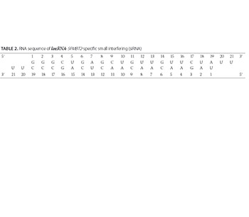
Long non-coding RNAs (lncRNAs) have been implicated in numerous biological processes, including epigenetic regulation, cell-cycle control, and transcriptional/translational regulation of gene expression. Differential expression of lncRNAs and disruption of the regulatory processes are recognized as critical steps in cancer development. The role of lncRNAs in hepatitis B virus (HBV) infection is not well understood. Here we analyzed the expression of 135 lncRNAs in plasma samples of 82 HBV patients (classified as chronic patients, inactive carriers, or resolved patients) at diagnosis and at 12 months of treatment in relation to control group (81 healthy volunteers). We also investigated the effect of small interfering RNA (siRNA)-mediated silencing of lincRNA-SFMBT2 on HBV-positive human liver cancer cell line. lncRNA expression was analyzed by quantitative reverse transcription-polymerase chain reaction (qRT-PCR). Chemically synthesized siRNAs were transfected into the cell lines using Lipofectamine 2000 Reagent (Thermo Fisher Scientific). HBV DNA and HBsAg and HBeAg were detected in transfected cultures by real-time PCR and ELISA, respectively, using commercial kits. We observed changes in lncRNA expression in all three HBV groups, compared to control group. Most notably, the expression of anti-NOS2A, lincRNA-SFMBT2, and Zfhx2as was significantly increased and expression of Y5 lncRNA was decreased in chronic HBV patients. A decreased Y5 expression and increased lincRNA-SFMBT2 expression were observed in inactive HBsAg carriers. The expression of HOTTIP, MEG9, and PCAT-32 was increased in resolved HBV patients, and no significant change in the expression of Y5 was observed, compared to control group. siRNA-mediated inhibition of lincRNA-SFMBT2 decreased the level of HBV DNA in human liver cancer cells. Further research is needed to confirm the prognostic as well as therapeutic role of these lncRNAs in HBV patients.

Downloads

Download data is not yet available.

Author Biographies

Sunde Yılmaz Susluer, Department of Medical Biology, Faculty of Medicine, Ege University, Izmir, Turkey

Department of Medical Biology

Cagla Kayabasi, Department of Medical Biology, Faculty of Medicine, Ege University, Izmir, Turkey

Department of Medical Biology

Besra Ozmen Yelken, Department of Medical Biology, Faculty of Medicine, Ege University, Izmir, Turkey

Department of Medical Biology

Aycan Asik, Department of Medical Biology, Faculty of Medicine, Ege University, Izmir, Turkey

Department of Medical Biology

Cigir Biray Avci, Department of Medical Biology, Faculty of Medicine, Ege University, Izmir, Turkey

Department of Medical Biology

Cumhur Gunduz, Department of Medical Biology, Faculty of Medicine, Ege University, Izmir, Turkey

Department of Medical Biology

References

Yuen MF, Hou JL, Chutaputti A, Asia Pacific Working Party on Prevention of Hepatocellular C. Hepatocellular carcinoma in the Asia pacific region. J Gastroenterol Hepatol 2009;24(3):346-53. https://doi.org/10.1111/j.1440-1746.2009.05784.x.

Kapranov P, Cheng J, Dike S, Nix DA, Duttagupta R, Willingham AT, et al. RNA maps reveal new RNA classes and a possible function for pervasive transcription. Science 2007;316(5830):1484-8. https://doi.org/10.1126/science.1138341.

Spizzo R, Almeida MI, Colombatti A, Calin GA. Long non-coding RNAs and cancer: A new frontier of translational research? Oncogene 2012;31(43):4577-87. https://doi.org/10.1038/onc.2011.621.

Ponting CP, Oliver PL, Reik W. Evolution and functions of long noncoding RNAs. Cell 2009;136(4):629-41. https://doi.org/10.1016/j.cell.2009.02.006.

Derrien T, Johnson R, Bussotti G, Tanzer A, Djebali S, Tilgner H, et al. The GENCODE v7 catalog of human long noncoding RNAs: Analysis of their gene structure, evolution, and expression. Genome Res 2012;22:1775-89. https://doi.org/10.1101/gr.132159.111.

Deng G, Sui G. Noncoding RNA in oncogenesis: A new era of identifying key players. Int J Mol Sci 2013;14(9):18319-49. https://doi.org/10.3390/ijms140918319.

Jeon Y, Sarma K, Lee JT. New and Xisting regulatory mechanisms of X chromosome inactivation. Curr Opin Genet Dev 2012;22(2):62-71. https://doi.org/10.1016/j.gde.2012.02.007.

Mattick JS, Amaral PP, Dinger ME, Mercer TR, Mehler MF. RNA regulation of epigenetic processes. Bioessays 2009;31(1):51-9. https://doi.org/10.1002/bies.080099.

Orom UA, Derrien T, Beringer M, Gumireddy K, Gardini A, Bussotti G, et al. Long noncoding RNAs with enhancer-like function in human cells. Cell 2010;143(1):46-58. https://doi.org/10.1016/j.cell.2010.09.001.

Rando TA, Chang HY. Aging, rejuvenation, and epigenetic reprogramming: Resetting the aging clock. Cell 2012;148(1-2):46-57. https://doi.org/10.1016/j.cell.2012.01.003.

Wapinski O, Chang HY. Long noncoding RNAs and human disease. Trends Cell Biol 2011;21(6):354-61. https://doi.org/10.1016/j.tcb.2011.04.001.

Gibb EA, Brown CJ, Lam WL. The functional role of long non-coding RNA in human carcinomas. Mol Cancer 2011;10:38. https://doi.org/10.1186/1476-4598-10-38.

Perz JF, Armstrong GL, Farrington LA, Hutin YJ, Bell BP. The contributions of hepatitis B virus and hepatitis C virus infections to cirrhosis and primary liver cancer worldwide. J Hepatol 2006;45(4):529-38. https://doi.org/10.1016/j.jhep.2006.05.013.

El-Serag HB. Hepatocellular carcinoma. N Engl J Med 2011;365(12):1118-27. https://doi.org/10.1056/NEJMra1001683.

Venook AP, Papandreou C, Furuse J, de Guevara LL. The incidence and epidemiology of hepatocellular carcinoma: A global and regional perspective. Oncologist 2010;15(Suppl 4):5-13. https://doi.org/10.1634/theoncologist.2010-S4-05.

Tsai WL, Chung RT. Viral hepatocarcinogenesis. Oncogene 2010;29(16):2309-24. https://doi.org/10.1038/onc.2010.36.

Feitelson MA, Reis HM, Tufan NL, Sun B, Pan J, Lian Z. Putative roles of hepatitis B x antigen in the pathogenesis of chronic liver disease. Cancer Lett 2009;286(1):69-79. https://doi.org/10.1016/j.canlet.2008.12.010.

Ringelhan M, Protzer U. Oncogenic potential of hepatitis B virus encoded proteins. Curr Opin Virol 2015;14:109-15. https://doi.org/10.1016/j.coviro.2015.08.015.

Tang H, Oishi N, Kaneko S, Murakami S. Molecular functions and biological roles of hepatitis B virus x protein. Cancer Sci 2006;97(10):977-83.

https://doi.org/10.1111/j.1349-7006.2006.00299.x.

Fu S, Li N, Zhou PC, Huang Y, Zhou RR, Fan XG. Detection of HBV DNA and antigens in HBsAg-positive patients with primary hepatocellular carcinoma. Clin Res Hepatol Gastroenterol 2017;41(4):415-23. https://doi.org/10.1016/j.clinre.2017.01.009.

Amaral PP, Clark MB, Gascoigne DK, Dinger ME, Mattick JS. lncRNAdb: A reference database for long noncoding RNAs. Nucleic Acids Res 2011;39(Database issue):D146-51. https://doi.org/10.1093/nar/gkq1138.

He H, Nagy R, Liyanarachchi S, Jiao H, Li W, Suster S, et al. A susceptibility locus for papillary thyroid carcinoma on chromosome 8q24. Cancer Res 2009;69(2):625-31. https://doi.org/10.1158/0008-5472.CAN-08-1071.

Korneev SA, Korneeva EI, Lagarkova MA, Kiselev SL, Critchley G, O'Shea M. Novel noncoding antisense RNA transcribed from human anti-NOS2A locus is differentially regulated during neuronal differentiation of embryonic stem cells. RNA 2008;14(10):2030-7. https://doi.org/10.1261/rna.1084308.

Guidotti LG, McClary H, Loudis JM, Chisari FV. Nitric oxide inhibits hepatitis B virus replication in the livers of transgenic mice. J Exp Med 2000;191(7):1247-52. https://doi.org/10.1084/jem.191.7.1247.

Mourtada-Maarabouni M, Pickard MR, Hedge VL, Farzaneh F, Williams GT. GAS5, a non-protein-coding RNA, controls apoptosis and is downregulated in breast cancer. Oncogene 2009;28(2):195-208. https://doi.org/10.1038/onc.2008.373.

Chang L, Li C, Lan T, Wu L, Yuan Y, Liu Q, et al. Decreased expression of long non-coding RNA GAS5 indicates a poor prognosis and promotes cell proliferation and invasion in hepatocellular carcinoma by regulating vimentin. Mol Med Rep 2016;13(2):1541-50. https://doi.org/10.3892/mmr.2015.4716.

Ariel I, Miao HQ, Ji XR, Schneider T, Roll D, de Groot N, et al. Imprinted H19 oncofetal RNA is a candidate tumour marker for hepatocellular carcinoma. Mol Pathol 1998;51(1):21-5. https://doi.org/10.1136/mp.51.1.21.

Kim KS, Lee YI. Biallelic expression of the H19 and IGF2 genes in hepatocellular carcinoma. Cancer Lett 1997;119(2):143-8. https://doi.org/10.1016/S0304-3835(97)00264-4.

Matouk IJ, DeGroot N, Mezan S, Ayesh S, Abu-lail R, Hochberg A, et al. The H19 non-coding RNA is essential for human tumor growth. PLoS One 2007;2(9):e845. https://doi.org/10.1371/journal.pone.0000845.

Iizuka N, Oka M, Yamada-Okabe H, Mori N, Tamesa T, Okada T, et al. Comparison of gene expression profiles between hepatitis B virus- and hepatitis C virus-infected hepatocellular carcinoma by oligonucleotide microarray data on the basis of a supervised learning method. Cancer Res 2002;62(14):3939-44.

Berteaux N, Aptel N, Cathala G, Genton C, Coll J, Daccache A, et al. A novel H19 antisense RNA overexpressed in breast cancer contributes to paternal IGF2 expression. Mol Cell Biol 2008;28(22):6731-45. https://doi.org/10.1128/MCB.02103-07.

Jiang YJ, Bikle DD. LncRNA profiling reveals new mechanism for VDR protection against skin cancer formation. J Steroid Biochem Mol Biol 2014;144(Pt A):87-90. https://doi.org/10.1016/j.jsbmb.2013.11.018.

Hao Y, Crenshaw T, Moulton T, Newcomb E, Tycko B. Tumour-suppressor activity of H19 RNA. Nature 1993;365:764-7. https://doi.org/10.1038/365764a0.

Pollard KS, Salama SR, Lambert N, Lambot MA, Coppens S, Pedersen JS, et al. An RNA gene expressed during cortical development evolved rapidly in humans. Nature 2006;443(7108):167-72. https://doi.org/10.1038/nature05113.

Liu H, Li J, Koirala P, Ding X, Chen B, Wang Y, et al. Long non-coding RNAs as prognostic markers in human breast cancer. Oncotarget 2016;7(15):20584-96. https://doi.org/10.18632/oncotarget.7828.

Chen X, Yan CC, Zhang X, You ZH. Long non-coding RNAs and complex diseases: From experimental results to computational models. Brief Bioinform 2017;18(4):558-76. https://doi.org/10.1093/bib/bbw060.

Johnson R, Richter N, Jauch R, Gaughwin PM, Zuccato C, Cattaneo E, et al. Human accelerated region 1 noncoding RNA is repressed by REST in Huntington's disease. Physiol Genomics 2010;41(3):269-74. https://doi.org/10.1152/physiolgenomics.00019.2010.

Zhou G, Du T, Roizman B. The role of the CoREST/REST repressor complex in herpes simplex virus 1 productive infection and in latency. Viruses 2013;5(5):1208-18. https://doi.org/10.3390/v5051208.

Wu L, Zhang L, Zheng S. Role of the long non-coding RNA HOTAIR in hepatocellular carcinoma. Oncol Lett 2017;14(2):1233-9. https://doi.org/10.3892/ol.2017.6312.

Zhang X, Lian Z, Padden C, Gerstein MB, Rozowsky J, Snyder M, et al. A myelopoiesis-associated regulatory intergenic noncoding RNA transcript within the human HOXA cluster. Blood 2009;113(11):2526-34. https://doi.org/10.1182/blood-2008-06-162164.

Wan L, Kong J, Tang J, Wu Y, Xu E, Lai M, et al. HOTAIRM1 as a potential biomarker for diagnosis of colorectal cancer functions the role in the tumour suppressor. J Cell Mol Med 2016;20(11):2036-44. https://doi.org/10.1111/jcmm.12892.

Wang KC, Yang YW, Liu B, Sanyal A, Corces-Zimmerman R, Chen Y, et al. A long noncoding RNA maintains active chromatin to coordinate homeotic gene expression. Nature 2011;472(7341):120-4. https://doi.org/10.1038/nature09819.

Quagliata L, Matter MS, Piscuoglio S, Arabi L, Ruiz C, Procino A, et al. Long noncoding RNA HOTTIP/HOXA13 expression is associated with disease progression and predicts outcome in hepatocellular carcinoma patients. Hepatology 2014;59(3):911-23. https://doi.org/10.1002/hep.26740.

Sonkoly E, Bata-Csorgo Z, Pivarcsi A, Polyanka H, Kenderessy-Szabo A, Molnar G, et al. Identification and characterization of a novel, psoriasis susceptibility-related noncoding RNA gene, PRINS. J Biol Chem 2005;280(25):24159-67. https://doi.org/10.1074/jbc.M501704200.

Vincent JB, Petek E, Thevarkunnel S, Kolozsvari D, Cheung J, Patel M, et al. The RAY1/ST7 tumor-suppressor locus on chromosome 7q31 represents a complex multi-transcript system. Genomics. 2002;80(3):283-94. https://doi.org/10.1006/geno.2002.6835.

Malik KT, Wallace JI, Ivins SM, Brown KW. Identification of an antisense WT1 promoter in intron 1: Implications for WT1 gene regulation. Oncogene 1995;11(8):1589-95.

Dallosso AR, Hancock AL, Malik S, Salpekar A, King-Underwood L, Pritchard-Jones K, et al. Alternately spliced WT1 antisense transcripts interact with WT1 sense RNA and show epigenetic and splicing defects in cancer. RNA 2007;13(12):2287-99. https://doi.org/10.1261/rna.562907.

Du T, Zhang B, Zhang S, Jiang X, Zheng P, Li J, et al. Decreased expression of long non-coding RNA WT1-AS promotes cell proliferation and invasion in gastric cancer. Biochim Biophys Acta 2016;1862(1):12-9. https://doi.org/10.1016/j.bbadis.2015.10.001.

Lerner MR, Boyle JA, Hardin JA, Steitz JA. Two novel classes of small ribonucleoproteins detected by antibodies associated with lupus erythematosus. Science 1981;211(4480):400-2. https://doi.org/10.1126/science.6164096.

Zhang AT, Langley AR, Christov CP, Kheir E, Shafee T, Gardiner TJ, et al. Dynamic interaction of Y RNAs with chromatin and initiation proteins during human DNA replication. J Cell Sci 2011;124(Pt 12):2058-69. https://doi.org/10.1242/jcs.086561.

Christov CP, Trivier E, Krude T. Noncoding human Y RNAs are overexpressed in tumours and required for cell proliferation. Br J Cancer 2008;98(5):981-8. https://doi.org/10.1038/sj.bjc.6604254.

Dhahbi JM, Spindler SR, Atamna H, Boffelli D, Mote P, Martin DI. 5'-YRNA fragments derived by processing of transcripts from specific YRNA genes and pseudogenes are abundant in human serum and plasma. Physiol Genomics 2013;45(21):990-8. https://doi.org/10.1152/physiolgenomics.00129.2013.

Dhahbi JM, Spindler SR, Atamna H, Boffelli D, Martin DI. Deep sequencing of serum small RNAs identifies patterns of 5' tRNA half and YRNA fragment expression associated with breast cancer. Biomark Cancer 2014;6:37-47. https://doi.org/10.4137/BIC.S20764.

Victoria Martinez B, Dhahbi JM, Nunez Lopez YO, Lamperska K, Golusinski P, Luczewski L, et al. Circulating small non-coding RNA signature in head and neck squamous cell carcinoma. Oncotarget 2015;6(22):19246-63. https://doi.org/10.18632/oncotarget.4266.

Subramanian M, Jones MF, Lal A. Long non-coding RNAs embedded in the Rb and p53 pathways. Cancers (Basel) 2013;5(4):1655-75. https://doi.org/10.3390/cancers5041655.

Loewer S, Cabili MN, Guttman M, Loh YH, Thomas K, Park IH, et al. Large intergenic non-coding RNA-RoR modulates reprogramming of human induced pluripotent stem cells. Nat Genet 2010;42(12):1113-7. https://doi.org/10.1038/ng.710.

Takahashi K, Yan IK, Kogure T, Haga H, Patel T. Extracellular vesicle-mediated transfer of long non-coding RNA ROR modulates chemosensitivity in human hepatocellular cancer. FEBS Open Bio 2014;4:458-67. https://doi.org/10.1016/j.fob.2014.04.007.

Analysis of long non-coding RNA (lncRNA) expression in hepatitis B patients

Downloads

Additional Files

Published

2018-05-20

How to Cite

1.
Yılmaz Susluer S, Kayabasi C, Ozmen Yelken B, Asik A, Celik D, Balci Okcanoglu T, Serin Senger S, Biray Avci C, Kose S, Gunduz C. Analysis of long non-coding RNA (lncRNA) expression in hepatitis B patients. Biomol Biomed [Internet]. 2018May20 [cited 2023Mar.28];18(2):150-61. Available from: https://www.bjbms.org/ojs/index.php/bjbms/article/view/2800

Issue

Section

Molecular Biology

INTRODUCTION

Hepatitis B virus (HBV) is a double-stranded DNA virus and a member of the Hepadnavirus family. HBV infection can lead to acute and chronic diseases of the liver and represents a global public health problem. Liver diseases associated with HBV are among the most common causes of liver transplantation [1].

Long non-coding RNAs (lncRNAs) are defined as transcribed RNA molecules not translated into proteins and longer than 200 nucleotides. Initially, the length of 200 nucleotides was set as a cut-off in RNA purification protocols to separate long from short ncRNAs, however, this length does not fully explain the functional properties of lncRNAs [2,3]. The majority of the identified lncRNAs are transcribed by RNA polymerase II. Although not necessarily, these transcripts may be polyadenylated, and are localized in the nucleus or cytosol [4]. It is assumed that lncRNAs are more numerous than protein-coding genes in the genome, however, accurate/complete classification and identification schemes for lncRNAs are still not available. Based on their location in relation to protein-coding genes, lncRNAs can be classified as intergenic, sense and antisense (exonic, intronic or overlapping), and bidirectional [5]. According to different functions, lncRNAs may be classified as RNAs that: 1) regulate gene expression, 2) act as microRNA (miRNA) decoys to free target mRNAs, 3) regulate mRNA translation, and 4) regulate protein activities [6].

lncRNAs have been implicated in numerous biological processes, including epigenetic regulation of gene expression (e.g., genomic imprinting), apoptosis, cell-cycle control, transcriptional, post-transcriptional, and translational regulation of gene expression, as well as in the development, differentiation and senescence of cells [6-11]. Differential expression of lncRNAs and disruption of such regulatory processes have been recognized as critical steps in cancer development. Moreover, different studies indicated the use of lncRNAs for diagnostic and therapeutic purposes, i.e., as biomarkers for specific cancers, candidates for therapeutic interventions, as well as targets in terms of regulation of lncRNA expression [12].

A popular technique that is used to suppress the expression of specific lncRNAs is RNA interference (RNAi) with small interfering RNAs (siRNAs). siRNAs can selectively target lncRNAs leading to their degradation. In experimental studies, the stability of siRNAs may be improved by different chemical modifications, to achieve prolonged inhibition of targeted lncRNAs.

The role of lncRNAs in hepatitis B infection is not clear. Perz et al. [13] investigated fractions of cirrhosis and hepatocellular carcinoma (HCC) related to chronic HBV or hepatitis C virus (HCV) infection in 11 World Health Organization (WHO)-based regions and reported that, globally, about three quarters of HCCs were attributed to HBV or HCV infection [13]. Worldwide, chronic HBV infection explains around 50% of all cases of HCC including all childhood cases [14]. Chronic HBV infection was estimated to be the main risk factor for HCC in countries with developing economies [15].

The HBV genome is a partially double-stranded DNA molecule containing four overlapping open reading frames (ORFs) encoding the envelope, core, polymerase, and HBx proteins. X ORF encodes the small regulatory protein HBx of 154 amino acids in length [16]. HBx promotes the expression and replication of viral genes through the transactivation of cellular promoters and enhancers important for continuous viral infection [17]. HBx also affects cell survival, proliferation, migration and transformation by interrupting several cell signal transduction pathways [18,19]. Current evidence supports an important role of HBx in the pathogenesis of HBV-mediated HCC [20].

In this study, we analyzed the expression of 135 lncRNAs in three HBV groups (chronic, inactive carriers, and resolved patients) at diagnosis and at 12 months of treatment in relation to control group. We also investigated the effect of siRNA-mediated silencing of lincRNA-SFMBT2 on HBV-positive human liver cancer cell line.

MATERIALS AND METHODS

Patient and control groups

The patient group included 82 randomized patients with HBV who had been referred to the Tepecik Training and Research Hospital for Infectious Diseases and Clinical Microbiology Clinic. Patients with decompensated cirrhosis, concomitant hepatitis A virus (HAV), HCV, hepatitis D virus (HDV), hepatitis E virus (HEV), human immunodeficiency virus (HIV), and any other viral infection or cause of liver disease were excluded. According to the treatment/follow-up response at 6 months and the American Association for the Study of Liver Diseases (AASLD) guidelines, the patients in HBV group were classified as follows: 27 patients (14 females, 13 males) with chronic HBV infection, 27 (23 F, 4 M) inactive hepatitis B surface antigen (HBsAg) carriers, and 28 (11 F, 17 M) resolved HBV patients. The control group included age-matched 81 healthy volunteers (41 F, 40 M) with no history of infectious diseases (including HBV), psychiatric, neurological, or metabolic disorder. Demographic and clinical features of HBV and control groups are given in Table 1.

TABLE 1: Demographic and clinical features of patients with hepatitis B virus (HBV) and control group

All procedures performed in studies involving human participants were in accordance with the ethical standards of the institutional and/or national research committee and with the 1964 Helsinki declaration and its later amendments or comparable ethical standards. Informed consent was obtained from all participants included in the study.

lncRNA expression profiling

lncRNA expression profiling included the following steps: collection of plasma samples from HBV patients and controls, total RNA isolation, cDNA synthesis, and quantitative reverse transcription-polymerase chain reaction (qRT-PCR) analysis. Changes in lncRNA expression were analyzed at diagnosis and at 12 months after treatment/follow-up in the three HBV groups, in relation to control group.

Collection of plasma samples

Peripheral blood samples (4 mL) of patients, at diagnosis and 12 months after treatment, and of controls were collected, transferred to microcentrifuge tubes, and centrifuged at 200 × g for 10 minutes at 40°C. The supernatant was then transferred to a new microcentrifuge tube and centrifuged at 12,000 × g for 10 minutes at 4°C to remove all cellular components. The upper plasma layer was collected for total RNA isolation.

lncRNA isolation

Total RNA, including lncRNAs, was isolated from plasma samples using the RNeasy mini kit (Qiagen, Germany). The purity and concentration of RNAs were determined by measuring the absorbance at 260/280 nm and 230/260 nm on a Nanodrop instrument (Thermo Scientific, USA). RNA samples with A260/A280 and A230/A260 absorbance ratios >2.0 were considered “pure” and used in further analysis.

cDNA synthesis

Total RNA was converted into cDNA using the RNA-Quant cDNA Synthesis Kit (System Biosciences, USA) which allows the detection of lncRNAs, following manufacturer’s instructions.

qRT-PCR analysis

Changes in lncRNA expression levels in the three HBV groups and control group were analyzed for a panel of 135 lncRNAs. These lncRNAs were selected from a lncRNA database (http://www.lncrnadb.org/), which provides a comprehensive list of lncRNAs that have been associated with biological functions in eukaryotes [21]. lncRNA expression was analyzed by qRT-PCR using the lncRNA Profiler qPCR Array Kit (Human) and Disease-Related lncProfiler 96-well Primer Sets (System Biosciences, USA) and Maxima SYBR Green qPCR Master Mix (Thermo Scientific, USA). Amplifications were performed on a LightCycler 480 II (Roche Life Science, Germany). The average values of reference gene expressions (7SL, small conditional [scRNA], 5.8S ribosomal RNA [rRNA], U87 Small Cajal body-specific RNA [scaRNA], U6 non-coding small nuclear RNA (snNA), ACTB, B2M, PGK1, GAPDH, HPRT1, RPL1A, and RPL13A) were used for normalization. The relative expression of lncRNAs in HBV groups at diagnosis and 12 months of treatment/follow-up was determined according to the 2-ΔΔCT method in relation to control group.

The expression profiles of the following 135 lncRNAs were analyzed: 21A, 7SK, 7SL, AAA1, aHIF, Air, AK023948, Alpha 250, Alpha 280, ANCR, ANRIL, anti-NOS2A, antiPeg11, BACE1AS, BC017743, BC043430, BC200, BCMS, BIC, CAR Intergenic 10, CCND1, CMPD, DD3, DGCR5, DHFR upstream transcript, DISC2, Dio3os, DLG2AS, E2F4 antisense, EGO, EGO B, EgoA, Emx2os, Evf1, EVF2, GAS5, GOMAFU, H19, H19 antisense, H19 upstream conserved 1 & 2, H19-AS, HAR1A, HAR1B, HOTAIR, HOTAIRM1, HOTTIP, HOXA11AS, HOXA1AS_AA489505, HOXA3as, HOXA3AS_BE873349, HOXA3AS_BI823151, HOXA6as, HOXA6AS_AK092154, HULC, IGF2A, IPW, Jpx, Kcnq1ot1, KRASP1, L1PA16, LIT, lincRNA-p21, lincRNA-RoR, LincRNA-SFMBT2, LincRNA-VLDLR, LOC285194, LUST, MALAT1, mascRNA, MEG3, MEG9, MER11C, NCRMS, ncR-uPAR, NDM29, NEAT1, Nespas, NRON, NTT, p53 mRNA, PANDA, PAR5, PCAT-1, PCAT-14, PCAT-29, PCAT-32, PCAT-43, PCGEM1, PCGEM1, PR antisense transcript, PR-AT2, PRINS, PRINS, PSF repressor RNA, PTENP1, RMRP, RNCR3, ROR, SAF, SCA8, snaR, SNHG1, SNHG3, SNHG4, SNHG5, SNHG6, Sox2OT, SRA, ST7OT, ST7OT1, ST7OT2, ST7OT3, ST7OT4, TEA ncRNAs, Telomeraz_RNA, TMEVPG1, Tmevpg1, TncRNA, Tsix, TU_0017629, TUG1, TUG1, UCA1, UCA1, UM9-5, WT1-AS, Xist, Y RNA-1, Y1, Y3, Y4, Y5, ZEB2NAT, Zeb2NAT, Zfas1, and Zfhx2as.

Analysis of lncRNA function and potential mechanism of action

lincRNA-SFMBT2 lncRNA, which showed significant changes in its expression at diagnosis and at 12 months of treatment/follow-up in HBV groups in relation to control group, was analyzed in vitro using HBV-positive human liver cancer cell line (Celprogen, USA). The cell line was cultured with Human Liver Cancer Cell Culture Full Growth Medium (Celprogen, USA) on flasks coated with Extracellular Matrix of Human Hepatic Cancer Cell Culture (Celprogen, USA). The cells were incubated at 37°C with 95% humidity and 5% CO2 until they reached the proliferation stage.

siRNA-mediated suppression of lncRNA

To investigate the effect of siRNA-mediated suppression of lncRNA on HBV-positive human liver cancer cell line, we selected siRNA for lincRNA-SFMBT2 using the online siDESIGN program (Dharmacon, USA). The designed siRNA sequence is given in Table 2. siRNAs were synthesized on a 10 nmol scale. The chemically synthesized siRNAs were transfected into HBV-positive human liver cancer cell lines using Lipofectamine 2000 Reagent (Thermo Fisher Scientific, USA), according to the manufacturer’s protocol. At 48, 96 and 144 hours after the transfection, the level of lncRNA was determined by qPCR. HBV DNA was isolated from the culture supernatant with the QIAamp DNA mini kit (Qiagen, Germany) and quantified with the ready-to-use artus HBV PCR Kit (Qiagen, Germany) on a Rotor-Gene cycler (Qiagen, Germany). The detection of HBsAg and hepatitis B envelope antigen (HBeAg) in the culture supernatants was performed with enzyme-linked immunosorbent assay (ELISA) kit (Beijing Wantai Biological Pharmacy, Beijing, China), according to the manufacturer’s protocol.

TABLE 2: RNA sequence of lincRNA-SFMBT2-specific small interfering (siRNA)

Statistical analysis

Categorical variables were expressed as frequencies and compared with chi-squared test between the groups. Quantitative variables were expressed as mean ± standard deviation (SD) and median (range) and compared with one-way analysis of variance (ANOVA) between the groups. Log2 transformation was applied to the 2-ΔΔCt values of lncRNA expression in control and HBV groups. The expression of 135 lncRNAs in each HBV group was determined at the initial diagnosis and at 12 months of treatment/follow-up in relation to control group and the values between HBV groups and control group, as well as between the two time points in each HBV group, were compared using Student’s t-test and false discovery rate (FDR)-corrected p values. The change in lncRNA expression of ± 2-fold in relation to control group and FDR-corrected p values <0.05 were considered significant. lncRNA expression data analysis was performed using the CLC Main Workbench software (Qiagen Bioinformatics, USA). A p value <0.05 was considered significant.

RESULTS

Analysis of lncRNA expression in patients with chronic HBV infection

In the group of 27 patients with chronic HBV infection, the expression of 15 lncRNAs increased by 2 fold from the time of initial diagnosis to the 12th month of treatment/follow-up, in relation to control group (p < 0.05). The expression of 7 lncRNAs was decreased more than 2 fold at 12 months of treatment/follow-up compared to the expression values at the initial diagnosis, and in relation to control group (p < 0.05). A significantly increased expression of anti-NOS2A, lincRNA-SFMBT2, and Zfhx2as and decreased expression of Y5 lncRNA in patients with chronic HBV infection compared to control group, indicate their potential use as biomarkers for monitoring the course of disease and response to treatment (Table 3, Figure 1).

TABLE 3: Long non-coding RNAs (lncRNAs) with ±2 fold changes in the expression between two time points (initial diagnosis/12 months of treatment) in hepatitis B virus (HBV) patients compared to control group
FIGURE 1: In chronic hepatitis B virus (HBV) patients (n = 27), the expression of 15 long non-coding RNAs (lncRNAs) increased by 2 fold from the time of initial diagnosis to the 12th month of treatment/follow-up, compared to control group (p < 0.05). The expression of 7 lncRNAs was decreased more than 2 fold at 12 months of treatment/follow-up compared to the expression values at the initial diagnosis, in relation to control group (p < 0.05). A significantly increased expression of anti-NOS2A, lincRNA-SFMBT2, and Zfhx2as and decreased expression of Y5 lncRNA was observed in this group. The change in lncRNA expression of ± 2-fold in relation to control group and false discovery rate (FDR)-corrected p values <0.05 were considered significant.

Analysis of lncRNA expression in inactive HBsAg carriers

In 27 inactive HBsAg carriers, the expression of lincRNA-SFMBT2 lncRNA increased by 2 fold from the initial diagnosis to the 12th month of treatment/follow-up, compared to control group (p < 0.05). The expression of 21 lncRNAs was significantly higher at the initial diagnosis and at 12 months of treatment/follow-up, compared to control group (p < 0.05). A decreased expression of Y5 and increased expression of lincRNA-SFMBT2 lncRNA in inactive HBsAg carriers suggest the role of those lncRNAs as prognostic biomarkers in patients with HBV (Table 3, Figure 2).

FIGURE 2: In inactive HBsAg carriers (n = 27), the expression of lincRNA-SFMBT2 increased by 2 fold at the initial diagnosis and the 12th month of treatment/follow-up, compared to control group, while the expression of Y5 was decreased 2.83 and 5.3 fold at initial diagnosis and at 12 months of treatment/follow-up, respectively (p < 0.05). The expression of 21 long non-coding RNAs (lncRNAs) was significantly higher at the initial diagnosis and at 12 months of treatment/follow-up, compared to control group (p < 0.05). The change in lncRNA expression of ± 2-fold in relation to control group and false discovery rate (FDR)-corrected p values <0.05 were considered significant.

Analysis of lncRNA expression in resolved HBV patients

In 28 resolved HBV patients, the expression of HOTTIP, MEG9, and PCAT-32 lncRNAs increased more than 2 fold from the initial diagnosis until 12 months of treatment/follow-up, compared to control group (p < 0.05). The expression of 20 lncRNAs was decreased more than 2 fold at 12 months of treatment/follow-up compared to the control (p < 0.05). No significant change was observed in the expression of Y5 between resolved HBV patients and control group, confirming the potential use of Y5 lncRNA as prognostic marker (Table 3, Figure 3).

FIGURE 3: In resolved hepatitis B virus (HBV) patients (n = 28), the expression of HOTTIP, MEG9, and PCAT-32 increased more than 2 fold from the initial diagnosis until 12 months of treatment/follow-up, compared to control group (p < 0.05). The expression of 20 long non-coding RNAs (lncRNAs) was decreased more than 2 folds at 12 months of treatment/follow-up compared to the initial diagnosis, and in relation to control group (p < 0.05). No significant change was observed in the expression of Y5 between resolved HBV patients and control group. The change in lncRNA expression of ± 2-fold in relation to control group and false discovery rate (FDR)-corrected p values <0.05 were considered significant.

siRNA-mediated inhibition of lncRNA expression

The expression of lincRNA-SFMBT2 in HBV-positive human liver cancer cell line transfected with the specific siRNA decreased 5 times until day 4 compared to control cells, and it was decreased 1.59 times at day 6 of transfection (Figure 4). These results indicate that the siRNA-mediated inhibition of lincRNA-SFMBT2 in the HBV-positive human liver cancer cell line was successfully achieved.

FIGURE 4: The expression of lincRNA-SFMBT2 in hepatitis B virus (HBV)-positive human liver cancer cell line transfected with the specific small interfering RNA (siRNA) decreased 5 times until day 4 compared to control cells, and it was decreased 1.59 times at day 6 of transfection.

HBsAg and HBeAg levels in siRNA-treated HBV-positive human liver cancer cell line

HBsAg and HBeAg were detected in the culture supernatant of HBV-positive human liver cancer cell line transfected with lincRNA-SFMBT2-specific siRNA and with negative control siRNA, at 48, 96 and 144 hours after transfection. These findings indicate that the replication of HBV DNA was continuously present in the cell line.

Quantification of HBV DNA in siRNA-treated HBV-positive human liver cancer cell line

The amount of HBV DNA in the culture supernatant of HBV-positive human liver cancer cell line transfected with lincRNA-SFMBT2-specific siRNA was decreased 1.55, 1.66 and 1.22 times at 48, 96, and 144 hours respectively, and the difference was significant compared to the cells treated with negative control siRNA (Figure 5). These results suggest that the siRNA-mediated inhibition of lincRNA-SFMBT2 affected the level of HBV DNA in the liver cancer cells.

FIGURE 5: The amount of hepatitis B virus (HBV) DNA in the culture supernatant of HBV-positive human liver cancer cell line transfected with lincRNA-SFMBT2 specific small interfering RNA (siRNA) was decreased 1.55, 1.66 and 1.22 times at 48, 96, and 144 hours respectively, and the difference was significant compared to the cells treated with negative control siRNA.*p < 0.05, ***p < 0.001, ****p < 0.0001.

DISCUSSION

The gene for AK023948 lncRNA was mapped to the 8q24 region, which also contains two overlapping protein-coding genes, thyroglobulin (TG) and Src-like adaptor (SLA). In the same study, the expression of AK023948 was significantly downregulated in majority of papillary thyroid carcinomas (PTC) [22]. In the group of resolved HBV patients, we observed a 45-fold reduction in the expression of AK023948 at diagnosis compared to control group, and a 7-fold reduction at 12 months of treatment/follow-up. Despite the increase in the expression of AK023948 during the 12-month period, AK023948 expression was still lower in resolved HBV patients compared to control group. Moreover, a significant reduction in the expression of AK023948 lncRNA was not observed in the other two groups of HBV patients, suggesting a role of AK023948 in the recovery from HBV.

anti-NOS2A is a ~1.9 kb intronless polyadenylated lncRNA transcribed from a locus located on 17q23.2, which has a high sequence similarity with the NOS2A gene that encodes the inducible isoform of nitric oxide synthase. Korneev et al. [23] reported several observations for the anti-NOS2A locus: the average sequence identity between the anti-NOS2A and the corresponding regions of the NOS2A gene was ~80%; this indicates that the anti-NOS2A is the result of gene duplication and subsequent internal DNA inversion; anti-NOS2A RNA was expressed in brain tumors such as meningiomas and glioblastomas; finally, anti-NOS2A likely regulates the neuronal differentiation of human embryonic stem cells by modulating the expression of NOS2A gene [23].

NO is a free radical produced by the inducible NO synthase (iNOS) in the liver. It was indicated that NO mediates the antiviral activity of interferon γ (IFN-γ) in HBV transgenic mice heterozygous or homozygous for the iNOS null mutation, and decreases the expression of viral antigens in the cell by inhibiting viral replication. Moreover, the absence of NO increased the severity of liver disease in those animals [24]. The expression of anti-NOS2A in our patients with chronic HBV infection was significantly increased both at diagnosis and at 12 months of treatment/follow-up, compared to control group, which may have affected the NO synthesis in these patients. On the other hand, anti-NOS2A expression was significantly lower in the inactive HBsAg carriers compared to control group, confirming the potential prognostic and therapeutic role of this lncRNA.

Growth arrest specific 5 (GAS5) is lncRNA involved in the regulation of cell cycle progression. The overexpression of several GAS5 transcripts induced growth arrest and apoptosis in mammalian cell lines, including human breast cancer cells. Moreover, the GAS5 expression was significantly downregulated in the breast cancer cells compared with adjacent healthy tissue [25]. Chang et al. [26] suggested that low expression of GAS5 in HCC indicates a poor prognosis. In our study, GAS5 expression was significantly lower at both time points (initial diagnosis and 12 months of treatment/follow-up) in all three patient groups compared to control group, suggesting the role of GAS5 in HBV infection.

H19 is an imprinted, maternally expressed gene (i.e., monoallelic expression) that transcribes to lncRNA. H19 RNA is normally expressed only during embryonic development, but may be re-activated in some processes such as adult tissue regeneration and tumorigenesis [27-29]. Biallelic expression due to loss of imprinting (LOI) of H19 and the associated paternally expressed insulin-like growth factor 2 gene (IGF2) gene, was demonstrated in HCC and several other tumor types, indicating the role of this process in epigenetic mechanism of tumor development [28]. Matouk et al. [29] showed that H19 RNA is upregulated in HCC and bladder carcinoma cell lines in response to hypoxic stress, and suggested that H19 acts as an oncogene, promotes tumorigenesis and affects tumor growth. Similarly, Iizuka et al. [30] showed that the H19 and IGF2 genes, as well as genes involved in signal transduction, transcription and tumor metastasis were upregulated in HBV-associated HCC [30]. Moreover, a 120-kb-long transcript (91H) antisense to the H19 gene, was characterized within the human and mouse H19/IGF2 locus. 91H RNA, preferentially expressed from the maternal chromosome, did not affect imprinting of the H19/IGF2 locus in human cells, but it did downregulate IGF2 expression in trans on the paternal allele [31]. In another study, lncRNA array analysis of 90 well-annotated mouse lncRNAs showed that the level of 91H (also named H19-as) was reduced in cultured vitamin D receptor (VDR)-deleted mouse keratinocytes [32]. The expression of H19 in our chronic HBV patients and inactive HBsAg carriers at diagnosis was significantly lower compared to control group. At 12 months of treatment/follow-up, we observed a 2-fold decrease in H19 expression in inactive HBsAg carriers and a 2-fold increase in chronic HBV group, compared to control group. The expression of H19-as in chronic HBV patients and inactive HBsAg carriers was lower both at the initial diagnosis and at 12 months of treatment/follow-up compared to control group. Lower H19 and H19-as expressions observed in our patient groups compared to controls may be associated with a suggested tumor-suppressor role of H19 [33].

Highly Accelerated Region 1A (HAR1A or HAR1F) is a 2.8 kb-long 2-exon transcript, it overlaps with HAR1B (HAR1R), and both of these genes contain the Human Accelerated Region (HAR1) [34]. Liu et al. [35] analyzed alterations in lncRNA expression in the breast invasive carcinoma dataset of the Cancer Genome Atlas (TCGA), containing ~1,000 cases. According to their results, the upregulation of HAR1A (and 8 other lncRNAs) could predict breast cancer recurrence [35]. HAR1A and HAR1B have also been associated with Alzheimer’s disease [36]. Moreover, transcriptional repression of HAR1 by RE1-Silencing Transcription factor (REST) has been shown in the striatum of patients with Huntington’s disease [37]. Zhou et al. [38] indicated the role of the REST corepressor 1 (CoREST)/REST complex in the suppression of Herpes Simplex Virus 1 gene expression, during its productive as well as latent infection in sensory neurons. The expression of HAR1A and HAR1B lncRNAs was significantly decreased in our resolved HBV patients both at the initial diagnosis and at 12 months following the treatment/follow-up, compared to control group. The inhibition of lncRNA as well HBV gene expression might be associated with the epigenetic mechanisms of the CoREST/REST complex.

HOX Transcript Antisense RNA (HOTAIR) is a 2.2 kb lncRNA involved in transcriptional modulation. By recruiting the polycomb repressive complex 2 (PRC2) and the lysine-specific demethylase 1 (LSD1)/coREST/REST complex, HOTAIR mediates the trimethylation of histone H3 at lysine 27 and the demethylation of histone H3 dimethyl Lys4 at promoters of target genes, leading to gene silencing. A number of studies reported that the overexpression of HOTAIR in HCC was associated with poor prognosis in those patients [39]. In our study, the expression of HOTAIR in resolved HBV patients was significantly decreased at the initial diagnosis and 12 months following the treatment/follow-up, compared to control group.

HOX antisense intergenic RNA myeloid 1 (HOTAIRM1) was first identified as a 483 nt long transcript located between the human HOXA1 and HOXA2 genes, and expressed specifically in the myeloid lineage. Short hairpin (shRNA)-mediated knockdown of HOTAIRM1 downregulated the expression of 3¢ HOXA neighboring genes and affected the expression of the alpha-M beta-2 integrin subunits, CD11B and CD18 genes, indicating the regulatory role of HOTAIRM1 in myelopoiesis [40]. Similarly, a tumor suppressor role of HOTAIRM1 was demonstrated in colorectal cancer (CRC) [41]. HOTAIRM1 lncRNA was significantly decreased in our inactive HBsAg carrier and resolved HBV patient groups at the initial diagnosis compared to control group, and it showed a 2-fold increase at 12 months of treatment/follow-up. Considering the tumor suppressor function of HOTAIRM1 in other cancer types, our results suggests that HOTAIRM1 may be used as a positive prognostic marker in HBV patients.

HOXA transcript at the distal tip (HOTTIP) is an antisense lncRNA located at the 5¢ end of the HOXA cluster. HOTTIP lncRNA is involved in the activation of several 5¢ HOXA genes by binding to WD repeat-containing protein 5 (WDR5) and targeting the WDR5/MLL complex to the HOXA locus, which further mediates trimethylation of histone H3 lysine 4 (H3K4me3) and thus maintains the active state of transcription [42]. Deregulation of HOX genes was indicated in hepatocarcinogenesis, and the upregulation of HOTTIP and HOXA13, in snap-frozen needle HCC biopsies prior to any treatment, was associated with increased metastasis and decreased overall survival in HCC patients [43]. In our resolved HBV patients, the expression of HOTTIP was significantly higher at both time points (initial diagnosis/12 months of treatment) compared to control group, which might serve as a good prognostic indicator in HBV patients.

Psoriasis susceptibility-related RNA Gene Induced by Stress (PRINS) is a 3.6 kb long transcript located on 10p12.1, comprised of two exons and both harbor Alu elements. Increased expression of PRINS was demonstrated under stress conditions, such as ultraviolet-B (UV-B) irradiation, HSV infection, and translational inhibition [44]. In our study group, chronic HBV patients had significantly higher PRINS expression at the initial diagnosis/12 months of follow-up compared to control group. In contrast, the expression of PRINS was significantly decreased in resolved HBV patients at both time points. These findings suggest, for the first time, that HBV infection may lead to increased PRINS expression and PRINS may be useful in distinguishing between chronic and resolved HBV patients.

The RAY1/ST7 locus is located at 7q31 and its complex multi-transcript system has been implicated in disease states, such as autism and cancer. The multigene system of the RAY1/ST7 locus is comprised of two noncoding sense strand genes (ST7OT3 and ST7OT4) overlapping with many alternative forms of the coding RAY1/ST7 transcript, and two noncoding antisense strand genes (ST7OT1 and ST7OT2) [45]. Our study is the first to provide information on the expression of ST7OT1, ST7OT2, ST7OT3 and ST7OT4 transcripts in HBV. The expression of ST7OT3 was significantly higher in chronic HBV compared to control group. Moreover, a significant suppression of ST7OT3 in resolved HBV group suggests it has an oncogenic function in HBV. Similarly, the expression of ST7OT1 and ST7OT2 was significantly decreased in inactive HBsAg carrier group.

WT1 (Wilms’ tumor 1) antisense transcript (WT1-AS), transcribed from an antisense promoter within WT1 intron 1 [46], is a 0.4-3 kb transcript present in multiple splicing isoforms [47]. Reduced expression of WT1-AS has been associated with increased cell proliferation and invasion in gastric cancer [48]. However, our results indicate that WT1-AS has an oncogenic role in HBV, i.e., the expression of WT1-AS was increased in chronic HBV group and decreased in resolved HBV patients, compared to control group.

Y RNAs are family of highly conserved small ncRNAs, 83-112 nt long. In humans, the family includes four types of ncRNAs, Y1, Y3, Y4 and Y5, which are transcribed by RNA polymerase III from individual genes. Y RNAs have a highly conserved stem-loop structure, where the stem (a double stranded region formed by the terminal 5¢- and 3¢-sequences of Y RNA) contains a sequence-specific binding site for Ro protein, forming together a ribonucleoprotein (RNP). These RNAs have an essential role in the initiation of DNA replication in mammalian cells [49,50]. In carcinomas and adenocarcinomas of the urinary bladder, cervix, colon, kidney, lung and prostate the relative expression levels of the four Y RNAs were significantly higher compared to normal nonmalignant tissues. Also, degradation of Y1 and Y3 RNAs by siRNAs in human cell lines led to the inhibition of cell proliferation [51]. It was also demonstrated that multiple small RNA fragments (including those derived from the 5¢-ends of specific Y RNA) circulate in the blood. These RNAs may serve as signaling molecules in various cellular processes, however, their targets as well as functions are still not completely understood [52]. The possibility of their use as markers of specific disease states, particularly cancer, has been indicated in breast cancer [53], lung cancer [51], and head and neck squamous cell carcinoma [54]. Overall the expression of Y5 was low in our HBV groups. Furthermore, Y5 expression was decreased in chronic HBV patients and inactive HBsAg carriers compared to control, while it was close to normal (control group) in resolved HBV patients.

Together with 7 other lncRNAs, lincRNA-SFMBT2, lincRNA-VLDLR and lincRNA-ST8SIA3 (also named lincRNA-RoR), were identified to have higher expression in induced pluripotent stem cells (iPSCs) compared to embryonic stem cells (ESCs), indicting their role in the development of iPSCs. Moreover, knock-down or overexpression of lincRNA-ST8SIA3 directly correlated with the formation of iPSCs colonies. Colocalization of transcription factors OCT4, SOX2 and NANOG near the lincRNA-ST8SIA3 promoter suggested that its expression is induced by those pluripotency factors. Also, lincRNA-ST8SIA3 negatively regulated TP53 expression, thus affecting cell cycle arrest and apoptosis [55,56]. In our study, the expression of lincRNA-SFMBT2 was significantly higher in chronic HBV patients and inactive HBsAg carriers. In addition, lincRNA-VLDLR expression was high in chronic HBV patients. lincRNA-ST8SIA3 expression was significantly different between the three HBV groups. Expression of lincRNA-SFMBT2 has been shown to increase in HepG2 cells 9.58 fold compared to malignant human hepatocytes (HepG2 cells) and non-malignant human hepatocytes (HH cells) [57].

Overall, different lncRNAs showed changes in the expression between HBV groups and control group, between initial diagnosis and 12 months of treatment in each HBV group, and between the three HBV groups (chronic HBV patients, inactive HBsAg carriers and resolved HBV patients). Most notably, Y5 expression was decreased in chronic HBV patients and inactive HBsAg carriers, and the level was close to normal (control group) in resolved HBV patients, suggesting its use in monitoring the course of disease. lincRNA-SFMBT2 expression was increased in chronic HBV patients and inactive HBsAg carriers, and siRNA-mediated inhibition of lincRNA-SFMBT2 decreased the level of HBV DNA in human liver cancer cells. In addition, AK023948, H19, H19-As and HOTAIRM1, as potential tumor suppressor lncRNAs, as well as HOTTIP may be important in the prognosis of HBV infection, while ST7OT3 and WT1-AS may have an oncogenic role. Similarly, GAS5 could play an important role in HBV infection. In addition to lincRNA-SFMBT2, WT1-AS might have an effect on HBV replication. The changes in the expression of HAR1A, HAR1B, and HOTAIR might indicate their involvement in epigenetic suppression of HBV genes via the CoREST/REST complex. Further research is needed to confirm the prognostic as well as therapeutic role of these lncRNAs in HBV patients.

Acknowledgements

ACKNOWLEDGMENTS

This work was supported by TUBITAK (the Scientific and Technological Research Council of Turkey) grant (Project No: 215S225).

DECLARATION OF INTERESTS

The authors declare no conflict of interests.

REFERENCES

  1. , , (). Asia Pacific Working Party on Prevention of Hepatocellular C. Hepatocellular carcinoma in the Asia pacific region. J Gastroenterol Hepatol. https://doi.org/10.1111/j.1440-1746.2009.05784.x
  2. , , , , , (). RNA maps reveal new RNA classes and a possible function for pervasive transcription. Science. https://doi.org/10.1126/science.1138341
  3. , , , (). Long non-coding RNAs and cancer: A new frontier of translational research?. Oncogene. https://doi.org/10.1038/onc.2011.621
  4. , , (). Evolution and functions of long noncoding RNAs. Cell. https://doi.org/10.1016/j.cell.2009.02.006
  5. , , , , , (). The GENCODE v7 catalog of human long noncoding RNAs: Analysis of their gene structure, evolution, and expression. Genome Res. https://doi.org/10.1101/gr.132159.111
  6. , (). Noncoding RNA in oncogenesis: A new era of identifying key players. Int J Mol Sci. https://doi.org/10.3390/ijms140918319
  7. , , (). New and Xisting regulatory mechanisms of X chromosome inactivation. Curr Opin Genet Dev. https://doi.org/10.1016/j.gde.2012.02.007
  8. , , , , (). RNA regulation of epigenetic processes. Bioessays. https://doi.org/10.1002/bies.080099
  9. , , , , , (). Long noncoding RNAs with enhancer-like function in human cells. Cell. https://doi.org/10.1016/j.cell.2010.09.001
  10. , (). Aging, rejuvenation, and epigenetic reprogramming: Resetting the aging clock. Cell. https://doi.org/10.1016/j.cell.2012.01.003
  11. , (). Long noncoding RNAs and human disease. Trends Cell Biol. https://doi.org/10.1016/j.tcb.2011.04.001
  12. , , (). The functional role of long non-coding RNA in human carcinomas. Mol Cancer. https://doi.org/10.1186/1476-4598-10-38
  13. , , , , (). The contributions of hepatitis B virus and hepatitis C virus infections to cirrhosis and primary liver cancer worldwide. J Hepatol. https://doi.org/10.1016/j.jhep.2006.05.013
  14. (). Hepatocellular carcinoma. N Engl J Med. https://doi.org/10.1056/NEJMra1001683
  15. , , , (). The incidence and epidemiology of hepatocellular carcinoma: A global and regional perspective. Oncologist. https://doi.org/10.1634/theoncologist.2010-S4-05
  16. , (). Viral hepatocarcinogenesis. Oncogene. https://doi.org/10.1038/onc.2010.36
  17. , , , , , (). Putative roles of hepatitis B x antigen in the pathogenesis of chronic liver disease. Cancer Lett. https://doi.org/10.1016/j.canlet.2008.12.010
  18. , (). Oncogenic potential of hepatitis B virus encoded proteins. Curr Opin Virol. https://doi.org/10.1016/j.coviro.2015.08.015
  19. , , , (). Molecular functions and biological roles of hepatitis B virus x protein. Cancer Sci. https://doi.org/10.1111/j.1349-7006.2006.00299.x
  20. , , , , , (). Detection of HBV DNA and antigens in HBsAg-positive patients with primary hepatocellular carcinoma. Clin Res Hepatol Gastroenterol. https://doi.org/10.1016/j.clinre.2017.01.009
  21. , , , , (). lncRNAdb: A reference database for long noncoding RNAs. Nucleic Acids Res. https://doi.org/10.1093/nar/gkq1138
  22. , , , , , (). A susceptibility locus for papillary thyroid carcinoma on chromosome 8q24. Cancer Res. https://doi.org/10.1158/0008-5472.CAN-08-1071
  23. , , , , , (). Novel noncoding antisense RNA transcribed from human anti-NOS2A locus is differentially regulated during neuronal differentiation of embryonic stem cells. RNA. https://doi.org/10.1261/rna.1084308
  24. , , , (). Nitric oxide inhibits hepatitis B virus replication in the livers of transgenic mice. J Exp Med. https://doi.org/10.1084/jem.191.7.1247
  25. , , , , (). GAS5, a non-protein-coding RNA, controls apoptosis and is downregulated in breast cancer. Oncogene. https://doi.org/10.1038/onc.2008.373
  26. , , , , , (). Decreased expression of long non-coding RNA GAS5 indicates a poor prognosis and promotes cell proliferation and invasion in hepatocellular carcinoma by regulating vimentin. Mol Med Rep. https://doi.org/10.3892/mmr.2015.4716
  27. , , , , , (). Imprinted H19 oncofetal RNA is a candidate tumour marker for hepatocellular carcinoma. Mol Pathol. https://doi.org/10.1136/mp.51.1.21
  28. , (). Biallelic expression of the H19 and IGF2 genes in hepatocellular carcinoma. Cancer Lett. https://doi.org/10.1016/S0304-3835(97)00264-4
  29. , , , , , (). The H19 non-coding RNA is essential for human tumor growth. PLoS One. https://doi.org/10.1371/journal.pone.0000845
  30. , , , , , (). Comparison of gene expression profiles between hepatitis B virus- and hepatitis C virus-infected hepatocellular carcinoma by oligonucleotide microarray data on the basis of a supervised learning method. Cancer Res.
  31. , , , , , (). A novel H19 antisense RNA overexpressed in breast cancer contributes to paternal IGF2 expression. Mol Cell Biol. https://doi.org/10.1128/MCB.02103-07
  32. , (). LncRNA profiling reveals new mechanism for VDR protection against skin cancer formation. J Steroid Biochem Mol Biol. https://doi.org/10.1016/j.jsbmb.2013.11.018
  33. , , , , (). Tumour-suppressor activity of H19 RNA. Nature. https://doi.org/10.1038/365764a0
  34. , , , , , (). An RNA gene expressed during cortical development evolved rapidly in humans. Nature. https://doi.org/10.1038/nature05113
  35. , , , , , (). Long non-coding RNAs as prognostic markers in human breast cancer. Oncotarget. https://doi.org/10.18632/oncotarget.7828
  36. , , , (). Long non-coding RNAs and complex diseases: From experimental results to computational models. Brief Bioinform. https://doi.org/10.1093/bib/bbw060
  37. , , , , , (). Human accelerated region 1 noncoding RNA is repressed by REST in Huntington’s disease. Physiol Genomics. https://doi.org/10.1152/physiolgenomics.00019.2010
  38. , , (). The role of the CoREST/REST repressor complex in herpes simplex virus 1 productive infection and in latency. Viruses. https://doi.org/10.3390/v5051208
  39. , , (). Role of the long non-coding RNA HOTAIR in hepatocellular carcinoma. Oncol Lett. https://doi.org/10.3892/ol.2017.6312
  40. , , , , , (). A myelopoiesis-associated regulatory intergenic noncoding RNA transcript within the human HOXA cluster. Blood. https://doi.org/10.1182/blood-2008-06-162164
  41. , , , , , (). HOTAIRM1 as a potential biomarker for diagnosis of colorectal cancer functions the role in the tumour suppressor. J Cell Mol Med. https://doi.org/10.1111/jcmm.12892
  42. , , , , , (). A long noncoding RNA maintains active chromatin to coordinate homeotic gene expression. Nature. https://doi.org/10.1038/nature09819
  43. , , , , , (). Long noncoding RNA HOTTIP/HOXA13 expression is associated with disease progression and predicts outcome in hepatocellular carcinoma patients. Hepatology. https://doi.org/10.1002/hep.26740
  44. , , , , , (). Identification and characterization of a novel, psoriasis susceptibility-related noncoding RNA gene, PRINS. J Biol Chem. https://doi.org/10.1074/jbc.M501704200
  45. , , , , , (). The RAY1/ST7 tumor-suppressor locus on chromosome 7q31 represents a complex multi-transcript system. Genomics.. https://doi.org/10.1006/geno.2002.6835
  46. , , , (). Identification of an antisense WT1 promoter in intron 1: Implications for WT1 gene regulation. Oncogene.
  47. , , , , , (). Alternately spliced WT1 antisense transcripts interact with WT1 sense RNA and show epigenetic and splicing defects in cancer. RNA. https://doi.org/10.1261/rna.562907
  48. , , , , , (). Decreased expression of long non-coding RNA WT1-AS promotes cell proliferation and invasion in gastric cancer. Biochim Biophys Acta. https://doi.org/10.1016/j.bbadis.2015.10.001
  49. , , , (). Two novel classes of small ribonucleoproteins detected by antibodies associated with lupus erythematosus. Science. https://doi.org/10.1126/science.6164096
  50. , , , , , (). Dynamic interaction of Y RNAs with chromatin and initiation proteins during human DNA replication. J Cell Sci. https://doi.org/10.1242/jcs.086561
  51. , , (). Noncoding human Y RNAs are overexpressed in tumours and required for cell proliferation. Br J Cancer. https://doi.org/10.1038/sj.bjc.6604254
  52. , , , , , (). 5’-YRNA fragments derived by processing of transcripts from specific YRNA genes and pseudogenes are abundant in human serum and plasma. Physiol Genomics. https://doi.org/10.1152/physiolgenomics.00129.2013
  53. , , , , (). Deep sequencing of serum small RNAs identifies patterns of 5’ tRNA half and YRNA fragment expression associated with breast cancer. Biomark Cancer. https://doi.org/10.4137/BIC.S20764
  54. , , , , , (). Circulating small non-coding RNA signature in head and neck squamous cell carcinoma. Oncotarget. https://doi.org/10.18632/oncotarget.4266
  55. , , (). Long non-coding RNAs embedded in the Rb and p53 pathways. Cancers (Basel). https://doi.org/10.3390/cancers5041655
  56. , , , , , (). Large intergenic non-coding RNA-RoR modulates reprogramming of human induced pluripotent stem cells. Nat Genet. https://doi.org/10.1038/ng.710
  57. , , , , (). Extracellular vesicle-mediated transfer of long non-coding RNA ROR modulates chemosensitivity in human hepatocellular cancer. FEBS Open Bio. https://doi.org/10.1016/j.fob.2014.04.007