Immunization with 3-oxododecanoyl-L-homoserine lactone-r-PcrV conjugate enhances survival of mice against lethal burn infections caused by Pseudomonas aeruginosa

Authors

  • Isar Dejban Golpasha Department of Biology, Science and Research branch, Islamic Azad University, Tehran, Iran
  • Seyed Fazlollah Mousavi Microbiology Research Center and Department of Microbiology, Pasteur Institute of Iran, Tehran, Iran
  • Parviz Owlia Molecular Microbiology Research Center, Shahed University, Tehran, Iran
  • Seyed Davar Siadat Microbiology Research Center and Department of Microbiology, Pasteur Institute of Iran, Tehran, Iran
  • Shiva Irani Department of Biology, Science and Research branch, Islamic Azad University, Tehran, Iran

DOI:

https://doi.org/10.17305/bjbms.2015.292

Keywords:

3-oxo-C12-HSL, Homoserine lactone (HSL), Immunization, PcrV, Pseudomonas aeruginosa, Vaccine

Abstract

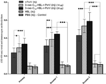
Quorum Sensing and type III secretion system play an important role in the virulence of Pseudomonas (P.) aeruginosa in burn wound infections. We aimed to explore the feasibility of using 3-oxo-C12-HSL-r-PcrV conjugate as a candidate vaccine against P. aeruginosa caused infections. 3-oxo-C12-HSL-r-PcrV conjugate was prepared and used for immunization of mice (10 μg, subcutaneous, three times, at 2-week intervals). Mice were divided into five groups: I: PcrV; II: 3-oxo-C12-HSL-r-PcrV (10 μg); III: 3-oxo-C12-HSL-r-PcrV (20 μg); IV: 3-oxo-C12-HSL; and V: PBS receiving groups.  After each shot of immunization, total and isotype antibody responses against corresponding antigen were measured to determine the immunization efficacy. One month after the last immunization, all groups were burned and challenged subeschar with P. aeruginosa PAO1. Survival rate and bacterial quantity in the skin and internal organs (liver and spleen) were evaluated 25-hr after burn infection. Immunization with 3-oxo-C12-HSL-r-PcrV significantly increased total IgG and specific subclass antibodies (IgG1, IgG2a, IgG2b, and IgM) in the serum of the groups II and III compared to the control group (p<0.001). While all the control mice (PBS injected group) died within 2 days after bacterial challenge, 64% of the group I, 78% of group II, and 86% of group III, survived within 14 days after challenge. Interestingly, bacterial burden in the liver and spleen of 3-oxo-C12-HSL-r-PcrV injected group (III) was significantly lower than the control group (p<0.001).  The present study proposed two-component vaccine to inhibit Pseudomonas infections in burned mouse. 

Downloads

Download data is not yet available.

Author Biography

Isar Dejban Golpasha, Department of Biology, Science and Research branch, Islamic Azad University, Tehran, Iran

Department of Biology, Science and Research branch, Islamic Azad University, Tehran, Iran

References

: Richards M J, Edwards J R, Culver D H, Gaynes R P. Nosocomial infections in medical intensive care units in the United States. National Nosocomial Infections Surveillance System. Crit Care Med 1999;27:887–892.

http://dx.doi.org/10.1097/00003246-199905000-00020

:Church D, Elsayed S, Reid O, Winston B, Lindsay R. Burn wound infections. Clinical microbiology reviews 2006;19(2):403-34.

http://dx.doi.org/10.1128/CMR.19.2.403-434.2006

:Erol S, Altoparlak U, Akcay MN, Celebi F, Parlak M. Changes of microbial flora and wound colonization in burned patients. Burns 2004;30(4):357-61.

http://dx.doi.org/10.1016/j.burns.2003.12.013

: Poole K, Srikumar R. Multidrug efflux in Pseudomonas aeruginosa: components, mechanisms and clinical significance. Curr Top Med Chem 2001;1:59-71.

http://dx.doi.org/10.2174/1568026013395605

: Schweizer HP. Efflux as a mechanism of resistance to antimicrobials in Pseudomonas aeruginosa and related bacteria: unanswered questions. Genet Mol Res. 2003;2(1):48-62.

: Erol S, Altoparlak U, Akcay M N, Celebi F, Parlak M. Changes of mi¬crobial flora and wound colonization in burned patients. Burns. 2004; 30(4):357-61.

http://dx.doi.org/10.1016/j.burns.2003.12.013

: Fuqua WC, Winans SC, Greenberg EP. Quorum sensing in bacteria: the LuxR-LuxI family of cell density-responsive transcriptional regulators. J of bacteriol. 1994;176(2):269.

: Van Delden C, Iglewski B H. Cell-to-cell signaling and Pseudomonas aeruginosa infections. Emerg Infect Dis 1998;4:551–560.

http://dx.doi.org/10.3201/eid0404.980405

: Pearson J P, Gray K M, Passador L, Tucker K D, Eberhard A, et al. Structure of the autoinducer required for expression of Pseudomonas aeruginosa virulence genes. Proc Natl Acad Sci USA 1994; 91:197–201.

http://dx.doi.org/10.1073/pnas.91.1.197

: Pearson JP, Passador L, Iglewski BH, Greenberg E. A second N-acylhomoserine lactone signal produced by Pseudomonas aeruginosa. PNAS. 1995;92(5):1490-4.

: Miller M B, Bassler B L. Quorum sensing in bacteria. Annu Rev Microbiol 2001;(55):165–199.

http://dx.doi.org/10.1146/annurev.micro.55.1.165

: Smith RS, Iglewski BH. Pseudomonas aeruginosa quorum sensing as a potential antimicrobial target. J Clin Invest 2003;(112):1460–1465.

http://dx.doi.org/10.1172/JCI200320364

: Whitehead NA, Barnard AM, Slater H, Simpson NJ, Salmond GP. Quorum-sensing in Gram-negative bacteria. FEMS Microbiol Rev 2001;(25):365–404.

http://dx.doi.org/10.1111/j.1574-6976.2001.tb00583.x

:Tateda K, Ishii Y, Horikawa M, Matsumoto T, Miyairi S, Pechere J C., et al. Pseudomonas aeruginosa autoinducer N-3-oxododecanoylhomoserine lactone accelerates apoptosis in macrophages and neutrophils. Infect Immun 2003;(71):5785–5793.

http://dx.doi.org/10.1128/IAI.71.10.5785-5793.2003

: DiMango E, Zar H J, Bryan R, Prince A. Diverse Pseudomonas aeruginosa gene products stimulate respiratory epithelial cells to produce interleukin-8. J Clin Invest 1995;(96)2204–2210.

http://dx.doi.org/10.1172/JCI118275

: Saleh A, Figarella C, Kammouni W, Marchand-Pinatel S, Lazdunski A, Tubul A, et al. Pseudomonas aeruginosa quorum-sensing signal molecule N-(3-oxododecanoyl)-L-homoserine lactone inhibits expression of P2Y receptors in cystic fibrosis tracheal gland cells. Infect. Immun. 1999;67(10):5076-82.

: Telford G, Wheeler D, Williams P, Tomkins P, Appleby P, Sewell H, et al. The Pseudomonas aeruginosaQuorum-Sensing Signal MoleculeN-(3-Oxododecanoyl)-l-Homoserine Lactone Has Immunomodulatory Activity. Infect. Immun. 1998;66(1):36-42.

: Suga H, Smith K M. Molecular mechanisms of bacterial quorum sensing as a new drug target. Curr Opin Chem Biol 2003;(7)586–591.

http://dx.doi.org/10.1016/j.cbpa.2003.08.001

: Smith RS, R Kelly, Iglewski BH, Phipps RP. The Pseudomonas autoinducer N-(3-oxododecanoyl) homoserine lactone induces cyclo-oxygenase-2 and prostaglandin E2 production in human lung fibroblasts: Implications for inflammation. J Immunol 2002;(169):2636–2642.

http://dx.doi.org/10.4049/jimmunol.169.5.2636

: Rumbaugh KP, Griswold JA, Hamood AN. The role of quorum sensing in the in vivo virulence of Pseudomonas aeruginosa. Microbes Infect 2000(2):1721-1731.

http://dx.doi.org/10.1016/S1286-4579(00)01327-7

: Sperandio V, Torres AG, Jarvis B, Nataro JP, Kaper JB. Bacteria–host communication: the language of hormones. PNAS. 2003;100(15):8951-6.

: Barman A, Savel R H, S. Racine,et al. Type III protein secretion is associated with death in lower respiratory and systemic Pseudomonas aeruginosa infections. J.Infect. Dis. 2001; (183):1767-1774.

http://dx.doi.org/10.1086/320737

: Vallis AJ, Yahr TL, Barbieri JT, Frank DW. Regulation of ExoS Production and Secretion by Pseudomonas aeruginosa in Response to Tissue Culture Conditions. Infect. Immun. 1999;67(2):914-20.

: Imamura Y, Yanagihara K, Fukuda Y, Kaneko Y, Seki M, Izumikawa K, et al. Effect of anti-PcrV antibody in a murine chronic airway P.aeruginosa infection model. European Respiratory Journal 2007;(5):965-8.

http://dx.doi.org/10.1183/09031936.00147406

: Holder IA, Neely AN, Frank DW. PcrV immunization enhances survival of burned P. aeruginosa -infected mice. Infect. Immun.2001;(9):5908-10.

http://dx.doi.org/10.1128/IAI.69.9.5908-5910.2001

: Sawa T, Yahr TL, Ohara M, Kurahashi K, Gropper MA, Wiener-Kronish JP, et al. Active and passive immunization with the Pseudomonas V antigen protects against type III intoxication and lung injury. Nat. Med. 1999;5(4):392-8.

: Miyairi S,Tateda K. Immunization with 3-oxododecanoyl-L-homoserine lactone–protein conjugate protects mice from lethal Pseudomonas aeruginosa lung infection. J.Med.Microbiol 2006;(55):1381–1387

http://dx.doi.org/10.1099/jmm.0.46658-0

: Kyd JM, Cooley M. Conjugates of acyl homoserine lactone and catalase a from pseudomonas aeruginosa .U.S. Patent Application 2011; 13/997,409

Horikawa M, Tateda K, Tuzuki E, Ishii Y, Ueda C, Takabatake T, et al. Synthesis of Pseudomonas quorum-sensing autoinducer analogs and structural entities required for induction of apoptosis in macrophages. Bioorg Med Chem Lett. 2006;16(8):2130-3.

: Hosoda H, Sakai Y, Yoshida H, Miyairi S, Ishii K, Nambara T. The preparation of steroid N-hydroxysuccinimide esters and their reactivities with bovine serum albumin. Chem Pharm Bull (Tokyo) 1979;(3):742-746.

http://dx.doi.org/10.1248/cpb.27.742

: Faezi S, Sattari M, Mahdavi M, Roudkenar MH. Passive immunisation against P.aeruginosa recombinant flagellin in an experimental model of burn wound sepsis. Burns 2011;37(5):865-72.

http://dx.doi.org/10.1016/j.burns.2010.12.003

: Haghighat S, Siadat SD, Sorkhabadi SMR, Sepahi AA, Mahdavi M. Cloning, Expression and Purification of Penicillin Binding Protein2a (PBP2a) from Methicillin Resistant Staphylococcus aureus: A Study on Immunoreactivity in Balb/C Mouse. Avicenna J Med Biotechnol 2013;5(4):204.

: Rumbaugh K P, Griswold J A, Iglewski B H, Hamood A N. Contribution of quorum sensing to the virulence of Pseudomonas aeruginosa in burn wound infections, Infect. Immun. 1999;(67):5854–5862.

: Holder IA. Pseudomonas aeruginosa virulence-associated factors and their role in burn wound infections, Pseudomonas aeruginosa: the opportunist. CRC Press, Boca Raton, FL .1993; 235-245.

: Dacheux D, Epaulard O, DeGroot A, et al. Activation of the Pseudomonas aeruginosa type III secretion system requires an intact pyruvate dehydrogenase aceAB operon. Infect. Immun. 2002; (70):3973-3977.

http://dx.doi.org/10.1128/IAI.70.7.3973-3977.2002

: Galan J E, Collmer A. Type III secretion machines: bacterial devices for protein delivery into host cells. Science 1999(284):1322–1328.

: El-Zaim HS, Chopra AK, Peterson JW, Vasil ML, Heggers JP. Protection against Exotoxin A (ETA) and Pseudomonas aeruginosa Infection in Mice with ETA-Specific Antipeptide Antibodies. Infect. Immun. 1998;66(11):5551-4.

:Pollack M, Anderson S.E. Toxicity of pseudomonas aeruginosa exotoxin A for human macrophages. Infect.Immun 1978;(19):1092.

PMid:417028 PMCid:PMC422301

: : Furuya N, Hiralata Y, Matsumoto T, Kaker M, Yamaguchi K. Mortality rates amongst mice with endogenous septicaemia caused by Pseudomonas aeruginosa isolates from various clinical sources. J Med Microbiol. 1993(39):141.

http://dx.doi.org/10.1099/00222615-39-2-141

:Gang R K, Bang R L, Sanyal S C, Mokaddas E, Lari A R. Pseudomonas aeruginosa septicemia in burns. Burns 1999;(25): 611-612

http://dx.doi.org/10.1016/S0305-4179(99)00042-X

:Frank DW, Vallis A, Wiener-Kronish JP, Roy-Burman A, Spack EG, Mullaney BP, et al.Generation and characterization of a protective monoclonal antibody to P.aeruginosa PcrV. J.INFECT.DIS. 2002;186(1):64-73.

http://dx.doi.org/10.1086/341069

: Van Delden C, Iglewski B H. Cell-to-cell signaling and Pseudomonas aeruginosa infections. Emerg Infect Dis 1998;(4):551–560.

http://dx.doi.org/10.3201/eid0404.980405

Immunization with 3-oxododecanoyl-L-homoserine lactone-r-PcrV conjugate enhances survival of mice against lethal burn infections caused by Pseudomonas aeruginosa

Downloads

Additional Files

Published

2015-05-25

How to Cite

1.
Golpasha ID, Mousavi SF, Owlia P, Siadat SD, Irani S. Immunization with 3-oxododecanoyl-L-homoserine lactone-r-PcrV conjugate enhances survival of mice against lethal burn infections caused by Pseudomonas aeruginosa. Biomol Biomed [Internet]. 2015May25 [cited 2023Jun.2];15(2):15-24. Available from: https://www.bjbms.org/ojs/index.php/bjbms/article/view/292

Issue

Section

Immunology

INTRODUCTION

Burns are one of the most common and debilitating forms of trauma that predispose patients to infection with Pseudomonas (P.) aeruginosa, as a result of damage to the protective skin barrier and suppression of the host immune system [1]. In most cases, these infections may lead to systemic sepsis, which is often associated with a high degree of mortality. Statistical studies have indicated that approximately 10% of burned patients are infected with P. aeruginosa and up to 80% of them die from the consequent septicemia. Therefore, patients with severe burn wounds require immediate intensive care [2]. In most cases, the treatment of burn wounds infections caused by P. aeruginosa is very difficult even under prolonged antibiotic therapy [3]. Resistance to treatment is mainly attributed to intrinsic resistance of this pathogen against a wide range of antibiotics and its ability to survive under harsh conditions [4, 5]. This fact has promoted use of serum therapy instead of antibiotics for prevention and treatment of post-burn infections caused by P. aeruginosa among high-risk populations like welders and firefighters [6]. During the last decades, various studies have been conducted on the pathogenesis of P. aeruginosa infections in burn wounds, in order to discover efficient virulence-associated factor as an immunogenic agent against this pathogen. P. aeruginosa produces a wide variety of virulence factors, such as pili, flagella, alginate, pigments, proteases, and exotoxins. Synthesis of these factors is regulated by a cell-to-cell signaling mechanism referred to as quorum sensing [7]. This mechanism enables bacteria to sense their environment and turn on/turn off the genes coordinately, in a density-dependent fashion through the production of small diffusible molecules called autoinducers [7, 8].

P. aeruginosa primarily produces two autoinducers: N-butanoyl-L-homoserine lactone (C4-HSL) and N-3-oxododecanoyl-L-homoserine lactone (3-oxo-C12-HSL) [9]. Expression of autoinducers along with the proper functioning of quorum sensing system components (LasRI and RhlRI) leads to synthesis and regulation of virulence factors that directly contribute to the colonization and dissemination of the Pseudomonas in burn/wound infections. Microarray analysis shows that expression of more than 616 genes involved in the pathogenesis of this bacterium are directly or indirectly regulated by these systems [10-13].

Among these, the genes associated with lipopolysaccharides (LPS), cell wall, multi-drug efflux pumps, protease, exotoxin A, hemolysin, type II secretion system, attachment, motility, chemotaxis and biofilm formations, are more interesting [12, 13].

Recent progress in quorum sensing research has demonstrated that 3-oxo-C12-HSL induces apoptosis in macrophages and neutrophils [14]. These findings suggest that autoinducer molecules are not only important in the expression and regulation of bacterial virulence genes, but also interact with eukaryotic cells and modulate immune responses [15-17]. Other studies have shown that quorum sensing mutant strains of P. aeruginosa significantly lose their pathogenic potential compared to wild strains in burn wound infections [12].

Quorum sensing systems have introduced a new area in designing and developing novel types of vaccines against P. aeruginosa [18, 19]. Specifically, blocking autoinducer molecules by using active and passive immunization has been identified as a promising strategy for the treatment of P. aeruginosa. Anti-autoinducers antibodies (anti-HSLs) can bind to these molecules and prevent their reaction with eukaryotic cells or contact with their receptors and then re-enter into the bacterial cells for activating transcription regulators of the virulence genes [20]. On the other hand, it seems that quorum sensing system is also involved in regulation and expression of the type III secretion system (T3SS) [21]. T3SS allows direct delivery of several toxic proteins (effectors) into the cytosol of the eukaryotic target cell [22, 23]. The PcrV protein (also known as V antigen) is a part of this system that plays a unique role in type III secretion system. Previous studies have revealed that active and passive immunization against PcrV can increase survival in animal models of the burn wound and lung infections induced by P. aeruginosa [24, 25]. In this study, for the first time, we developed a bivalent antigenic composition by using PcrV protein and 3-oxo-C12-HSL and investigated the feasibility of using this conjugate molecule as a prophylactic vaccine against Pseudomonas-induced burned/wound infections. We also examined the potential of immunization with 3-oxo-C12-HSL-r-PcrV protein conjugate in promoting the immune response against Pseudomonas infection in comparison r-PcrV protein alone. In addition, we explored the protective effect of specific antibody against 3-oxo-C12-HSL-r-PcrV conjugate on local and systemic spread of P. aeruginosa in skin, liver and spleen of burned mouse models following burn/wound infection.

MATERIALS AND METHODS

Bacterial strains and culture conditions

P. aeruginosa strain PAO1 was used for the challenge and in vivo experiments (Pasteur Institute, Iran). Escherichia (E.) coli strains DH5a and Bl21 (DE3) (Novagen Co., Wisconsin, USA) were used for clone and expression of recombinant PcrV protein (r-PcrV), respectively. Luria-Bertani (LB) broth, agar (Merck Co., Germany) and Pseudomonas Selective Agar were used for the bacterial culturing.

Recombinant PcrV expression and purification

First, recombinant PcrV was cloned and expressed as a histidine-tagged protein. The following primers were designed and used in the cloning process: Forward primer, containing a restriction site for BamHI (5´-ATGGATCCGAAGTCAGAAACCTTAATGC-3´) and reverse primer with HidIII restriction site (5´-GGCAAGCTTGTAGATCGCGCTGAG-3´).

The purified pcrV fragment was cloned into the pET24a plasmid, expressed in E.coli Bl21 (DE3) and product protein purified by nickel affinity chromatography according to the manufacturer’s instructions. The protein was extracted into the phosphate buffer and stored at -70 ºC until the use. The absence of detectable levels of endotoxin in the final solution was verified by a Limulus amoebocyte lysate assay, following the purification process [26].

Preparation of 3-oxo-C12-HSL-r-PcrV conjugate

Conjugation was carried out and confirmed according to the previous studies [27,28]. Functionalized carboxyl-HSL (N-(12-(3-Carboxypropionyl) oxy-3-oxododecanoyl) HSL) was synthesized as a hapten according to the method described by Horikawa et al. [29]. The hapten was then conjugated to the recombinant PcrV (carrier protein) using activated ester method by Hosoda et al., [30]. In this regard, hapten (1 mg) was dissolved in 250 µl of dimethyl formamide (DMF). Solutions of both N-hydroxysuccinimide (NHS) in DMF (1 mg/mL) and 1-ethyl- 3-(3-dimethylaminopropyl) carbodiimide hydrochloride (EDC) (2 mg/mL) in 100 mM carbonate buffer (pH 8.2) were then added to the dissolved hapten solution and mixed well. The resulting solution was stirred at 18–24 °C for 6 hours. The PcrV was diluted to a concentration of 2.2 mg/mL in 100 mM carbonate buffer with pH 8.2. The hapten solution was then added to the protein solution and incubated at 25 °C for 3-4 hrs.

Afterward, the resulting mixture was subjected to dialysis against the saline solution and stored in -20 °C until use. The hapten was also conjugated with bovine serum albumin (BSA) in a similar manner to the immobilized antigen [27-30].

Mice and bacteria

Six to eight weeks old female BALB/c mice were obtained from Pasteur Institute of Iran. All mice were housed (one week) in specific pathogen-free conditions within the animal care facility. Following that, mice were divided into five different groups (n=14) with the following characteristics: Group I: r-PcrV- injected group: (10 μg in 0.1 mL of Complete Freund’s adjuvant and PBS 1:1 - Sigma, Saint Louis, MO, USA); Group II: 3-oxo-C12-HSL-r-PcrV conjugate, injected group: (10 μg in 0.1 mL of Complete Freund’s adjuvant and PBS 1:1); Group III: 3-oxo-C12-HSL-r-PcrV conjugate, injected group: (20 μg in 0.1 mL of Complete Freund’s adjuvant and PBS 1:1); Group IV: 3-oxo-C12-HSL injected group: (10 μg in 0.1 mL of Complete Freund’s adjuvant and PBS 1:1); Group 5: Control group or PBS injected group: (10 μg in 0.1 mL of Complete Freund’s adjuvant).

Immunization

A concentration of 0.2 mg/mL, of a relevant immunogen in PBS, (0.4 mg/mL for group III) was prepared and mixed with equivalent ratio (1:1) of Complete Freund’s adjuvant (Sigma, Saint Louis, MO, USA). Finally, 0.1 mL of this solution (containing 10 μg of relevant immunogen in all group and 20 μg for group III), was used for each injection. On day zero, the groups of mice were immunized subcutaneously, behind the neck, with the relevant immunogen. Immunization procedure was performed according to the specific schedule (Figure 1). Booster doses were prepared, the same way as the primary injection condition in PBS, and mixed with an equivalent ratio (1:1) of incomplete Freund’s adjuvant. Two weeks after each shot of immunization, mice were bled via the retro-orbital sinus and the sera were separated and stored at -20 ºC for future analysis (Figure 1).

Figure 1: Schematic diagram of immunization protocol.

IgG total antibodies

Optimized indirect enzyme-linked immune sorbent assay (ELISA) was used two weeks after each shot of immunization to examine the presence and alteration of total specific antibodies against relevant immunogen in sera of each group. ELISA immunoplates, were coated with 100 μL of 10 μg/mL of relevant immunogen diluted in PBS buffer (1 μg/well) and incubated overnight at 4°C (For group IV, immunoplate was coated with 3-oxo-C12-HSL-BSA conjugate).

After washing and blocking process, the plates were washed and then 100 μL of (1/100 to 1/204800) diluted sera in blocking buffer (PBS-Tween 20 containing 5% BSA) added into the each well as primary antibody, followed by incubation at 37 °C for 2 hr. In the next step, wells were washed three times and incubated with the HRP-conjugated anti mouse-IgG antibody (Sigma, USA) (diluted 1:7000 as the secondary antibody) at 37°C for 2 hr. After five times washing, the wells incubated 30 min with 100 μL TMB (tetramethylbenzidine) as the substrate in darkness. The enzymatic reaction was stopped by adding 100 µl of H2SO4 (2N). Optical density was read at 450 nm (OD 450). All tests were performed in triplicate for each serum sample [31, 32].

IgG isotypes and IgM

Two weeks after the last immunization, groups of mice were bled and individual sera were tested for existence of anti-immunogen specific subclass IgG1, IgG2a, IgG2b and IgM antibodies by using indirect-ELISA, as described by the manufacturer instructions (R&D Systems, USA) [31,32].

Antigenic effect of 3-oxo-C12-HSL after conjugation

We designed the experiment to confirm the antigenic effect of 3-oxo-C12-HSL after conjugation. ELISA Immunoplates were coated with 100 μL of 10 μg/mL of 3-oxo-C12-HSL-BSA peptide diluted in PBS buffer (1 μg/well) and incubated overnight at 4°C. After washing and blocking process, 100 μL of 1:100 diluted sera (second booster) from each group (in blocking buffer) were added to each well and incubated at 37°C for 2 hrs. After washing step, HRP-conjugated anti mouse-IgG antibody (Sigma, USA) diluted 1:7000 was added and incubated at 37 °C for 2 hrs. After five times washing, immunoplates were incubated 30 min with 100 μL TMB (tetramethylbenzidine) as the substrate in darkness. The enzymatic reaction was stopped by adding 100 μl of H2SO4 (2N). Optical density assessment was performed at 450 nm (OD 450). This method can confirm and reveal the efficacy of conjugation process [27, 28].

Thermal injury model

Thermal injury model was induced according to the method developed by Stieritz and Holder [25, 31] with slight modifications. Accordingly, 30 days after the last immunization, female BALB/c mice were anesthetized (IP injection) with a mixture of ketamine hydrochloride (100 mg/mL, Alfasan Woerden-Holand) and xylazine (20 mg/mL Alfasan Woerden-Holand) in distilled water. The back hair of mice was shaved (2.5×2.5 cm), then a flame resistant plate was put on the unshaved area, and uncovered area was subjected to an ethanol flame for 20 s to complete thermal injury process (12-15% of total body). Immediately, after burning process, 0.5 mL of sterile saline solution was administered intraperitoneally into the burned skin. The burn model was used in this experiment is of grade 3, partial-thickness and non-lethal [25, 31, 33, 34].

Bacterial challenge and survival assessments

The survival rate of a burned mouse model against a lethal dose of P. aeruginosa PAO1 was investigated to evaluate the protective efficacy of produced antibodies in each group against P. aeruginosa infection. Previous studies revealed that thermal injury causes immune suppression and reduces the lethal dose of P. aeruginosa from 106 CFU to 102 -103 CFU in these models [31]. For the bacterial challenge, P. aeruginosa PAO1 was grown overnight on brain-heart infusion broth (BHI). Fifty-one hundred μl of 24 h culture was transferred into the 5 mL BHI broth under agitation (200 rpm) at 37 ºC and incubated for 3-4 hrs to reach favorite optical density (OD of 0.2 at 620 nm). Next, bacterial cells were harvested by centrifugation and resuspended in 10 mL of sterile saline solution, followed by preparation of serial dilutions (10-1-10-10 fold). The number of bacteria in injected inoculate, was counted by plating 100 μL of each dilution onto MacConckey agar plates. Inoculums of 5-6×10² CFU (3 folds higher than LD50) were used and administered in 100 μL volume subeschar immediately after thermal injury process in burned site. A new group was defined in this section that contained only burned models without the challenge as witness group. Animals were monitored for survival twice a day up to 14 days [25,33,34].

Bacterial burden in skin and internal organs

This test was done to examine the efficacy and effectiveness of the produced antibodies, in inhibiting the distribution of P. aeruginosa PAO1 from the infected wound to internal organs. To this end, 25 hrs after challenge (post burn/infection) four mice of each defined groups were selected arbitrary and sacrificed. Following that, a biopsy of their burned skin and surrounding edges of non-burned skin (2×2 cm), spleen, and liver were prepared in aseptic conditions. Afterwards, each biopsy sample was weighed and homogenized in PBS (2 mL), by using a Sorvall Omni mixer (Ivan Sorvall, Inc, Norwalk, Conn.). For bacterial counting, serial dilutions (10-1 -10-4 fold) of homogenates were prepared in PBS. Next, 100 µl of each diluted suspension were cultured in triplicate on Pseudomonas selection agar plates (Merck, Germany). The plates were then incubated at 37ºC and the number of CFUs was counted after 24-48 hrs after the culturing process. Results were expressed as log10 CFUg-1 of wet weight for infected organs [25, 33, 34].

Statistical analysis

All data were expressed as the mean ±S.D. Data were analyzed using one-way analysis of variation (ANOVA) and student’s t-test. A p<0.05 was considered statistically significant [31, 32]. All statistical analyses were carried out using SPSS Version 17 Software Package.

RESULTS

Antibody responses

The total antibody titration results revealed that all experimental groups except group IV and V were able to produce specific antibodies against relevant immunogen (Figure 2) and the highest antibody titer were detected after third immunization (second booster) in all of them (Figure 3). As it is shown, the serum of mice treated with 3-oxo-C12-HSL alone showed no meaningful variation in antibody titers compared to control mice serum. On the other hand, as expected, immunization with 3-oxo-C12- HSL–r-PcrV conjugate (groups II and III) strongly induced high level of antibody response Compared with the control group (p<0.001)(Figure 3).

Figure 2: The total antibody titration. ELISA was done to reveal total IgG antibody at the dilution of 1/100 to 1/204800. Values presented as mean ±S.D of experimental groups (n=14).
Figure 3: The total antibody titration. Variation of Total IgG antibodies in sera of each defined group, after each course of immunization. Data presented as mean ±S.D of experimental groups (n=14).

In this investigation, 3-oxo-C12- HSL-r-PcrV was used with different concentration: 10 and 20 μg/mouse for immunization of mice in group II and III, respectively. Obtained results indicate that there was a remarkable increase in titers of specific anti-3-oxo-C12- HSL-r-PcrV antibodies in sera of group III in contrast to group II.

Identification of antibody isotypes

To determine the type of immune responses (cellular or humoral immunity), the existence of specific antibody isotypes (IgG1/IgG2a/IgG2b) and total IgM were evaluated. The isotype responses in each group are represented in Figure 4. These data display a functional response of the humoral immune system in all immunized groups, except group IV, compared with the control group (Figure 4).

Figure 4: Poly-isotypic anti-immunogen antibody titers. Isotype antibody analysis showed that the IgG1 was produced more than IgG2a in groups I-III. This pattern of humoral immune responses (IgG1/IgG2a) indicate that, although both branches of the immune system are activated but humoral immunity is dominant immune response against P. aeruginosa in burn wound sepsis model. Experiments carried out triplicate and values are shown as the mean ± S.D.

Antigenic effect of 3-oxo-C12-HSL after conjugation

This experiment was done to confirm that conjugation process could convert hapten (HSL) to an active immunogen. As shown in Figure 5 there were significant differences in titers of total IgG antibodies against 3-oxo-C12-HSL between groups II and III (AHL-conjugated groups) and other groups (p<0.001). Interestingly, in our results immunization with HSL alone (group IV, AHL injected group) was not able to induce antibodies against hemoserine lactone (Figure 5).

Figure 5: Total IgG antibodies titer against 3-oxo-C12 –HSL in sera of experimental groups of mice, two week after second booster.

Survival rate

To evaluate the protective efficacy of the induced anti-sera against P. aeruginosa in each group, the survival rate of burned mouse model against the lethal dose of P. aeruginosa PAO1 infection was investigated and mortality in all groups were followed-up for 14 days. As shown in the Figure 6 the highest survival rate among the defined groups belongs to the Group III, and this level of survival was significantly greater than the survival rate observed for the group I (none conjugated) (p<0.001).

Figure 6: Survival rate after challenge with P. aeruginosa PAO1. It must be noted that all mice in witness group (only burned without challenge) survived.

Finally, as shown in Figure 6, all mice in group IV and V died, and there was no protection effect observed in these groups, following the immunization.

Bacterial burden

This part of the experiment was designed to evaluate the efficiency of produced antibodies in each group to inhibit the local and systemic spread of bacteria from infected burn wound to internal organs in burned/infected mice.

Our results indicate that the produced antibodies against 3-oxo-C12-HSL-r-PcrV significantly decrease bacterial titer in the liver and spleen of the immunized mice in groups II and III compared to the control group. By contrast, no significant difference was identified in bacterial load between these groups in skin samples (Figure 7).

Figure 7: Bacterial titer in skin, liver, and spleen: effects of produced specific antibodies on local and systemic spread of P. aeruginosa PAO1 following burn/wound infection. Data are presented in mean ± SD.

Finally, these data suggests that anti 3-oxo-C12-HSL-r-PcrV antibodies play a crucial role in inhibiting the systemic spread of P. aeruginosa PAO1 from the infection site into the internal organs.

DISCUSSION

Severe burns cause damage to the protective skin barrier and overall immune system, and replace healthy skin with moist, protein-rich, avascular eschar. These conditions facilitate colonization of P. aeruginosa in burned wounds to high densities, potentially inducing high rate of infection-related morbidity and mortality [25].

After the initial colony formation, P. aeruginosa multiplies rapidly within the necrotic tissue and spreads locally within the burned skin. Once the number of bacteria reaches a threshold level within the burned skin, they disseminate into the bloodstream, causing septicemia [33, 34].

Despite the recent advances in surgical care and introduction of a wide variety of antimicrobial agents with anti-Pseudomonal activities, life threatening infection caused by this bacterium is still a common complication among burned patients [34].

Statistical studies indicate that this infection is associated with a high rate of mortality in burned patients. Therefore, these patients require immediate intensive care [2,25,31].

The main purpose of this study was to evaluate the feasibility of using r-PcrV and 3-oxododecanoyl-L-homoserine lactone conjugate as a candidate vaccine against P. aeruginosa induced infections. In addition, it investigated the role of r-PcrV-HSL conjugate in promoting poly-isotypic humoral immune response in burned mouse models. Our designed immunogen is composed of two parts: r-PcrV and 3-oxododecanoyl-L-homoserine lactone. Both virulence factors are essential for Pseudomonas pathogenesis cycle in burned/wound infections. Hence, this study attempted to demonstrate the protective effect of anti-PcrV and anti-HSL antibodies against P. aeruginosa PAO1 in the burn/wound infections.

Previous studies have revealed that type III secretion system plays a vital role in Pseudomonas burn infection. This system can help P. aeruginosa to escape from phagocytes. Moreover, type III secretion system injects effector proteins and toxins directly into the adjacent eukaryotic cytosol across the eukaryotic plasma membrane and facilitates the spread of this bacterium into the bloodstream, causing septicemia [35, 36].

So far, four type III secretory effector toxins (ExoS, ExoT, ExoU and ExoY) have been identified in P. aeruginosa. PcrV protein plays a crucial role in transposition of these toxins into the eukaryotic cells [34, 37, 38]. These toxins contribute to the dissemination of P. aeruginosa throughout the patient’s body and its horizontal spread within the burned skin [39, 40]. As a result, protective antibodies against PcrV can enhance P. aeruginosa clearance in burned wound and survival of infected burn model.

Our study revealed that PcrV is one of the most effective immunologic stimulating agents of the immune system. The results of the similar study by Sawa et al., showed that PcrV immunization inhibits the chronic infection in mouse lung model [26].

In addition, we demonstrated that immunization with r-PcrV significantly enhances the survival of mice in the burn wound sepsis model and results in a long-term protection against P. aeroginosa PAO1 strain (64% in group I, the r-PcrV injected group). Similarly, Frank et al. showed that PcrV bonded polyclonal antibodies inhibit the delivery of type III toxins and enhance the clearance of highly cytotoxic strain of P. aeruginosa during acute lung infection. We obtained similar results in burning infections [41]. Our findings showed that our designed vaccine can promote anti-PcrV antibodies and prevent the normal action of type III secretion system. Finally, various vaccines based on PcrV have shown high efficacy in prevention and treatment of P. aeruginosa infections in different mice models.

The second part of our designed vaccine is 3-oxododecanoyl-L-homoserine lactone. It is clear that P. aeruginosa produces a variety of virulence factors. The expression of the vast majority of these factors is delicately regulated by quorum sensing system [42].

Previous studies have demonstrated that mutations in this system limit the ability of P. aeruginosa to cause systemic and local damage in the burn wound infections [43]. Obtained results in this area revealed that the bacterial quorum-sensing system, especially autoinducer molecules, can be considered as a novel candidate for vaccine development against P. aeruginosa infections.

In addition, some studies have revealed that 3-oxo-C12-HSL specific antibodies play a protective role in acute pulmonary infections caused by P. aeruginosa [43]. Other studies have demonstrated that 3-oxo-C12-HSL can act as an immunomodulator, interfering with the function of multiple different eukaryotic cells. Therefore, this molecule has a pivotal role in the pathogenesis of P. aeruginosa [12]. As molecular mass of 3-oxo-C12-HSL is approximately 300 Da, the natural antibodies against this molecule may not be produced in animals and humans, even the infected hosts (We observed this fact in the serum of mice treated with 3-oxo-C12-HSL alone; group IV). Thus, a carrier-protein-conjugated 3-oxo-C12-HSL is needed to produce antibodies against this molecule. In this study, we selected PcrV protein as a carrier protein. It is expected that immunization with 3-oxo-C12-HSL-r-PcrV can lead to a production of specific antibody against 3-oxo-C12-HSL in the immunized models. This new idea suggests a production of a bivalent candidate vaccine that can induce protective antibodies against both PcrV and 3-oxo-C12-HSL to interfere pathogenesis cycle of P. aeruginosa, by inhibiting normal function of Type III secretion system, and quorum-sensing system, respectively.

Type III secretion system and quorum-sensing system are required for the optimal virulence of P. aeruginosa in the burn infection model, which may result in sepsis and systemic inflammatory response, multiple-organ failure, and death.

The conjugation process was performed and confirmed based on the previous methods described by Miyairi et al [27, 28]. We used acyl side chain of 3-oxo-C12-HSL to conjugate this HSL to the carrier protein. Conjugation position is an important factor to reach the maximal activity of this bivalent immunogen. In this regard, additional experiments using a variety of techniques (e.g. direct conjugation of carrier protein to the HSL ring) are required to develop vaccine products with optimal effects.

In our experiments, specific antibodies were produced after the primary injection of 3-oxo-C12-HSL-r-PcrV, and the highest titers of antibodies were observed following the second, booster injection. This observation suggests that this immunogen may be considered a strong inducer of humoral immunity.

Based on the previous studies, in our study we used 3-oxo-C12-HSL-r-PcrV concentrations of 10 and 20 μg/mouse for immunization. We found that immunization with 20 μg/mouse of 3-oxo-C12-HSL-r-PcrV resulted in a higher titer of specific antibody compared to 10 μg/mouse. It is recommended to investigate different concentrations of this conjugate to assess its effects on the immune system.

In a similar study conducted by Miyairi et al [27], 3-oxo-C12-HSL was conjugated with BSA to produce specific antibodies against 3-oxo-C12-HSL and protect mice from lethal P. aeruginosa lung infection. They indicated that to 3-oxo-C12-HSL specific antibody plays a protective role against acute P. aeruginosa infection, and concluded that these antibodies did not influence the bacterial titers in internal organs. On the contrary, our findings show that antibodies (group II and III) induced by our vaccine decrease bacterial titers in internal organs of the burn/wound infected mouse [27]. This observation implies that the specific antibodies against the r-PcrV in designed immunogen can reduce the amount of bacteria in internal organs.

According to previous studies, PcrV protein is a strong inducer for humoral immunity [25,26]. Here we used this protein as carrier to convert a non-immunogen compound (3-oxo-C12-HSL) to an efficient immunogen. The antigenic feature of 3-oxo-C12-HSL after conjugation process was confirmed by ELISA method. In this method immunoplates were coated with 3-oxo-C12-BSA and the sera of all experimental groups were examined. Using this method we only detected the existence of specific antibodies against 3-oxo-C12-HSL, the second component of bivalent immunogen. Results revealed that these antibodies merely exist in 3-oxo-C12-HSL-r-PcrV conjugated groups (II and III). Obtained results revealed the antigenic effect of 3-oxo-C12-HSL following conjugation.

On the other hand, based on our results, the existence of specific antibodies against r-PcrV in sera of group I, and results from previous studies [25], r-PcrV is a strong inducer for humoral immunity. According to the terms of conjugation and feature of this protein, it is clear that its antigenic effect was preserved during the conjugation process. The existence of specific antibodies against the first part of bivalent immunogen (r-PcrV) in sera of group II and III was confirmed by ELISA method (data not shown).

Isotype antibody analysis showed that the IgG1 was produced in higher amounts in the immunized groups as compared to IgG2a (groups I, II and III). This pattern of IgG1/IgG2a response indicates that although both branches of the immune system are activated, humoral immunity is the dominant immune response against P. aeruginosa in burn wound sepsis model. These data represent functional responses of the humoral immune system in all immunized groups, except group IV (HSL injected group) compared with the control group.

To study the protective effect of anti-immunogen antibodies against P. aeruginosa, survival of burn wound infected mice against the lethal dose of the P. aeruginosa PAO1 was investigated.

Survival studies demonstrated that active immunization with 10 or 20 μg/mouse of 3-oxo-C12-HSL-r-PcrV conjugate, enhances protection in these groups by 78% or 86%, respectively, when compared to the control group (PBS injected group). The highest survival rate was observed in the group III, and this confirmed that immunization with 3-oxo-C12-HSL-r-PcrV provides better protection against the lethal dose of P. aeroginosa PAO1 in the burned mice. Baseline mortality rates in the groups I and II indicate that conjugated immunogen (3-oxo-C12-HSL-r-PcrV) decreased the mortality rate by a factor of 14% when compared with the non-conjugate immunogen (r-PcrV) with the same dose of vaccine. Similar results were obtained when comparing the group I with group III (22%). These results clearly indicate that conjugation of 3-oxo-C12-HSL to r-PcrV enhances the protective effect of this protein against the lethal doses of P. aeruginosa PAO1.

Obtained results also revealed that systemic spread of P. aeruginosa within the liver and spleens of burned/infected mice in the group II was significantly lower than that in the group I. This observation suggests that produced antibodies against both parts of 3-oxo-C12-HSL-r-PcrV immunogen have synergistic effects in preventing the spread of bacteria from the infection site to internal organs.

Our results are consistent with the findings of Rumbagh et al. who indicated that quorum-sensing systems are probably involved in the initial spread of P. aeruginosa within the burned tissue as well as its systemic spread into the internal organs [43]. The field of quorum sensing is still in its infancy. Therefore, there is a need for continued studies on the role of quorum sensing system in mediating infections and affecting host immune responses. Such studies can improve our understanding of host-bacterial interactions, and thus help developing novel treatments to these infections [43].

CONCLUSION

Taking all together, we demonstrated that each part of bivalent conjugated immunogen can stimulate humoral immune responses to produce specific antibodies against them. These specific antibodies can prevent P. aeruginosa pathogenesis cycle in different ways and show synergistic effects (Figure 8). Autoinducer molecules offer valuable means to produce effective immunotherapeutic agents against P. aeruginosa.

Figure 8: Effect of designed vaccine on pathogenesis cycle of Pseudomonas in burn/wound infection.

Acknowledgements

ACKNOWLEDGEMENTS

The authors thank Dr. Setareh Haghighat, Dr. Mehdi Shafiee Ardestani and Dr. Sobhan Faezi for their technical comments regarding this work. This work was supported by the Microbiology Departments of Pasteur Institute of Iran.

DECLARATION OF INTERESTS

The authors declare no conflict of interests.

REFERENCES

  1. , , , (). Nosocomial infections in medical intensive care units in the United States. National Nosocomial Infections Surveillance System. Crit Care Med. http://dx.doi.org/10.1097/00003246-199905000-00020
  2. , , , , (). Burn wound infections. Clinical microbiology reviews. http://dx.doi.org/10.1128/CMR.19.2.403-434.2006
  3. , , , , (). Changes of microbial flora and wound colonization in burned patients. Burns. http://dx.doi.org/10.1016/j.burns.2003.12.013
  4. , (). Multidrug efflux in Pseudomonas aeruginosa: components, mechanisms and clinical significance. Curr Top Med Chem. http://dx.doi.org/10.2174/1568026013395605
  5. (). Efflux as a mechanism of resistance to antimicrobials in Pseudomonas aeruginosa and related bacteria: unanswered questions. Genet Mol Res.
  6. , , , , (). Changes of mi¬crobial flora and wound colonization in burned patients. Burns. http://dx.doi.org/10.1016/j.burns.2003.12.013
  7. , , (). Quorum sensing in bacteria: the LuxR-LuxI family of cell density-responsive transcriptional regulators. J of bacteriol.
  8. , (). Cell-to-cell signaling and Pseudomonas aeruginosa infections. Emerg Infect Dis. http://dx.doi.org/10.3201/eid0404.980405
  9. , , , , (). Structure of the autoinducer required for expression of Pseudomonas aeruginosa virulence genes. Proc Natl Acad Sci USA. http://dx.doi.org/10.1073/pnas.91.1.197
  10. , , , (). A second N-acylhomoserine lactone signal produced by Pseudomonas aeruginosa. PNAS.
  11. , (). Quorum sensing in bacteria. Annu Rev Microbiol. http://dx.doi.org/10.1146/annurev.micro.55.1.165
  12. , (). Pseudomonas aeruginosa quorum sensing as a potential antimicrobial target. J Clin Invest. http://dx.doi.org/10.1172/JCI200320364
  13. , , , , (). Quorum-sensing in Gram-negative bacteria. FEMS Microbiol Rev. http://dx.doi.org/10.1111/j.1574-6976.2001.tb00583.x
  14. , , , , , (). Pseudomonas aeruginosa autoinducer N-3-oxododecanoylhomoserine lactone accelerates apoptosis in macrophages and neutrophils. Infect Immun. http://dx.doi.org/10.1128/IAI.71.10.5785-5793.2003
  15. , , , (). Diverse Pseudomonas aeruginosa gene products stimulate respiratory epithelial cells to produce interleukin-8. J Clin Invest. http://dx.doi.org/10.1172/JCI118275
  16. , , , , , (). Pseudomonas aeruginosa quorum-sensing signal molecule N-(3-oxododecanoyl)-L-homoserine lactone inhibits expression of P2Y receptors in cystic fibrosis tracheal gland cells. Infect. Immun.
  17. , , , , , (). The Pseudomonas aeruginosaQuorum-Sensing Signal MoleculeN-(3-Oxododecanoyl)-l-Homoserine Lactone Has Immunomodulatory Activity. Infect. Immun.
  18. , (). Molecular mechanisms of bacterial quorum sensing as a new drug target. Curr Opin Chem Biol. http://dx.doi.org/10.1016/j.cbpa.2003.08.001
  19. , , , (). The Pseudomonas autoinducer N-(3-oxododecanoyl) homoserine lactone induces cyclo-oxygenase-2 and prostaglandin E2 production in human lung fibroblasts: Implications for inflammation. J Immunol. http://dx.doi.org/10.4049/jimmunol.169.5.2636
  20. , , (). The role of quorum sensing in the in vivo virulence of Pseudomonas aeruginosa. Microbes Infect. http://dx.doi.org/10.1016/S1286-4579(00)01327-7
  21. , , , , (). Bacteria–host communication: the language of hormones. PNAS.
  22. , , (). Type III protein secretion is associated with death in lower respiratory and systemic Pseudomonas aeruginosa infections. J.Infect. Dis. http://dx.doi.org/10.1086/320737
  23. , , , (). Regulation of ExoS Production and Secretion by Pseudomonas aeruginosa in Response to Tissue Culture Conditions. Infect. Immun.
  24. , , , , , (). Effect of anti-PcrV antibody in a murine chronic airway P.aeruginosa infection model. European Respiratory Journal. http://dx.doi.org/10.1183/09031936.00147406
  25. , , (). PcrV immunization enhances survival of burned P. aeruginosa -infected mice. Infect. Immun. http://dx.doi.org/10.1128/IAI.69.9.5908-5910.2001
  26. , , , , , (). Active and passive immunization with the Pseudomonas V antigen protects against type III intoxication and lung injury. Nat. Med.
  27. , (). Immunization with 3-oxododecanoyl-L-homoserine lactone–protein conjugate protects mice from lethal Pseudomonas aeruginosa lung infection. J.Med.Microbiol. http://dx.doi.org/10.1099/jmm.0.46658-0
  28. , (). Conjugates of acyl homoserine lactone and catalase a from pseudomonas aeruginosa. U.S. Patent Application. 13/997,409
  29. , , , , , (). Synthesis of Pseudomonas quorum-sensing autoinducer analogs and structural entities required for induction of apoptosis in macrophages. Bioorg Med Chem Lett.
  30. , , , , , (). The preparation of steroid N-hydroxysuccinimide esters and their reactivities with bovine serum albumin. Chem Pharm Bull (Tokyo). http://dx.doi.org/10.1248/cpb.27.742
  31. , , , (). Passive immunisation against P.aeruginosa recombinant flagellin in an experimental model of burn wound sepsis. Burns. http://dx.doi.org/10.1016/j.burns.2010.12.003
  32. , , , , (). Cloning, Expression and Purification of Penicillin Binding Protein2a (PBP2a) from Methicillin Resistant Staphylococcus aureus: A Study on Immunoreactivity in Balb/C Mouse. Avicenna J Med Biotechnol.
  33. , , , (). Contribution of quorum sensing to the virulence of Pseudomonas aeruginosa in burn wound infections. Infect. Immun.
  34. (). Pseudomonas aeruginosa virulence-associated factors and their role in burn wound infections. Pseudomonas aeruginosa: the opportunist.
  35. , , (). Activation of the Pseudomonas aeruginosa type III secretion system requires an intact pyruvate dehydrogenase aceAB operon. Infect. Immun. http://dx.doi.org/10.1128/IAI.70.7.3973-3977.2002
  36. , (). Type III secretion machines: bacterial devices for protein delivery into host cells. Science.
  37. , , , , (). Protection against Exotoxin A (ETA) and Pseudomonas aeruginosa Infection in Mice with ETA-Specific Antipeptide Antibodies. Infect. Immun.
  38. , (). Toxicity of pseudomonas aeruginosa exotoxin A for human macrophages. Infect. Immun. PMid:417028 PMCid:PMC422301
  39. , , , , (). Mortality rates amongst mice with endogenous septicaemia caused by Pseudomonas aeruginosa isolates from various clinical sources. J Med Microbiol. http://dx.doi.org/10.1099/00222615-39-2-141
  40. , , , , (). Pseudomonas aeruginosa septicemia in burns. Burns. http://dx.doi.org/10.1016/S0305-4179(99)00042-X
  41. , , , , , (). Generation and characterization of a protective monoclonal antibody to P.aeruginosa PcrV. J.INFECT.DIS. http://dx.doi.org/10.1086/341069
  42. , (). Cell-to-cell signaling and Pseudomonas aeruginosa infections. Emerg Infect Dis. http://dx.doi.org/10.3201/eid0404.980405