Detection of neurovascular structures using injection pressure in blockade of brachial plexus in rat
DOI:
https://doi.org/10.17305/bjbms.2005.3276Keywords:
regional anesthesia, block of brachial plexus, intraneural injection, intravascular injectionAbstract
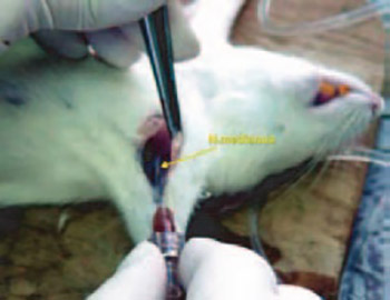
In the last few decades there has been a great development of regional anesthesia; all the postulates are defined and all the techniques of usage are perfected. However, like any other medical procedure, the block of brachial plexus carries a risk of certain unwanted complications, like possible intraneural and intravascular injections. The reason for great discrepancy between the injury of brachial plexus and other periphery nerves while performing the nerve blockade is the frequent usage of this block, but also the specific proximity of neurovascular structures in axilla. The purpose of this work is to determine the values of pressures which appear in paraneural, intraneural and intravascular injection applications of local anesthetic, and to compare those values in order to avoid cases of intraneural and in-travascular injections in clinical practice with consequential complications. In experimental study there have been used 12 Wistar rats of both genders. After anesthesia with ether and midhumeral access to the neurovascular structures in axilla, the injection of 2% lidocain with epinephrine was performed with the help of automatic syringe charger. The needle was at first placed paraneural, and then also intraneural and intravascular. During every application the pressure values were monitored using the manometer, and then they were analyzed by special software program. All paraneural injections resulted with the pressure between 13,96-27,92 kPa. The majority of intraneural injections were combined with the injection pressure greater than 69,8 kPa, while the intravascular injections were combined with injection pressure less than 6,98 kPa. Based on the available data it can be noticed that so far none of the methods of prevention from unwanted complications of regional anesthesia can insure the avoidance of intraneural and intravascular injection of local anesthetic. Based on our research it is obvious that the measuring of pressure during the nerve blockade is very important in order to decrease the risk of neurological and possible systematic complications. It is also clear that a small, mobile, and financially quite available apparatus for pressure measurement can help in differentiation between paraneural, intra-neural and intravascular injection. Avoiding high injection pressure prevents from lodging the needle into intraneural space, while avoiding a very low injection pressure prevents from lodging the needle into intravascular space followed by consequential complications. The usage of this apparatus can find its application in other blockades of periphery nerves, and in other branches of medicine as well.
Citations
Downloads
Downloads
Published
Issue
Section
Categories
How to Cite
Accepted 2018-03-02
Published 2005-08-20


