Contribution to the knowledge of position, flow and arterial distribution of cerebral blood vessels in foetuses 4 to 9 months of age
DOI:
https://doi.org/10.17305/bjbms.2004.3363Keywords:
fetus, brain, arteries, veinsAbstract
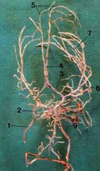
We studied cerebral blood vessels in 25 fetuses of gestational age 16-36 weeks and in 10 cadavers of still-born babies by injection-corrosive method. In the early fetal life, arteries are thin with the straight flow, which is directly connected with the brain development. Progressive changes are observed in all the three cerebral arteries in 28-week old fetus, which straight flow becomes more and more tortuous. As in the 32nd week the brain develops faster and gyri and sulci are being formed, the arteries assume wavy flow and number of their rami increases. In a still-born baby, arteries are of rather bigger caliber; they branch abundantly; and due to their relatively broad cerebral sulci, it can be said that their flow is partly tortuous. Our results show evidently that position, flow and relation of cerebral arteries change concurrently with the brain development and appearance of cerebral gyri and sulci.
Citations
Downloads
Downloads
Published
How to Cite
Accepted 2018-03-14
Published 2004-11-20


