Prediction of breast cancer metastasis risk using circulating tumor markers: A follow-up study

Authors

  • Sibel Cetintas Radiation Oncology Department, Faculty of Medicine, Uludag University, Bursa, Turkey
  • Gulcin Tezcan Institute of Fundamental Medicine and Biology, Kazan Federal University, Kazan, Russia
  • Berrin Tunca Medical Biology Department, Faculty of Medicine, Uludag University, Bursa, Turkey
  • Unal Egeli Medical Biology Department, Faculty of Medicine, Uludag University, Bursa, Turkey
  • Mustafa Sehsuvar Gokgoz General Surgery Department, Faculty of Medicine, Uludag University, Bursa, Turkey
  • Gulsah Cecener Medical Biology Department, Faculty of Medicine, Uludag University, Bursa, Turkey

DOI:

https://doi.org/10.17305/bjbms.2018.3371

Keywords:

Breast cancer, circulating tumor cell markers, metastasis

Abstract

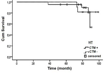
Distant organ tumor dissemination is a major cause of breast cancer-related deaths. In 2010, we analyzed the prognostic importance of the circulating tumor markers (CTMs) cytokeratin 19 (CK19), CK20, epidermal growth factor receptor (EGFR) and human epidermal growth factor receptor 2 (HER2) in relation to the clinical and pathological characteristics of patients with breast cancer (BC). To assess the clinical utility of CK19, CK20 and EGFR in predicting distant metastasis in BC, here we report 7-year follow-up results of 77 patients. The patients with at least one positive CTM were classified as CTM(+) and those negative for all CTMs were assigned to CTM(-) group. In patients who received no treatment following CTM analysis, 25.0% had metastasis in CTM(+) and 10.0% in CTM(-) group. In patients who received one of the following therapies: chemotherapy, radiotherapy or hormone therapy, or the combinations of these therapies, the rate of metastasis was 33.3% in CTM(+) and 20.0% in CTM(-) group. Disease-free time was shorter in CTM(+) patients compared to CTM(-) group (28.83 ± 10.76 and 41.38 ± 9.5 months, respectively). According to multivariate Cox proportional hazard regression analysis, the presence of regional lymph node metastasis, Ki-67 expression, higher tumor grade and CTM expression status were predictors of poor prognosis associated with distant metastasis (p < 0.05). Also, CTM positivity was a factor associated with metastasis-related poor prognosis (HR = 0.492, p = 0.026). The mean survival for CTM(+) patients was shorter than that for CTM(-) patients (90.671 ± 2.66 and 101.23 ± 3.92 months, respectively; p > 0.05). Our findings demonstrate that CTM positivity may indicate a high metastasis risk; however, CTM negativity does not guarantee low metastasis risk. These results may encourage further preclinical investigation of CTMs, to evaluate the possible implications of these findings to the clinical setting.

Downloads

Download data is not yet available.

Author Biographies

Sibel Cetintas, Radiation Oncology Department, Faculty of Medicine, Uludag University, Bursa, Turkey

Radiation Oncology Department

Berrin Tunca, Medical Biology Department, Faculty of Medicine, Uludag University, Bursa, Turkey

Medical Biology Department

Unal Egeli, Medical Biology Department, Faculty of Medicine, Uludag University, Bursa, Turkey

Medical Biology Department

Mustafa Sehsuvar Gokgoz, General Surgery Department, Faculty of Medicine, Uludag University, Bursa, Turkey

General Surgery Department

Gulsah Cecener, Medical Biology Department, Faculty of Medicine, Uludag University, Bursa, Turkey

Medical Biology Department
Prediction of breast cancer metastasis risk using circulating tumor markers: A follow-up study

Downloads

Additional Files

Published

2019-05-20

How to Cite

1.
Cetintas S, Tezcan G, Tunca B, Egeli U, Gokgoz MS, Cecener G. Prediction of breast cancer metastasis risk using circulating tumor markers: A follow-up study. Biomol Biomed [Internet]. 2019May20 [cited 2023Nov.30];19(2):172-9. Available from: https://www.bjbms.org/ojs/index.php/bjbms/article/view/3371

Issue

Section

Translational and Clinical Research

INTRODUCTION

Breast cancer (BC) is the most common cancer affecting women worldwide. The majority of BC-related deaths result from unsuccessful treatment of metastases [1,2]. Therefore, it is important to develop therapies that prevent dissemination of tumor cells at an early stage. Over the past decade, we have gained a better understanding of the role of circulating tumor cells (CTCs) in metastasis and recurrence [3]. Accordingly, different techniques have been developed to detect CTCs in the peripheral blood of cancer patients. One of the approaches is to measure mRNA expression of biomarkers associated with CTCs, which can help advance precision medicine in oncology and prevent metastasis [4]. Numerous CTC biomarkers are demonstrated to have clinical validity, however, many have not undergone rigorous testing to validate clinical utility, which is necessary to integrate these biomarkers into clinical care [5].

In a previous study [6], we analyzed the prognostic importance of the circulating tumor markers (CTMs) cytokeratin 19 (CK19), CK20, epidermal growth factor receptor (EGFR) and human epidermal growth factor receptor 2 (HER2) in BC. We showed that C20 positivity was more frequent in BC patients with aggressive tumors compared to other BC patients. On the other hand, mRNA expression of CK19, EGFR and HER2 was not significantly associated with the clinical and pathological characteristics of BC patients [6]. Although our previous findings indicate a potential relationship between these CTMs and aggressive tumor characteristics in BC, two facets related to the clinical utility of CTMs remained unaddressed as follows: 1) the predictive ability of CTMs for both local recurrence and distant metastasis and 2) the effect of therapy on CTM ability to predict distant metastasis. To assess the clinical utility of CK19, CK20 and EGFR in predicting distant metastasis in BC, here we report 7-year follow-up results of 77 patients.

MATERIALS AND METHODS

Patients

In 2010, we analyzed EGFR, CK19 and CK20 mRNA expression profiles in peripheral blood from 84 female patients with invasive ductal BC compared to 20 healthy female volunteers using SYBR green-based quantitative polymerase chain reaction (qPCR). In addition, we assessed HER2 mRNA expression in 46/84 patients with HER2-positive BC compared to 30 healthy women, to determine the cutoff level for positive detection. The clinical and pathological characteristics of 84 patients were summarized previously [6]. Briefly, the median patient age at diagnosis was 46 years (range, 18–82 years), 42 patients had locally advanced cancer and 46.4% of the advanced cases were characterized as stage III cancer. CK19, CK20 and EGFR mRNA expression was detected in 5.95% (5/84), 28.57% (24/84) and 20.23% (17/84) cases, respectively. HER2 mRNA expression was detected in 2.17% cases (1/46). Presence of CK20 expression was more frequent among patients who had a family history of BC, regional lymph node metastasis, high-grade primary tumor or who were progesterone-receptor-positive (PR+). The patients were followed up every six months for seven years, until December 2017. The study was approved by the local Ethics Committee (2011-6/8) and conformed to the ethical standards of the Helsinki Declaration. The patients provided written informed consent for each follow-up as required by the Ethics Commission of the Medical Faculty Hospital, Uludag University, Bursa, Turkey.

During the follow-up period, seven of 84 patients developed a second primary tumor. Because the second tumor may also cause metastasis, we excluded these seven cases from further analysis. Therefore, for the current study, we evaluated 77 cases over the 7-year follow-up period.

Follow-up assessments

After completion of radiotherapy (RT), follow-up was performed every three months for one year and then every six months for six years or until death. The presence of recurrence and/or distant metastasis was determined using computed tomography (CT)/magnetic resonance imaging (MRI) and bone scans. Physical examinations, routine biochemical blood analyses, and liver function tests were conducted periodically. All therapies administered to patients, time to recurrence and/or distant metastasis, occurrence of a second primary tumor, and date of death were recorded.

Statistical analysis

Patients with at least one positive CTM were classified as CTM(+) and those negative for all CTMs were assigned to CTM(-) group. The predictive potential of CTMs for metastasis in BC and the effect of administered therapies on the predictive ability of CTMs were evaluated by Fisher’s exact test. The effect of CTM analysis time on the ability of CTMs to predict metastasis and the association between CTM expression and time to metastasis were analyzed by independent sample t-test. The efficiency of CTM expression and administered therapies on the survival from metastatic disease were analyzed using a Cox proportional hazard regression model. To calculate the efficiency of CTM expression we defined the Hazard Ratio (HR) using the following formula: [Expβ-β]. The probability of efficiency of CTM expression in percent was calculated as follows: [(1- HR)×100] [7]. The median survival time was calculated from the day of CTM analysis, and survival curves were plotted using the Kaplan-Meier method. The log-rank test was used to assess differences in mean survival between groups. The mean survival was defined as an interval between sampling and the last follow-up. Confidence intervals (CIs) of 95% were calculated using the associated estimated standard errors. A p value less than 0.05 was considered statistically significant. IBM SPSS Statistics for Windows, Version 20.0. (IBM Corp., Armonk, NY, USA) was used for all statistical analyses.

RESULTS

Study population

Out of 84 patients with invasive ductal BC analyzed in 2010 year [6], seven developed a second primary tumor during the follow-up period. Because the second tumor may also cause distant metastasis, we excluded these patients from further analysis. In the current study, we evaluated a total of 77 patients with BC over the 7-year follow-up period.

In 2010, 61.0% (47/77) of patients who had received the recommended chemotherapy (CT) and RT were evaluated for CTMs after a time interval elapsed since the last CT and/or RT. In addition, CTMs were tested in 39.0% (30/77) of patients who were still receiving CT or RT at the time SYBR Green qPCR was performed. After the first evaluation of CTMs in 2010, patients received therapy according to standard protocols; including chemotherapy and radiotherapy and hormone therapy, and independent of CTM expression status. The distribution of patients according to treatment was as follows: 18.2% (14/77) of patients did not receive any treatment; 44.2% (34/77) only received hormone therapies, such as tamoxifen, letrozole, zoladex, anastrozole, lucrin, aromasin and trastuzumab; 3.8% (3/77) only received RT; 15.6% (12/77) received hormone therapy and RT; 6.5% (5/77) received CT and RT; 1.3% (1/77) received CT and hormone therapy; and 10.4% (8/77) received CT, RT and hormone therapy. During the follow-up period, 20.7% (16/77) of patients developed distant metastasis and/or recurrence. Among these 16 patients, 50.0% (8/16) were diagnosed with bone metastasis, 31.3% (5/16) had metastasis in other tissues and organs such as the brain, lungs and pleura, and 18.7% (3/16) were diagnosed with recurrence.

Although in our first study the number of positive blood samples was higher for CK20 than for the other CTMs, there was no significant difference in metastasis prediction between CK19, CK20 and EGFR (p > 0.05). In the first study, we found that HER2 mRNA is overexpressed in only one patient (2.17%, 1/46), and she did not develop metastasis or recurrence during the 7-year follow-up.

Therefore, in the current study, we included CK19, CK20 and EGFR CTMs in statistical analyses, to evaluate their clinical utility in predicting distant metastasis in BC. The previous findings on CTMs and demographic data of BC patients with metastasis are shown in Table 1.

TABLE 1: Clinical features of patients with BC who were diagnosed with metastasis or recurrence after CTM analysis

Effect of therapies on CTM ability to predict metastasis in BC

The effect of therapies on the predictive ability of CTMs in BC was evaluated by Fisher’s exact test. In patients who received no treatment following CTM analysis 25.0% had metastasis in CTM(+) and 10.0% in CTM(-) group. Among patients who received therapy following CTM analysis, the type of therapy notably affected the ability of CTMs to predict metastasis. In patients who received chemotherapy, radiotherapy or hormone therapy, the metastasis occurrence rates were 33.3% and 20.0% for CTM (+) and CTM (-) patients, respectively. Among patients who received hormone therapies, 18.2% of patients in CTM(+) and 17.4% in CTM(-) group developed metastasis during the 7-year follow-up period (Table 2).

TABLE 2: Predictive ability of CTMs for metastasis in BC

Effect of evaluation time on CTM ability to predict metastasis in BC

The time interval between the date of the last CT and/or RT and date of CTM analysis was compared between CTM(+) and CTM(-) patients with metastasis. The mean time between the last treatment and CTM analysis was 19 months in patients who were positive for CTMs prior to developing metastasis, while it was 5.78 months in patients who were negative for CTMs before developing metastasis (independent sample t-test, p = 0.045; Table 3).

TABLE 3: Effect of time interval between the last therapy and CTM analysis on predictive ability of CTMs for metastasis in BC

Association between CTM expression status and time to metastasis

The time between the date of CTM analysis and date of metastasis diagnosis were compared between CTM(+) and CTM(-) groups in all patients by independent sample t-test. While the mean time was 28.83 ± 10.76 months in CTM(+) group, it was 41.38 ± 9.5 months in CTM(-) group (p = 0.402).

Association between CTM expression status and bone metastasis

Because bone is one of the most frequent sites of distant metastasis in BC, we evaluated the association between CTM expression status and bone metastasis using Fisher’s exact test. The frequency of bone metastasis was equal in CTM(+) and CTM(-) groups (37.5%; p = 1.000; Table 4). In this study, we did not statistically analyze the association between recurrence or distant metastasis in other locations and CTM expression status in BC patients.

TABLE 4: CTM positivity in relation to distant metastasis location in BC

Effect of CTM expression status and administered therapies on patient prognosis

Multivariate Cox hazard regression analysis was performed to evaluate the association between distant metastasis, pathological tumor characteristics (i.e., tumor size, regional lymph node metastasis, invasion, PR, estrogen receptor [ER], HER2, E-cadherin, Ki-67, and tumor grade), familial history of BC, CTM status, administered therapies and mean survival after CTM analysis. In our Cox regression model, dependent variables were metastasis-positive = 1 and metastasis-negative = 0. Tumor size, regional lymph node metastasis, invasion, PR, ER, HER2, E-cadherin, Ki-67, tumor grade, familial history of BC, CTM status, and therapies received after CTM analysis were independent variables (Table 5). Multivariate Cox hazard regression analysis indicated that the presence of regional lymph node metastasis, Ki-67 expression, high primary tumor grade and CTM expression status were predictors of poor prognosis associated with distant metastasis (regional lymph node metastasis: HR = 0.644 [35% efficiency], p = 0.002; Ki67: HR = 0.700 [30% efficiency], p = 0.013; and grade 3 primary tumor: HR = 0.161 [83% efficiency], p = 0.044; CTM: HR = 0.492 [50% efficiency], p = 0.026).

TABLE 5: Multivariate Cox regression analysis showing the association of survival time with tumor features and CTM expression status in BC. The time of CTM analysis and applied therapy affected metastasis occurrence

According to the Kaplan-Meier survival analysis, there were no statistically significant differences in OS time between CTM(-) and CTM(+) groups [log rank: Chi-square: 0.049, p = 0.824] (Figure 1A). However, the mean survival time for CTM(-) patients was longer than for CTM(+) patients; i.e., CTM(-) patients had a mean survival of 101.23 ± 3.92 months, while CTM(+) patients had a mean survival of 90.671 ± 2.66 months. Although we could not statistically analyze survival of patients in relation to therapies they received, CTM(-) patients who did not receive any therapy, who received hormone therapy, or who received CT, RT and hormone therapy had a longer survival time compared to CTM(+) patients, according to the Kaplan-Meier plots (Figure 1B-D).

FIGURE 1: Kaplan-Meier survival analysis of patients with BC in relation to CTM expression status. A) All patients; there was no statistically significant difference in survival time between CTM(-) and CTM(+) groups [p = 0.824]; B) patients who did not receive any therapy after CTM analysis; C) patients who received HT after CTM analysis; D) patients who received CT, RT and HT after CTM analysis. HT: Hormone therapy; CT: Chemotherapy; RT: Radiotherapy; CTM: Circulating tumor marker.

DISCUSSION

In our previous study, we analyzed mRNA expression of EGFR, CK19, CK20 and HER2 in the peripheral blood of 84 patients with invasive ductal BC [6]. Based on the findings of Gervasoni et al., these biomarkers can detect metastasis in 87.7% of cases [8]. In the study of Colzani et al. [9], the risk of developing distant metastasis varied over the course of 10 years, and was dependent on the clinical and pathological characteristics of patients and the type of therapies they received [9]. In our initial study, the median follow-up was 29.57 ± 21.76 months, which covered the period from pathological diagnosis of BC to approximately one year after CTM analysis [6]. To assess the predictive potential of CTMs for distant metastasis in BC, the patients were followed for additional five years, until December 2017. During the follow-up period, seven patients developed a second primary tumor. To avoid false positive results, these patients were excluded from statistical analyses and a total of 77 patients were finally enrolled in the present study.

In our previous study, positive detection rates of CK19, CK20, EGFR and HER2 mRNA in peripheral blood samples of BC patients were 28.57%, 20.23%, 5.95% and 2.17%, respectively [6]. Previous studies on predictive ability of circulating CK19, CK20, EGFR and HER2 showed controversial results. Gradilone et al. showed CK19 positivity in blood samples of 20 Italian patients with BC in 100 % frequency, however, they did not detected CK20 positivity for these patients [10]. In a similar study involving blood samples from 72 BC patients from Hong Kong, Hu and Chow demonstrated 2.78% positivity in CK20 expression and suggested that the expression of CTMs in blood samples of BC patients may differ depending on tumor stage [11]. In the previous study, we showed a high CK20 positivity rate in Turkish patients and, in contrast to other studies, 46.4% of our study population had advanced cancer. CK20 expression is often observed in colorectal cancer (CRC), and Haraldsson et al. showed a higher CK20 positivity rate in patients with familial colorectal cancer type X compared to patients with Lynch syndrome [12]. In the current study, 29.87% patients (out of 77 patients) had family history of BC and we determined a significant relationship between CK20 positivity and family history of BC [6]. Thus, we assume that differences in innate and adaptive immunity, ethnicity, family history of cancer, and inherited mutations between BC patients affect the detection of CTMs. To explain this variability, larger international studies that include various ethnic groups and different subtypes of BC and which account for mutation status and variability in immune response of patients are necessary. In the current study, although CK20 positivity rate was significantly higher compared to EGFR and CK19 positivity rates, there was no significant differences in the ability to predict metastasis between CK20 and the other two CTMs. The rate of metastasis was higher in our CTM(+) group (25.0%) compared to CTM(-) group (20.4%). Because new polychemotherapy regimens and combination hormonal therapies have decreased the mortality rate of BC by approximately 25% [13], we evaluated the prognostic value of CTMs in BC while taking into account the therapy regimens. Among patients who did not receive any therapy after CTM analysis, 25.0% of patients in CTM(+) and 10.0% in CTM(-) group developed distant metastasis during the follow-up period. In patients who received hormone therapies, both radiotherapy and hormone therapies, or all three of chemotherapy, radiotherapy and hormone therapies, the prediction rates for metastasis with CTM positivity were 18.2%, 20.0% and 33.3%, respectively. Although a follow-up study with a larger group is required to validate our findings on predictive ability of CTMs for metastasis in BC after therapy, our results indicate that, following combined CT, RT and hormone therapy, metastasis rate is higher in CTM(+) compared to CTM(-) patients. Due to the low number of patients that received therapy, we could not perform a separate statistical analysis for each therapeutic regimen, i.e., RT alone, CT and RT, and CT and hormone therapy.

Previous studies indicated that CTMs are not present in the peripheral circulation until metastasis occurs [14]. In our study, metastasis was observed in 17.4% of patients in CTM(-) group; however, they did not have any signs of metastasis at the time of CTM analysis. Among patients who received CT, RT and hormone therapy the metastasis rate was 20.0% in CTM(-) group. In patients who received both RT and hormone therapy the metastasis rate was 42.9% in CTM(-) group. Furthermore, we observed a significant difference in the meantime of CTM analysis between CTM(+) and CTM(-) BC patients with metastasis. In patients whose CTM findings accurately predicted metastasis, the mean time interval between the last CT and/or RT and CTM analysis was 19 months. In addition, 39.8% of our patients were still receiving CT or RT at the time of CTM testing. In patients whose CTM results did not predict development of metastasis, the mean time interval between the last CT and/or RT and CTM analysis was 5.78 months. According to recent studies, CTM concentrations may change after the initiation of CT, which is related to therapy-mediated apoptosis or necrosis of tumor cells [15-17]. Therefore, the “spiking” phenomena after CT may lead to false positive CTM findings. In addition, RT destroys cancer cells by exposing the cancer tissues to high-energy radiation. Radiation can either directly or indirectly (via free radicals) damage the genome of tumor cells. However, this has been challenged in recent years by a newly identified phenomenon known as the radiation-induced bystander effect [18]. High doses of radiation can sterilize tumors either alone or in combination with surgery and CT [19]. DNA damage in CTCs may affect the expression of CTMs. Therefore, evaluating CTM expression at least 1.5 years after the last CT and/or RT may provide more accurate data about the metastasis risk for patients compared to results obtained during active CT and/or RT treatments.

The time to diagnosis of distant metastasis after CTM analysis varied in our CTM(+) and CTM(-) groups. Nevertheless, while the average time to metastasis was 28.83 months in CTM(+) group, it increased to 41.38 months in CTM(-). The number of participants in our study was not sufficient for therapy-dependent statistical evaluation.

Bone is the most common site of metastasis in BC patients; accordingly, up to 75% of stage IV BC patients develop skeletal metastases [20-23]. In our study, 7.79% (6/77) of patients were diagnosed with bone metastasis, but there were no significant differences in the frequency of bone metastasis between CTM(+) and CTM(-) groups. Therefore, we suggest that the lack of expression of CTMs in the peripheral blood of BC patients alone may not be a good indicator of absence of distant metastasis but instead it may imply the time of metastasis occurrence. Although in 20.4% of CTM(-) patients the metastasis risk was not observed in the first evaluation, the mean survival time following CTM analysis was shorter for CTM(+) than for CTM(-) patients (90.671 ± 2.66 months and 101.23 ± 3.92 months, respectively). According to the multivariate Cox hazard regression analysis, regional lymph node metastasis, grade 3 primary tumor and Ki67 expression were related to distant metastasis with 35%, 83% and 30% efficiency, respectively. Previously, we determined an association between high CK20 expression and regional lymph node metastasis and advanced grade of primary tumors [6]. Supporting our previous data, in the current Cox regression model, CTM positivity was a factor associated with metastasis-related poor prognosis, with 50% efficiency. On the other hand, therapy reduced the risk of metastasis in both CTM(+) and CTM(-) patients (p = 0.001). Thus, we suggest that even if CTM positivity is not detected in the first evaluation, repeating CTM analysis annually may provide more reliable signs of metastasis.

CONCLUSION

In conclusion, our previous study demonstrated an association between CTM mRNA expression and tumor aggressiveness in BC. Accordingly, we discussed the potential of CTMs as novel biomarkers for predicting BC progression and metastasis [6]. In the current study, we evaluated the clinical utility of these markers in predicting distant metastasis in BC by analyzing the same population of patients over the 7-year follow-up period. Because the pathological features of patients with primary BC were highly variable, patients received different types of therapies during the follow-up period. This variation decreased the number of patients for each sample group and made it difficult to evaluate a homogeneous group with a high number of participants. However, this setup was also advantageous for evaluating the effect of different types of therapies on the metastasis risk in both the presence and absence of CTM expression. In the current study, we showed the diagnostic value of CK19, CK20 and EGFR CTMs for metastasis prediction in BC. In addition, we demonstrated that although CTM positivity may indicate a high risk for metastasis in BC, CTM negativity does not guarantee low risk. Repeating CTM analysis annually may increase the chance to detect mutations. Future studies should include a larger patient population and a higher number of molecular markers associated with tumor progression and metastasis in BC, to confirm their application in the clinical setting. The results presented in this study may serve as the basis for further preclinical investigation of CTMs in BC.

DECLARATION OF INTERESTS

The authors declare no conflict of interests.

REFERENCES

  1. (). Parallel progression of primary tumours and metastases. Nat Rev Cancer. https://doi.org/10.1038/nrc2627
  2. , , , , , (). Cancer incidence and mortality worldwide:Sources, methods and major patterns in GLOBOCAN 2012. Int J Cancer. https://doi.org/10.1002/ijc.29210
  3. , (). Metastasis:Recent discoveries and novel treatment strategies. Lancet. https://doi.org/10.1016/S0140-6736(07)60781-8
  4. , (). Molecular detection and characterisation of circulating tumour cells and micrometastases in solid tumours. Eur J Cancer. https://doi.org/10.1016/S0959-8049(00)00152-0
  5. (). The pathway to clinical use of a cancer biomarker. Scand J Clin Lab Invest Suppl. https://doi.org/10.1080/00365513.2016.1206441
  6. , , , , , (). CK19, CK20, EGFR and HER2 status of circulating tumor cells in patients with breast cancer. Tumori. https://doi.org/10.1177/030089161209800211
  7. (). Graphical methods for assessing violations of the proportional hazards assumption in cox regression. Stat Med. https://doi.org/10.1002/sim.4780141510
  8. , , , , , (). Molecular signature detection of circulating tumor cells using a panel of selected genes. Cancer Lett. https://doi.org/10.1016/j.canlet.2008.01.003
  9. , , , , , (). Time-dependent risk of developing distant metastasis in breast cancer patients according to treatment, age and tumour characteristics. Br J Cancer. https://doi.org/10.1038/bjc.2014.5
  10. , , , , , (). Detection of CK19, CK20 and EGFR mRNAs in peripheral blood of carcinoma patients:Correlation with clinical stage of disease. Oncol Rep. https://doi.org/10.3892/or.10.1.217
  11. , (). Detection of circulating breast cancer cells with multiple-marker RT-PCR assay. Anticancer Res.
  12. , , , , , (). Differential expression of CK20, β-catenin, and MUC2/5AC/6 in Lynch syndrome and familial colorectal cancer type X. BMC Clin Pathol. https://doi.org/10.1186/s12907-017-0052-1
  13. (). Effects of chemotherapy and hormonal therapy for early breast cancer on recurrence and 15-year survival:An overview of the randomised trials. Lancet. https://doi.org/10.1016/S0140-6736(05)66544-0
  14. , , , , , (). Identification of carcinoma cells in peripheral blood samples of patients with advanced breast carcinoma using RT-PCR amplification of CK7 and MUC1. Breast. https://doi.org/10.1016/S0960-9776(03)00126-7
  15. , , , , , (). CA15-3 and CEA spikes during chemotherapy for metastatic breast cancer. Proc Am Soc Clin Oncol.
  16. , , (). Tumor marker kinetics in the monitoring of breast cancer. Cancer. https://doi.org/10.1002/1097-0142(19900115)65:2<193:AID-CNCR2820650202>3.0.CO;2-E
  17. , , , , (). Utility of CA 15-3 and CEA in monitoring breast cancer patients with bone metastases:Special emphasis on “spiking”phenomena. Clin Biochem. https://doi.org/10.1016/S0009-9120(96)00133-6
  18. , , , , (). Biological response of cancer cells to radiation treatment. Front Mol Biosci. https://doi.org/10.3389/fmolb.2014.00024
  19. , , , , (). Past, present, and future of radiotherapy for the benefit of patients. Nat Rev Clin Oncol. https://doi.org/10.1038/nrclinonc.2012.203
  20. (). Metastatic bone disease:Clinical features, pathophysiology and treatment strategies. Cancer Treat Rev. https://doi.org/10.1053/ctrv.2000.0210
  21. , , , , , (). Incidence, consequences and treatment of bone metastases in breast cancer patients - Experience from a single cancer centre. J Bone Oncol. https://doi.org/10.1016/j.jbo.2013.09.001
  22. , (). Differences of osteoblastic bone metastases and osteolytic bone metastases in clinical features and molecular characteristics. Clin Transl Oncol. https://doi.org/10.1007/s12094-014-1247-x
  23. , , , , (). Breast cancer bone metastases:Pathogenesis and therapeutic targets. Int J Biochem Cell Biol. https://doi.org/10.1016/j.biocel.2018.01.003