Different digital imaging techniques in dental practice

Authors

  • Amela Lačević Department of Dental Pathology and Endodontics, Faculty of Stomatology, University of Sarajevo, Sarajevo, Bosnia and Herzegovina
  • Edina Vranić Department of Pharmaceutical Technology, Faculty of Pharmacy, University of Sarajevo, Sarajevo, Bosnia and Herzegovina

DOI:

https://doi.org/10.17305/bjbms.2004.3412

Keywords:

digital imaging, radiography, image processing

Abstract

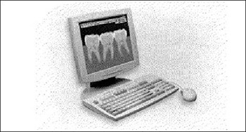
Different imaging techniques are used to pick up the signal of interest in digital sensors, including charge-coupled devices (CCD), complementary metal-oxide semiconductors (CMOS), photostimulable phosphors plates (PSP) and tuned-aperture computed tomography (TACT) Digital radiography sensors are divided into: storage phosphor plates (SPP) called photostimulable phosphor plates (PSP), silicon devices such as charge-coupled devices (CCD) or complementary metal oxide semiconductors (CMOS). Relatively new type of imaging that may hold advantage over current radiographic modalities is tuned-aperture computed tomography (TACT).

Downloads

Download data is not yet available.
Different digital imaging techniques in dental practice

Downloads

Published

2004-05-20

How to Cite

1.
Lačević A, Vranić E. Different digital imaging techniques in dental practice. Biomol Biomed [Internet]. 2004May20 [cited 2023Sep.22];4(2):37-40. Available from: https://www.bjbms.org/ojs/index.php/bjbms/article/view/3412

Issue

Section

Reviews

INTRODUCTION

Direct digital imaging was first presented in 1984 by Dr Frances Moujones. Since then, digital radiography, as a new technology in dental imaging practice, has been sue-cessfully advanced for almost last twenty years.

Different imaging techniques were used to pick up the signal of interest in digital sensors, including charge-coupled devices (CCD), complementary metal-oxide semiconductors (CMOS), photostimulable phosphors (PSP) and tuned-aperture computed tomography (TACT) (1,2). Digital radiography sensors are divided into: storage phosphor plates (SPP) called photostimulable phosphor plates (PSP), silicon devices such as charge-coupled devices (CCD) or complementary metal oxide semiconductors (CMOS).

Relatively new type of imaging that may hold advantage over current radiographic modalities, is tuned-aperture computed tomography (TACT)

There are two types of digital sensor array designs: area and linear. Area arrays are used for intraoral radiography, while linear arrays are used in extraoral imaging. Area arrays are available in sizes comparable to size 0, size 1, and size 2 film. The sensors are rigid and thicker than radiographic film and have smaller sensitive area for image capture. Area array CCDs have two primary formats: fiberoptically coupled sensors and direct sensors. Fiberoptically coupled sensors utilize scintillation screen coupled with CCD. X-rays interact with the screen material, light photons are generated, detected, and stored by CCD. Direct sensor CCD arrays capture the image directly

Figure 1: Digital radiography
Figure 2: Direct CCD sensor

Further development in direct digital sensor technology is introduction of complementary metal oxide semiconductor. CMOS sensors are less expensive to produce, use an active pixel technology and have low power requirements. CMOS sensors have more fixed pattern noise and a smaller active area of image acquisition.

Three basic types of operations are improved by digital imaging processing: analysis - that produce numeric information based on acquired image, enhancement - that subjectively, or objectively modify the appearance or qualities of the image and encoding - that code the image into a new format. The most common analysis operation is histogram. The image histogram is a graphic representation of the number of pixels with a specific gray value.

Brightness, contrast, and dynamic range data can readily be obtained from this analysis. This is the starting point for determining appropriate enhancement operations that will produce the desired result. Density analysis is the determination of the intensity of gray value at a specific point in the image. Dimensional analysis such as length, width, angle, area or perimeter are facilitated by digital imaging.

The most common used enhancement operations are: contrast manipulations, spatial filtering, subtraction, and pseudo-color.

Digital image subtraction reduces the structured noise of normal anatomic detail and therefore increases signal to noise ratio. Increasing signal to noise ratio makes the pathology more evident to human observer. Digital image subtraction has been applied to almost every disease in dental hard tissues. With this application it is possible to monitor, for example, the healing process of an apical radioluscens, marginal bone retraction or progress of caries decay. A prerequisite for digital subtraction is that projections are identical at different examinations, proper alignment of the two images, which is referred to as registration, and the ability to correct variations in exposure and processing that may obscure the changes in radiographic density associated with the pathology. These prerequisites limit the clinical application of the techniques.

Image coding permits either faster transmission of image data, or better utilization of a storage device. There are two basic types of encoding schemes. Those types where no information is lost are called lossless algorithms, while types where information is lost are called lossy algorithms.

Tuned-aperture computed tomography

The TACT presents a new method for creating three-dimensional radiographic displays. This system uses digital radiographic images. Software collates individual images of a subject and forms layering of images that can be viewed as slices. The result is reconstructed image, made from a series of eight digital radiographs that are assimilated into one. Preliminary studies show that this system may have advantages over conventional film in the visualization of root canals (3). It also proved to be an effective diagnostic tool for evaluation of dental caries and simulated osseous defects (4,5). This system consists of a standard radiographic unit, digital image acquisition device, and necessary software for reconstruction of the acquired images.

The TACT and digital subtraction radiography as more sensitive techniques might be recommended in cases of early bone changes detection, as important diagnostic procedure. The clinical application of these techniques is still explored.

DIGITAL RADIOGRAPHY IN DENTAL PRACTICE- RESEARCH FINDINGS

An imaging system that would allow better visualization of caries decay, marginal bone retraction, fine files at the apex, and enhance early detection of periapical lesions involving lamina dura, cancellous and cortical bone lesions, would be clinically desirable.

Digital radiography may offer some advantages over film radiography conventionaly used in diagnostics and monitoring of healing processes.

Advantages and Disadvantages

Advantages include 50-70% less radiation exposure to the patient, reduction in time between exposure and image generation, ability to manipulate and produce clear diagnostic image, elimination of chemical processing of radiographs, as well as ability to electronically store patient records.

Disadvantages include size, shape, thickness and rigidity of the sensor, lower image resolution, greater initial cost, unknown life expectancy of the sensor, inability to control possible infections present in direct digital imaging (6,7). CCD sensors cannot be sterilized. Direct saliva contact with the receptor and electrical cable must be avoided to prevent cross-contamination (8).

During CCD procedures, patient discomfort may result in greater number of retakes (9). Versteeg et al. demonstrated substantial horizontal placement errors, especially in molar areas, and vertical angulation errors, in the anterior regions where the incisal edges were cut off and not viewable. 28% CCD images were unacceptable and required retakes compared to 6% for films (9).

Direct digital image is the original image captured in a digital format, made of picture elements, called pixels. On the other hand, indirect digital imaging implies that an image is taken in analog format and then converted into digital one. This analog to digital conversion results in a loss and alteration of information. The original indirect digital imaging technique meant optical scanning of a conventional film image (analog) and generation of a digital image. This technique required an optical scanner for processing of transparent images, and software for production of the digital images. Later, more sophisticated conversion techniques were developed. Photostimulable phosphor radiographic systems were first introduced in 1981 by Fuji Corporation, in Tokyo (10). The image is captured on a phosphor plate as analog information, and then converted into digital format when the plate is processed. The PSP consist of polyester base coated with crystalline halide emulsion that converts X-radiation into stored energy. The crystalline emulsion is made of europium-activated barium flourohalide compound. The energy stored in these crystals is released in the form of blue fluorescent light when PSP is scanned with helium-neon laser beam. The emitted light is captured and intensified by a photomultiplier tube and then converted into digital data. PSP images have limited resolution of approximately 6 Ip/mm (line pairs per millimeter). This resolution is significantly smaller than can be achieved with conventional film (Č20 Ip/mm). The receptor of PSP is approximately the same in size as conventional film, somewhat flexible and easy for placement. PSP is used for intraoral and extraoral imaging techniques. (11)

In endodontics, researchers have examined the effects of enhancement on periapical lesion detection and application of measurment algoritms for dimensional assessment. Digital radiography research investigation did not reach consensus on the volume and type of bone loss that must be present for bony lesions to be detected. Some have concluded that lesions can be detected only if perforation or erosion of the bone cortex is present.(12,13) Other researchers reported that cancellous lesions or lesions that involved lamina dura were evident (14). Yokota at al. (15) found no difference between films and digital images of lesions that involved cortical bone. Kullendorff et al. (16) found no differences in diagnostic accuracy between conventional film and digitally acquired images. Wallace at al. (17) found that sensitivity and specificity calculations reveal that film had higher values, followed by digital images, both with high specificity and low sensitivity values. Film had the highest PSR score, followed by PSP-and CCD-based images in that order.

Use of statistical programs such as Receiver Operating Characteristic (ROC) curve analysis gives support for the visualization of bony lesions.

Enhancement of digital images through use of histogram equalization or contrast has proven to be valuable for the detection of periapical lesions in low density images (18). Others have shown that although contrast and brightness adjustments produce preferred images, image processing does not improve diagnostic accuracy (19, 20).

Recently, color-coding has been proposed for detecting differences between sequential images, in periodontics, by means of image addition to detect marginal bone changes. Color image displays may be superior to achromatic or monochromatic display as they provide perceptual dimension that enhances observer information processing and heightens the ability to interpret different types of data present in a particular image (21). William at al. (22) found that color-coded image processing applied to digital images had limited value in the estimation of periradicular lesional dimensions.

CONCLUSION

Using solid state sensors, Norwegian dental practitioners found preparation and placement of the sensors significantly more difficult than films (23).

They reported that technical problems and repairs were common. However, practitioners also reported that processing, viewing, and archiving were easier than for filmbased systems (23).

Sommers et al. found greater number of technical errors and unsatisfactory images in CCD imaging when compared to film (24).

The errors in periapical CCD imaging were vertical angulation and cone cutting, while errors in periapical film were placement and horizontal angulation (24). CMOS have more fixed pattern noise and smaller active area for image acquisition (10).

More sensitive techniques, such as TACT and digital subtraction radiography, might be recommended in cases of early bone changes detection. Clinical application of these techniques is still explored.

Generally, the findings are consistent and demonstrate that film and digital imaging modalities are not different in their ability to record dental disease conditions (1,10,17).

References

  1. , , , , (). Comparison of diagnostic accuracy of digital imaging by using CCD and CMOS-APS sensors with Е-speed film in detection of periapical bony lesion. Oral Surgery Oral Med Oral Path Oral Radiol Endod.
  2. , (). Endodontic measurments in digital radiographs acquired by a photostimulable storage phosphor system. Endod Dent Traumatol.
  3. , , , (). Idenjpgication of root canals in molars by tuned-aperture computed tomography. Int Endod J.
  4. , , , (). Tuned aperture computer tomography and detection of recurrent caries. Dentomaxillofac Radiol.
  5. , , , , (). A comparison of film, direct digital, and tuned aperture computed tomography images to idenjpgy the location of crystal defects around endosseus implants. Oral Surgery Oral Med Oral Pathol Oral Radiol Endod.
  6. , , , (). Computed dental radiography: evaluation of a new charge-coupled-devices-based intraoral radiographic system. Qintessence International.
  7. (). Imaging using solid-state detectors. Dent ClinN Am.
  8. , (). Digital Radiography: An Overview. J Contemp Dent Pract.
  9. , , (). An evaluation of periapical radiography with a charge couple devices. Dentomaxillofac Radiol.
  10. , , (). Image plate systems differ in physical performance. Oral Surg Oral Med Oral Pathol Oral Radiol Endod.
  11. , , (). Dental photostimulable phosphor radiography. Dent. Clin. North. Am.
  12. , (). Roentgenophic and direct observation of experimental lesion in bone. Int. J. Am. Dent. Assoc.
  13. (). Factor influencing the radiographic appearance of bony lesions. J. Endod.
  14. , (). Radiographic appearance of arjpgicially prepared periapical lesions confined to cancellous bone. Int. Endod J.
  15. , , , (). Interpretation of periapical lesions using radiovisiography. J. Endod.
  16. , , (). Diagnostic accuracy of direct digital dental radiography for detection of periapi-cal bone lesion: overall comparison between conventional and direct digital radiography. Oral Surg Oral Med Oral Pathol Oral Radiol Endod.
  17. , , , (). A comparative evaluation of the diagnostic efficacy of film and sensors for detection of simulated periapical lesion. Oral Surg Oral Med Oral Pathol Oral Radiol Endod.
  18. (). Effect of image enhancement for detectability of bone lesions in digitized intraoral radiographs. Scand. J. Dent. Res.
  19. , (). Diagnostic accuracy of direct digital dental radiography for the detection of periapical bone lesions, П: effects on diagnostic accuracy after application of image processing. Oral Surg Oral Med Oral Pathol Oral Radiol Endod.
  20. , , , (). Effects of calibration and automatic greyscale adjustment on detectability of simulated bone lesions using a storage phosphor system. Dentomaxillofac Radiol.
  21. (). . Psychovisual issues in the display of medical images.Proceedings of NATO: Advanced studies Institute on Pictorial Information Systems in Medicine.
  22. , , , , (). In vivo accuracy and reliability of color-coded image enhancement for the assessment of periradicular lesion dimensions. Oral Surg Oral Med Oral Pathol Oral Radiol Endod.
  23. , (). Experience of Norwegian general dental practitioners with solid state and storage phosphor detectors. Dentomaxillofac. Radiol.
  24. , , (). Pre-clinical performance comparing film and CCD-based systems. J. Dent. Hyg.