p16/Ki-67 dual staining has a better accuracy than human papillomavirus (HPV) testing in women with abnormal cytology under 30 years old

Authors

  • Laurențiu Pirtea Department of Obstetrics and Gynecology, University of Medicine and Pharmacy “Victor Babeş” Timişoara, Timișoara, Romania https://orcid.org/0000-0002-4398-2492
  • Cristina Secosan Department of Obstetrics and Gynecology, University of Medicine and Pharmacy “Victor Babeş” Timişoara, Timișoara, Romania https://orcid.org/0000-0003-1355-3790
  • Madalin Margan Department of Obstetrics and Gynecology, University of Medicine and Pharmacy “Victor Babeş” Timişoara, Timișoara, Romania https://orcid.org/0000-0003-2375-2793
  • Lavinia Moleriu Department of Informatics and Statistics, University of Medicine and Pharmacy “Victor Babeş” Timişoara, Timișoara, Romania
  • Oana Balint Department of Obstetrics and Gynecology, University of Medicine and Pharmacy “Victor Babeş” Timişoara, Timișoara, Romania https://orcid.org/0000-0003-4218-4189
  • Dorin Grigoras Department of Obstetrics and Gynecology, University of Medicine and Pharmacy “Victor Babeş” Timişoara, Timișoara, Romania
  • Ioan Sas Department of Obstetrics and Gynecology, University of Medicine and Pharmacy “Victor Babeş” Timişoara, Timișoara, Romania
  • Florin Horhat Department of Microbiology, University of Medicine and Pharmacy “Victor Babeş” Timişoara, Timișoara, Romania https://orcid.org/0000-0001-6133-0204
  • Adelina Jianu Department of Anatomy, University of Medicine and Pharmacy “Victor Babeş” Timişoara, Timișoara, Romania https://orcid.org/0000-0002-4238-7253
  • Răzvan Ilina Department of Surgery, University of Medicine and Pharmacy “Victor Babeş” Timişoara, Timișoara, Romania

DOI:

https://doi.org/10.17305/bjbms.2018.3560

Keywords:

Human papillomavirus, immunocytochemistry, p16/Ki-67 dual staining, HPV genotyping

Abstract

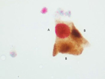
Due to a high rate of transient human papillomavirus (HPV) infection, HPV genotyping has a low specificity for high-grade cervical lesions, especially in young women. p16/Ki-67 dual immunocytochemical staining can also be used for the detection of oncogenic changes in cervical cells. Our aim was to compare the performance of p16/Ki-67 dual staining and HPV genotyping in the detection of high-grade cervical lesions in patients with atypical squamous cells of undetermined significance (ASCUS)/low-grade squamous intraepithelial lesion (LSIL) on Pap smear. We retrospectively analyzed 310 patients with ASCUS/LSIL on Pap smear, who underwent colposcopy. Among these, 161 patients with suspected lesions detected by colposcopy were referred for biopsy. HPV genotyping by LINEAR ARRAY HPV Genotyping Test (CE-IVD) and p16/Ki-67 dual staining by CINtec PLUS Cytology kit was performed prior to cervical biopsy. The overall sensitivity and specificity of HPV genotyping for the detection of cervical intraepithelial neoplasia (CIN) 2-3 was 79% and 72%, respectively in patients with ASCUS, and 85% and 64%, respectively in patients with LSIL. For p16/Ki-67 test, sensitivity and specificity rate was 66% and 93%, respectively in ASCUS and 59% and 79%, respectively in LSIL group. The specificity of p16/Ki-67 staining was significantly higher in both groups in patients aged <30 years compared to patients >30 years old (p < 0.001). Our results showed that p16/Ki-67 dual staining has a higher specificity compared to HPV genotyping, especially in patients under 30 years old. This indicates the usefulness of p16/Ki-67 testing in the triage of patients with ASCUS/LSIL and <30 years old, prior to referral for colposcopy and biopsy.

Downloads

Download data is not yet available.
p16/Ki-67 dual staining has a better accuracy than human papillomavirus (HPV) testing in women under 30 years old

Downloads

Additional Files

Published

2019-11-08

How to Cite

1.
Pirtea L, Secosan C, Margan M, Moleriu L, Balint O, Grigoras D, Sas I, Horhat F, Jianu A, Ilina R. p16/Ki-67 dual staining has a better accuracy than human papillomavirus (HPV) testing in women with abnormal cytology under 30 years old. Biomol Biomed [Internet]. 2019Nov.8 [cited 2023Sep.25];19(4):336-41. Available from: https://www.bjbms.org/ojs/index.php/bjbms/article/view/3560

Issue

Section

Pathology

INTRODUCTION

Cervical cancer represents one of the most common gynecological malignancies. Screening programs for cervical cancer are available in a majority of countries, and cervical cytology (Papanicolaou [Pap] smear) is the most frequently used screening tool. Still, it is estimated that almost 30% of patients with cervical cancer had at least one previous false-negative Pap smear, indicating that more accurate screening tests are required [1]. The important role of persistent human papillomavirus (HPV) infection in the etiopathogenesis of cervical cancer has drawn attention to HPV genotyping as a primary screening tool. However, because of a high incidence rate of transient HPV infections, HPV genotyping has a low specificity for high-grade cervical lesions, especially in young women. According to the American College of Obstetricians and Gynecologists (ACOG) guidelines, HPV testing is not currently recommended as a screening test for cervical cancer in women younger than 30 years [2].

p16/Ki-67 dual immunocytochemical staining (p16/Ki-67 dual-stained cytology) can be used for the detection of precancerous/cancerous cervical lesions. p16 or p16-INK4A is a tumor-suppressor involved in the regulation of cell cycle by inhibiting the S phase, and is expressed in low concentrations in normal cells. In high-risk (HR)-HPV-infected cervical cells, HPV E7 oncoprotein targets the p16/Rb pathway leading to the overexpression of p16. Under these conditions, p16 has oncogenic functions and is essential for cell survival. Ki-67 is a nuclear protein expressed in dividing cells, and is used as a cell proliferation marker. p16/Ki-67 dual staining is considered positive when the two proteins are co-expressed within the same cell, and can be performed with the same liquid-based cytology samples used for HPV genotyping and Pap smear.

The usefulness of p16/Ki-67 dual staining for the triage of patients with atypical squamous cells of undetermined significance (ASCUS) or low-grade squamous intraepithelial lesion (LSIL) prior to referral for colposcopy and biopsy has been evaluated in several studies. Bergeron et al. [3] and Possati-Resende et al. [4] compared the performance of p16/Ki-67 dual staining and HPV genotyping in the management of women with abnormal cervical cytology. They found that p16/Ki-67 dual staining had a higher specificity for high-grade cervical intraepithelial neoplasia (CIN2/CIN3) compared to HPV genotyping, in both ASCUS and LSIL groups [3,4]. Moreover, in patients with LSIL ≤30 years old, p16/Ki-67 dual staining had a higher accuracy in identifying precursor cervical lesions [4].

The objective of this study is to compare the performance of p16/Ki-67 dual staining and HPV genotyping in the detection of high-grade cervical lesions in patients with ASCUS or LSIL on cervical cytology (Pap smear).

MATERIALS AND METHODS

Patients

This retrospective study included patients with ASCUS or LSIL on cervical cytology who underwent colposcopy at the Department of Obstetrics and Gynecology of County Hospital Timişoara, between January 2015 and December 2016.

The inclusion criteria were ASCUS or LSIL on Pap smear and suspected lesions on colposcopy. The exclusion criterion was negative colposcopy.

Written informed consent was obtained from all patients before their inclusion in the study. All procedures were performed in accordance with the ethical standards laid down in the 1964 Declaration of Helsinki and its later amendments and were approved by the Institutional Review Board and Ethical Committee of the University of Medicine and Pharmacy “Victor Babeş” Timişoara.

The study was registered by the Institutional Review Board and Ethical Committee of “Victor Babeş” University of Medicine and Pharmacy, Timişoara under the registration number 17/2017.

Cervical cytology, colposcopy, and biopsy

All patients underwent Pap smear testing and colposcopy. Pap smear was performed and evaluated according to the 2001 Bethesda System for reporting cervical cytology. Colposcopy examinations were performed by the same team, with expertise in colposcopy, in all patients, and results were interpreted according to the 2011 International Federation for Cervical Pathology and Colposcopy (IFCPC) terminology system. Only patients with suspected lesions detected by colposcopy were referred for biopsy. Loop electrosurgical excision procedures (LEEPs) were performed in all cases by the same team of surgeons and biopsy specimens were evaluated by the same pathologist. All patients referred for biopsy were selected for HPV genotyping and p16/Ki-67 dual staining. Both tests were performed before cervical biopsy (Figure 1).

FIGURE 1: (A)Immunocytochemical reaction for p16/Ki-67, magnification 40×. A cell with a double signal and impaired cell cycle regulation; red nuclei positive for Ki-67 and brown cytoplasm and/or nuclei positive for p16. (B) Cells positive for high-risk-human papillomavirus (HPV) in which the cell cycle is still under control.

HPV testing

HPV genotyping was performed by LINEAR ARRAY HPV Genotyping Test [CE-IVD] (ROCHE, Germany). The HPV test is based on the amplification of target DNA (HPV L1 gene) by multiplex polymerase chain reaction (PCR) and reversed hybridization of the amplified products to a linear array of 37 immobilized probes which represent different HPV types (6, 11, 16, 18, 26, 31, 33, 35, 39, 40, 42, 45, 51, 52, 53, 54, 55, 56, 58, 59, 61, 62, 64, 66, 67, 68, 69, 70, 71, 72, 73, 81, 82, 83, 84, IS39, and CP6108). PCR was performed on a GeneAmp PCR System 9700 thermal cycler (Applied Biosystems, USA), according to the manufacturer’s instructions. Automated hybridization and detection of HPV-DNA were performed on a ProfiBlot 48 Western Blot processor (Tecan Trading AG, Zurich, Switzerland).

p16/Ki-67 dual staining

Immunocytochemistry analysis was performed using the CINtec PLUS Cytology kit (Roche MTM Laboratories, Heidelberg, Germany), according to the manufacturer’s instructions.

Statistical analysis

SPSS Statistics for Windows, Version 17.0. (SPSS Inc., Chicago), Epi Info v7 (Centers for Disease Control and Prevention [CDC], Atlanta, GA, USA), and Microsoft Excel 2007 were used for statistical analysis. All p values were obtained by applying a chi-square test for proportions between CinTest and HPV genotyping, and are presented in Table 1. Furthermore, we calculated the area under the receiver operating characteristic (ROC) curve which is a popular measure for the accuracy of a diagnostic test, where the value of 1 implies a perfect diagnostic test (Table 2 and Figure 2) [5,6].

TABLE 1: P values obtained by applying the Chi-square test for proportion between CINtec test and HPV genotyping
TABLE 2: The area under the ROC curve for HPV genotyping and p16/Ki-67 dual staining (CINtec test) specificity in ASCUS and LSIL patients, according to the age groups and in the total sample
FIGURE 2: ROC curve for specificity and sensitivity of CINtec test and HPV genotyping for: (A) patients with ASCUS under 30 years old, (B) patients with ASCUS over 30 years old, (C) all the patients with ASCUS regardless of age, (D) patients with LSIL under 30 years old, (E) patients with LSIL over 30 years old, (F) all the patients with LSIL regardless of age. The specificity of p16/Ki-67 dual staining (CINtec test) was significantly increased (p < 0.001) in the group of patients under 30 years old compared with patients over 30 years of age (p < 0.001) in both ASCUS and LSIL group. ROC: Receiver operating characteristic, ASCUS: Atypical squamous cells of undetermined significance, LSIL: Low-grade squamous intraepithelial lesion.

RESULTS

A total of 310 patients with ASCUS or LSIL on cervical cytology were referred for colposcopy at the Department of Obstetrics and Gynecology of County Hospital Timişoara. Patients with colposcopy negative for high-grade lesions were excluded from the study. The remaining 161 patients with ASCUS (67 patients; 42%) or LSIL (94 patients; 58%) were referred for biopsy.

Among 161 patients, 56 (35%) were <30 years and 105 patients (65%) were >30 years old.

Overall, 102/161 patients (63%) tested positive for HR-HPV and 70/161 patients (43%) were positive for p16/Ki-67. CIN2-3 was detected by biopsy in 99/161 patients (61%).

In ASCUS group, 38/67 patients (57%) were positive for HR-HPV, and 27/67 (40%) were positive for p16/Ki-67 test. In LSIL group, 64/94 patients (68%) were HR-HPV-positive and 43/94 (46%) were p16/Ki-67-positive.

In women over 30 years old, in ASCUS group, HR-HPV positivity rate was 63% (27/43 patients) and p16/Ki-67 positivity was 42% (18/43 patients). In LSIL patients over 30 years, HR-HPV positivity rate was 77% (48/62 patients) and p16/Ki-67 positivity was 37% (23/62 patients).

In women less than 30 years old, in ASCUS group, HR-HPV positivity rate was 45% (11/24 patients) and p16/Ki-67 positivity was 37% (9/24 patients). In LSIL group less than 30 years, the positivity rate for HR-HPV test was 50% (16/32 patients) and for p16/Ki-67 dual staining it was 62% (20/32 patients).

In women with CIN2-3 detected by biopsy and <30 years old HR-HPV positivity was 50% (7/14 patients) in ASCUS and 83% (15/18 patients) in LSIL group, p16/Ki-67 positivity rate was 57% (8/14) and 88% (16/18 patients) in the two groups respectively.

The overall sensitivity and specificity of HPV genotyping for the detection of CIN2-3 were 79% and 72%, respectively in the group of patients with ASCUS, and 85% and 64%, respectively in the group of patients with LSIL. The sensitivity and specificity rates of p16/Ki-67 dual staining for CIN2-3, were 66% and 93%, respectively in ASCUS group, and 59% and 79%, respectively in LSIL group (Table 3).

TABLE 3: The sensitivity and specificity values of HPV genotyping and p16/Ki-67 dual staining, according to the age groups and in the total sample

The specificity of p16/Ki-67 dual staining was significantly increased in the group of patients <30 years old compared to patients >30 years of age (p < 0.001) in both ASCUS and LSIL groups (Figure 2).

DISCUSSION

Persistent infection with HR-HPV is essential for the development of cervical cancer. Factors affecting HPV persistence include HPV type, patient age, and immune status [7,8]. This is the main reason why HPV genotyping has a high sensitivity for the detection of cervical disease, and according to recent studies, HPV testing is superior for the detection of high-grade cervical lesions compared to cervical cytology [9]. However, due to a high prevalence of transient HPV infections, especially in young women, the specificity of HPV genotyping as a screening method for cervical cancer is limited.

According to the clinical algorithms available in most countries, patients categorized as having ASCUS or LSIL on cytology are referred for further testing, such as colposcopy and biopsy, to exclude CIN2 or CIN3. However, many studies found that 70–90% of LSIL cases regress to normal spontaneously, especially in women under 30 years old [10]. This means that a large number of patients are referred for unnecessary colposcopy and possibly invasive diagnostic methods such as biopsy or large loop excision of the transformation zone (LLETZ) [11]. Furthermore, this suggests the need for a better method for triaging ASCUS and LSIL patients, where immunocytochemical methods could be used as an additional tool for the assessment of disease severity. Immunohistochemistry has already been used for ultrastaging of sentinel lymph node in cervical cancer [12] or for the assessment of other gynecological malignancies, such as ovarian and endometrial cancer [13-15].

p16 has been found to be overexpressed in CIN as a result of the inactivation of retinoblastoma protein (Rb), which is mediated by the action of HPV E7 oncoprotein [16]. This is considered to be a critical early event in cervical carcinogenesis. On the other hand, p16 can also be overexpressed in normal metaplastic or atrophic cells, so the specificity of p16 alone in the detection of cervical neoplasia is limited. Ki-67 represents a proliferation-associated protein which can be detected only in the nucleus of proliferating cells. The co-expression of the tumor-suppressor protein p16 and the proliferation marker Ki-67 does not occur in a normal healthy cell and, therefore, indicates altered cell cycle control [17]. Furthermore, Wentzensen et al. [18] found a strong correlation between p16/Ki-67 expression and the presence of HPV type 16, which is the most oncogenic HPV type and with the highest rate of persistence [7,8,18].

Due to the higher sensitivity for cervical neoplasia, HPV testing is considered more suitable for primary cervical screening, while p16/Ki-67 dual-staining has been evaluated as an alternative tool for the triage of patients with ASCUS or LSIL prior to referral for colposcopy and biopsy. Bergeron et al. [3] performed a prospective multicenter study, including 1,100 patients with ASCUS or LSIL, aiming to evaluate the performance of p16/Ki-67 dual-stained cytology and HPV genotyping in the management of women with abnormal cervical cytology. They found that p16/Ki-67 dual-stained cytology showed comparable (ASCUS: 94.4% for dual-stained cytology vs. 100% for HPV genotyping) or lower (LSIL: 85.7% for dual-stained cytology vs. 98.4% for HPV genotyping) sensitivity for CIN2-3, but higher specificity compared with HPV genotyping, in both ASCUS (78.7% vs. 60.4%; p < 0.001) and LSIL (53.3% vs. 15.6%; p < 0.001) group. Positive predictive values for CIN2-3 were substantially higher for dual-stained cytology versus Hybrid Capture 2 (HC2) HPV testing, especially in ASCUS and LSIL patients aged under 30 years [3].

Similar findings were reported by Possati-Resende et al. [4], who compared the performance of dual-stained cytology and HPV genotyping in a group of 200 patients with ASCUS or LSIL. They found that both p16/Ki-67 dual staining and the HR-HPV DNA test had a similar performance in predicting high-grade cervical lesions. They also found that in patients with LSIL under 30 years old, p16/Ki-67 test had a higher accuracy in identifying cervical lesions. Among women over 30 years old diagnosed with LSIL, the two methods showed a similar performance [4]. We obtained comparable results in this study, with the dual-stained cytology performing significantly better in terms of specificity in the group of patients under 30 years of age.

The fact that the same liquid-based sample can be used for cervical cytology, HPV genotyping, and immunocytochemistry simplifies the procedure, so that the patient is not obliged to pay another visit to the specialist to perform the test. This means that p16/Ki-67 dual staining could be offered as a “reflex test” for patients with abnormal results on liquid-based cervical cytology or with HR-HPV positive results.

The weakness of our study is the relatively small number of patients involved. More data are necessary to validate the use of p16/Ki-67 dual staining as part of a management algorithm for patients with abnormal Pap smear results.

CONCLUSION

In our study, p16/Ki-67 dual staining showed a higher specificity for CIN2-3 compared to the HPV genotyping test, especially in the group of patients under 30 years of age. Generally, HPV genotyping has a good sensitivity but poor specificity for high-grade cervical lesions in women under 30 years old. Our data indicate that p16/Ki-67 dual staining might be an option in the triage of patients younger than 30 years with ASCUS or LSIL on cytology test, prior to performing colposcopy and biopsy. Therefore, the major benefit of using p16/Ki-67 dual staining in patients with ASCUS or LSIL on Pap smear, and especially in the group less than 30 years old, would be a decreased number of unnecessary colposcopies and biopsies.

Acknowledgements

ACKNOWLEDGMENTS

This paper was supported by Grant PIV-CI-PDCC-02 2015/2016, University of Medicine and Pharmacy “Victor Babes”, Timisoara, Romania.

DECLARATION OF INTERESTS

The authors declare no conflict of interests.

REFERENCES

  1. , (). New technologies in cervical cytology screening:A word of caution. Obstet Gynecol. https://doi.org/10.1097/00006250-199908000-00029;|https://doi.org / 10.1016 /S0029 - 7844 (99) 00289- 6
  2. , , , , , (). American Cancer Society, American Society for Colposcopy and Cervical Pathology, and American Society for Clinical Pathology screening guidelines for the prevention and early detection of cervical cancer. Am J Clin Pathol. https://doi.org/10.1309/AJCPTGD94EVRSJCG
  3. , , , , , (). Prospective evaluation of p16/Ki-67 adual-stained cytology for managing women with abnormal Papanicolaou cytology:PALMS study results. Cancer Cytopathol. https://doi.org/10.1002/cncy.21542
  4. , , , , , (). The accuracy of p16/Ki-67 and HPV test in the detection of CIN2/3 in women diagnosed with ASC-US or LSIL. PLoS One. https://doi.org/10.1371/journal.pone.0134445
  5. , , , , , (). Insights into the mechanisms of thymus involution and regeneration by modeling the glucocorticoid-induced perturbation of thymocyte populations dynamics. J Theor Biol. https://doi.org/10.1016/j.jtbi.2014.01.020
  6. (). Determination of cutoff score for a diagnostic test. Internet J Laboratory Med.
  7. , , , , , (). Age and HPV type as risk factors for HPV persistence after loop excision in patients with high grade cervical lesions:An observational study. BMC Surg. https://doi.org/10.1186/s12893-016-0185-7
  8. , , , , , (). Human papilloma virus persistence after cone excision in women with cervical high grade squamous intraepithelial lesion:A Prospective study. Can J Infect Dis Med Microbiol 2016. 3076380https://doi.org/10.1155/2016/3076380
  9. , , , , , (). 2012 aupdated consensus guidelines for the management of abnormal cervical cancer screening tests and cancer precursors. Obstet Gynecol. https://doi.org/10.1097/AOG.0b013e3182883a34
  10. , , , (). Natural history of established low grade cervical intraepithelial (CIN 1) lesions. Anticancer Res.
  11. , , , , , (). HPV16 genotype, p16/Ki-67 dual staining and koilocytic morphology as potential predictors of the clinical outcome for cervical low-grade squamous intraepithelial lesions. Cytopathology. https://doi.org/10.1111/cyt.12121
  12. , , , (). Sentinel lymph node biopsy in uterine cervical cancer patients:Ready for clinical use?A review of the literature. ISRN Surg 2014. 841618https://doi.org/10.1155/2014/841618
  13. , , , , (). The immunohistochemical expression of endocrine gland-derived-VEGF (EG-VEGF) as a prognostic marker in ovarian cancer. Rom J Morphol Embryol.
  14. , , (). Endothelial progenitor cells contribute to the development of ovarian carcinoma tumor blood vessels. Oncol Lett. https://doi.org/10.3892/ol.2014.1917
  15. , , (). Endothelial cell activation and proliferation in ovarian tumors:Two distinct steps as potential markers for antiangiogenic therapy response. Mol Med Rep. https://doi.org/10.3892/mmr.2012.812
  16. , , , , , (). Overexpression of p16(INK4A) as a specific marker for dysplastic and neoplastic epithelial cells of the cervix uteri. Int J Cancer. https://doi.org/10.1002/ijc.1174
  17. , , , , , (). Interpretation of p16(INK4a)/Ki-67 dual immunostaining for the triage of human papillomavirus-positive women by experts and nonexperts in cervical cytology. Cancer Cytopathol. https://doi.org/10.1002/cncy.21511
  18. , , , , , (). Performance of p16/Ki-67 immunostaining to detect cervical cancer precursors in a colposcopy referral population. Clin Cancer Res. https://doi.org/10.1158/1078 0432.CCR-12-0270