Isolation and immunochemical characterization of human cystatin C

Authors

  • Lejla Begić Department of Biochemistry, Faculty of Medicine, University of Tuzla
  • Selma Berbić Department of Biochemistry, Faculty of Medicine, University of Tuzla

DOI:

https://doi.org/10.17305/bjbms.2002.3576

Keywords:

human cystatin C, cystatin C isoforms, ELISA test, chronic renal failure

Abstract

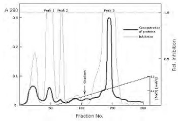
Cystatin C is a natural inhibitor of the cysteine proteinases papain, and mammalian lysosomal cathepsins B, H, L and S. This protein is thought to serve an important physiological role as a local regulator of enzyme activity. The changes of levels of cystatin C in extracellular fluids have shown themselves having potential clinical importance. We have purified cystatin C from urine of patients with chronic renal failure by procedure using affinity chromatography on CM-papain Sepharose, gel filtration on Sephacryl S-200, and ion exchange chromatography on CM-cellulose. After isolation we obtained three inhibitory peaks (pI's from 7.8 to 9.2) which represent isoforms of the same protein. These isoforms are immunologically identical and differ in N-terminal sequence of the molecule. The form with pI 9.2 represents the intact inhibitor form, whereas the form with pI 7.8 is shortened for 8 amino-acid residues at N-terminal end. Purified cystatin C pI 9.2 was used for immunization of rabbits. Polyclonal antibodies, produced in rabbits, were isolated from rabbit sera by affinity chromatography on Protein A Sepharose. Enzyme immunoassay (ELISA) for cystatin C is developed on the basis of purified antibodies. Using ELISA test we determined amount of cystatin C in urine and serum samples of patients with chronic renal failure. The concentration of the inhibitor in the urine of these patients was approximately 100-fold more than in normal urine. In the serum from the same patients we found concentrations of cystatin C to be five times higher in comparison with the serum of healthy individuals.

Downloads

Download data is not yet available.
Isolation and immunochemical characterization of human cystatin C

Published

2002-02-20

How to Cite

1.
Begić L, Berbić S. Isolation and immunochemical characterization of human cystatin C. Biomol Biomed [Internet]. 2002Feb.20 [cited 2023Jun.6];2(1-2):12-5. Available from: https://www.bjbms.org/ojs/index.php/bjbms/article/view/3576

Issue

Section

Biochemistry

Introduction

Lysosomal cysteine proteinases, the cathepsins, belong to the papain family of proteinases. Under normal physiological conditions proteinases participate in a variety of processes at tissue; they mediate intracellular and extracellular protein turnover, regulating the lifetime of proteins and other molecules critical for normal cell function. Beside protein turnover, proteinases are involved in specific processing step for smaller peptides, proenzymes and prohormones (1). The activation of cysteine proteinase requires acidic pH and may occur autocatalytically or by other lysosomal cathepsins, such as cathepsin D, in lysosomal vesicles, localized close to the plasma membrane (2).

Cysteine proteinases are regulated by endogenous cysteine proteinase inhibitors (CPIs), cystatins (3). Cystatin superfamily comprises families of closely homologous CPIs, stefins (family I), cystatins (family II), kininogens (family III) and non-inhibitory proteins (family IV) such as human histidine-rich glycoprotein and α2 HS glyco-protein (4).

Cystatins (family II) are single chain proteins stabilized by two disulfide bridges, with molecular weight of about 13.000. A presence of hydrophobic signal peptides at amino-terminal end of the cystatin molecule, which was determined by molecular cloning, points out to extracellular function of that protein (5). Human cystatin C is abundant in various tissues and body fluids. The highest levels have been determined in cerebrospinal fluid, seminal plasma and synovial fluid. It is the most potent inhibitor of cysteine proteinases with apparent inhibition constant in picomolar range (6).

Changes of levels of cystatin C in extracellular fluids have good potential for clinical use. Its level in blood plasma has been shown to be a good indicator of glomerular filtration rate (GFR), considerably better than the widely used creatinine (7, 8). Decreased levels of cystatin C in cerebrospinal fluid are associated with hereditary cystatin C amyloid angiopathy (HCCAA), an inherited disease characterized by deposition of cystatin C variants as amyloid fibrils in blood vessels, resulting in death from cerebral haemorrhage (9). Cystatin C has been suggested to play a role in several other diseases associated with alteration of proteolytic system, such as cancer (10, 11), inflammatory lung diseases (12), peri-odontal disease (13), multiple sclerosis (14), autoimmune diseases (15), HIV infection (16), and renal failure (17). The aim of our work was purification and characterization of cystatin C from urine of patients with chronic renal failure, and determination of cystatin C levels in urine and serum samples of the same patients by employing of ELISA method.

Materials and methods

Materials

Urine from patients with chronic renal failure was collected 24 hours, and 1 mol/L NaCl was added as preservative. Papain, bovine serum albumin (BSA), Tween 20 and horse radish peroxidase were from Sigma, 2,2’-Azino-Di-(3-ethylbenzthiazoline sulphonic acid) (ABTS) from Boehringer Mannheim. Sephacryl S-200 Superfine, CNBr-activated Sepharose 4B, ampholines, and molecular weight standard proteins were from Pharmacia. CM-cellulose was purchased from Serva. S-carboxymethylated papain Sepharose 4B was prepared as described by Barrett (18).

Purification of the inhibitor

Urine was centrifuged at 3000xg for 15 minutes and the supernatant was applied directly to a CM-papain Sepharose 4B column. Binding of the inhibitor to the affinity column and subsequent washing were performed at pH 8.0 with Tris/HCl buffer containing 0.5 mol/L NaCl. The inhibitor was eluted with 0.01 mol/L NaOH concentrated to 40 mL using YM-5 membrane and applied to a Sephacryl S-200 column (4x100 cm), equilibrated and eluted with 0.02 mol/L acetate buffer pH 6.1. The second papain inhibiting peak was collected and further chromatographed on a column of CM-cellulose (3x15 cm) using the same buffer as in the gel filtration step. After elution with starting buffer, a linear gradient of NaCl (0-0.2 mol/L) in a total volume of 400 mL of starting buffer was applied. The inhibitory peaks were eluted and concentrated using a Diaflo YM-2 membrane.

Inhibitor assay

The inhibitory activity was determined using the conditions of papain activity assay on benzoil-DL-arginine-2-naphtylamide. Prior to the assay, a known amount of inhibitor was pre-incubated wit the enzyme for 5 min at room temperature before adding the substrate. One inhibitory unit (I.U.) is defined as the amount of inhibitor which completely inhibits 1 μg of the test papain.

ELISA test

Antibodies against cystatin C pI 9.2, isolated from human urine (17) were raised in rabbits. Microtitre plates were coated with anti-cystatin C —IgG and incubated over night at 4°C. The samples were applied in the wells and after 2 hours of incubation at 37°C the anti-cystatin C—IgG peroxidase conjugate was added. The indicator reaction was carried out applying the substrate solution (ABTS and H2O2). The measured enzyme activities residing in the individual wells were plotted against the logarithm of the concentration of the antigen.

Other methods

SDS-polyacrilamide gel electrophoresis was performed according to Weber and Osborne (19). Analytical gel focusing was carried out on 1mm thick 5% polyacrylamide gel plates with the ampholines of the pH 2-10 range following the manufacturer’s instruction (Pharmacia). Double radial immunodiffusion test was performed according Ouchterlony (20). Proteins were determined by the modified method of Lowry (21).

Results

The first step of cystatin C isolation was CM-papain Sepharose affinity chromatography at pH 8.0. Inhibitory fractions from the affinity resin were concentrated and chromatographed on Sephacryl S-200. Two inhibitory peaks were resolved (Fig.1). The first one contained high molecular weight cysteine proteinase inhibitors of the kininogen family. The second peak, which contained low molecular weight inhibitors and kininogen cleavage products, was further purified on a CM-cellulose column, where three papain inhibiting peaks were resolved. The bulk of the inhibitor capacity was eluted in the third peak (Fig.2). The total of 9 mg of inhibitor protein from this peak was obtained in several preparations from 7 litres of urine. The peaks from ion exchange column were analyzed by isoelectric focusing on polyacrylamide gels. The protein from peak 3 focused at pH 9.2, and those from the minor peaks 1 and 2 focused at pH 7.8 and 8.4, respectively (not shown). Although the three purified isoelectric forms were immunologically identical (Fig.3), they differ in molecular weights. The difference was confirmed by SDS-PAGE (Fig.4). Isoelectric variant 9.2 is the longest form in the urine of patients with chronic renal failure and has Mr of about 13.260, whereas the shorter pI 7.8 form of cystatin C has Mr of about 12.500. Using ELISA test we determined the levels occystatin C in the urine and serum of patient suffering from chronic renal diseases. The concentration of the inhibitor in the urine of these patients was found to be 9mg/L, which is approximately 100-fold more than in normal urine. In the serum from the same patients we found concentrations of cystatin C to be five times higher in comparison with the serum of healthy individuals.

Figure 1: Figure 1
Figure 2: Figure 2
Figure 3: Figure 3
Figure 4: Figure 4

Discussion

Using the described procedure we have isolated cystatin C in 3 electrophoretically pure forms. Two isoforms were obtained in an amount sufficient to carry out their characterization. The intact polypeptide chain, pI 9.2, consists of 120 amino acid residues. Cleavage of the N-terminal octapeptide from the pI 9.2 form results in the formation of the 7.8 variant. Swiss-Prot Database (22) claims that cystatin C precursor consists of 146 amino acid residues, where residues from1 to 26 constitute signal sequence. That corresponds with our results. The relatively high concentration of cystatin C in cerebrospinal fluid, compared to plasma, indicates the production of cystatin C in central nervous system (23). Researches of gene’s structure for cystatin C and its expression, have confirmed synthesis of that inhibitor in many tissues, but highest level of mRNA for cystatin C has been observed in chorioidal plexus of rat’s brain (24). The protein is probably able to cross the blood brain barrier to the vascular space, and rapidly filtered in the glomeruli. Nowadays, a great attention is paid to cystatin C as a possible indicator of renal function, e.g. in cases of transplantation of kidneys (25) or in diabetes (26). Cystatin C has been advocated as a better marker of glomerular filtration rate than serum creatinine because, unlike creatinine, it is not secreted by the renal tubule, is not affected by muscle mass, and does not suffer the same problems with analytical interference (27).

Our results reveal that quantification of cystatin C in serum and urine by ELISA test can be used to detect and follow pathophysiological processes during chronic renal disease. The accurate screening of subjects for impairment in renal function requires either laborious collection of timed urine samples and processing by ELISA (or some other equally complicated test, e.g. immunotur-bidimetry), or the intravenous injection of radioactive substances. Thus, the development of simple serum test for detecting mild to moderate reductions in glomerular filtration rate would represent a major advance that would be of relevance to many fields of medicine.

Gel filtration on Sephacryl S-200 Superfine of the inhibitory fraction obtained after affinity chromatography on CM-papain Sephrose. Protein concentration (blue) is expressed (left ordinate) as absorbance at 280 nm. Inhibitory activity (green) 100 μL of fractions is represented as relative inhibition (right ordinate).

Ion exchange chromatography on CM-cellulose of inhibitory fraction obtained in second peak after gel filtration. Concentration of protein (blue) is represented on the left ordinate as absorbance at 280 nm, and relative inhibition, determined with papain using BANA test (green) is represented on the right ordinate. Bound proteins were eluted with gradient NaCl (0-0.2 mol/L).

Double radial immunodiffusion test of purified cystatin C isoforms pI 7.8 (I), pI 8.6 (II) and pI 9.2 (III) against anti-cystatin C antiserum (AS).

SDS-PAGE of the purified cystatin C isoforms. From the left to the right: standards, cystatin C pI 9.2(I), cystatin C pI 8.6 (II), cystatin C pI 7.8 (III). The samples were pre-incubated for 30 minutes at 370C in 2% SDS containing 10% μ-mercaptoethanol. Phosphorylase b, bovine serum albumin, ovalbumin, carbonic anhydrase, soya-bean rypsin inhibitor and α-lactalbumin were used as molecular weight standards.

References

  1. , , , , , (). Cell and tissue distribution of lysosomal cysteine proteinases, cathepsins B, H and L, and their biological roles. Acta Histochem Cytochem.
  2. , , , (). Activation of cathepsin B, secreted by a colorectal cancer cell line requires low pH and is mediated by cathepsin D. Int J Cancer.
  3. , (). The cystatins:protein inhibitors of cysteine proteinases. FEBS Lett.
  4. , (). Evolution of proteins of the cystatin superfamily. J Mol Evol.
  5. , , (). Human salivary cystatin S. Cloning, sequence analysis, hybridization in situ and immunocytochemistry. Biochem J.
  6. , , , , (). Characterisation by rapid-kinetic and equilibrium methods of the reaction between N-terminally truncated forms of chicken cystatin and the cysteine proteinases papain and actinidin. Biochem J.
  7. , , , , , , (). Is serum cystatin C a sensitive marker of glomerular filtration rate (GFR)?A preliminary study on renal transplant patients. Ren Fail.
  8. , , , (). Clinical value of cystatin C determination. Clin Chim Acta.
  9. , , , , , (). Abnormal metabolism of γ-trace alkaline micro-protein. The basic defection in hereditary cerebral haemorrhage with amyloidosis. N Engl J Med.
  10. , , (). Lysosomal enzymes and their endogenous inhibitors in neoplasia. Biochemical and Molecular Aspects of Selected Cancers.
  11. , , (). Cystatins in health and disease. Biol Chem.
  12. , , , , , , (). Human sputum cathepsin B degrades proteoglycan, is inhibited by a2-macroglobulin and is modulated by neutrophil elastase cleavage of cathepsin B precursor and cystatin C. Biochem J.
  13. , , , , (). Immunochemical quantisation of cysteine proteinase inhibitor cystatin C of inflamed human gingiva. Arch Oral Biol.
  14. , (). Alias post g globulin:a marker for multiple sclerosis?. J Clin Chem Clin Biochem.
  15. , , , , , (). Cystatin C (post γglobulin) in serum of patients with autoimmune diseases. Cysteine Proteinases and their Inhibitors.
  16. , , , , , , , (). Cystatin C levels in sera of patients with human immunodeficiency virus infection. J Immunoassay.
  17. , , , , (). Human cystatin, a new protein inhibitor of cysteine proteinases. Biochem Biophys Res Commun.
  18. (). Cystatin, the egg white inhibitor of cysteine proteinases. Methods in Enzymol.
  19. , (). The reliability of molecular weight determination by dodecilsulphate polyacralamide gel electrophoresis. J Biol Chem.
  20. , (). Immunodiffusion and immunoelectrophoresis. Handbook of Experimental Immunology.
  21. , , , (). Protein measurement with Folin phenol reagent. J Biol Chem.
  22. , (). Quantisation of γ-trace in human biological fluids:indications for production in the central nervous system. Scand J Clin Lab Invest.
  23. , , , , , , (). The cDNA structure and expression analysis of the genes for the cysteine proteinase inhibitor cystatin C and for B2-microglobulin in rat brain. Eur J Biochem.
  24. , , , , , (). Cystatin C and creatinine after successful kidney transplantation in children. Clin Nephrol.
  25. , , , , , (). Evaluation of plasma cystatin C as a marker for glomerular filtration rate in patients with type 2 diabetes. Clin Nephrol.
  26. , , , (). Biological variation of cystatin C:implications for the assessment of glomerular filtration rate. Clin Chem.