Stereological Analysis of the Mammary Gland in Primiparous Lactating Rats during the Lead Acetate Intoxication

Authors

  • Zakira Mornjaković Institute of histology and embryology, Medical faculty, Sarajevo
  • Muhamed Kadić Institute of histology and embryology, Medical faculty, Sarajevo

DOI:

https://doi.org/10.17305/bjbms.1998.3605

Keywords:

Stereological Analysis, Mammary Gland, Acetate Intoxication

Abstract

The present study was undertaken to investigate the changes in composition of the mammary gland volume unit through all phases of lactation in rats subjected to lead acetate administration via drinking water during the first pregnancy and lactation. Stereological analysis was performed on serial slices of the organs. The point of support for our study was the fact that lead, apart from being a poison of wide range, is mostly poisonous for gonads and to their supervisory neuroendocrine structures and the mammary gland, in morpho-functional sense, should be admitted as their integral part.

On the 7th day of lactation there was significant difference of the alveolar and ductal epithelium phase, which was significantly reduced in animals treated by lead. In the same animals there were larger lumens of the alveoli and ducti, more abundant connective tissue and greater number of the adipocytes but these differences in comparison to control group were not significant.

On the 14th day of lactation there was significantly greater presence of the adipocytes phase and stromal tissue while the volume of the epithelium of alveoli and ducti was significantly reduced in study group. There was also an increase of the alveoloductal lumen phase but this was not significant.

On the 21st day of lactation there were significant difference in epithelial and stromal tissue phases in two groups, having a significant decrease in the epithelium, and significant increase of stromal tissue in glands of lactating rats treated by lead acetate. There were non-significant differences as far as the presence of the adipocytes and alveoloductal lumen volume were concerned although the values for both phases in study group were above the control values.

On the basis of given results we concluded that lead changes the quantitative characteristics of the mammary gland, i.e. the composition of the volume unit of the organ through all phases of lactation.

Downloads

Download data is not yet available.
Stereological Analysis of the Mammary Gland in Primiparous Lactating Rats during the Lead Acetate Intoxication

Published

1998-02-20

How to Cite

1.
Mornjaković Z, Kadić M. Stereological Analysis of the Mammary Gland in Primiparous Lactating Rats during the Lead Acetate Intoxication. Biomol Biomed [Internet]. 1998Feb.20 [cited 2023Jun.4];1(1):4-7. Available from: https://www.bjbms.org/ojs/index.php/bjbms/article/view/3605

Issue

Section

Pathology

INTRODUCTION

In order to make the morphological observations and the estimation of changes of various constituents of the marrenary gland in rats during the pregnancy and lactation numerous methods of quantification have been used/the changes in number of the alveoli and the alveolar cells (Munford, 1963), the grading of the changes according to, inadvance, subjectively determined criteria (Warner and Warner, 1977), DN A concentration detenninations for the quantification of theparenchyma (Nagasawa et al.,1964), the planimetry (Meites and Nicoll, 1959, Munford, 1963) and Chalkley’s method of the hits (Nickerson et al.,1978). Steven et al.(1989) suggested the use of the stereology as a method of choice in the analysis of the mammary gland which, due toits high sensitivity, enabled the detection of the relatively small changes of the fractions within the organ that couldn’t be obtained through the other imprecise methods.

Following this suggestion we decided to estimate the influence oflead acetate on the lactating mammary gland through the quantification of its structure using the stere-ological methods. The goal seemed tobe justified because of complete shortage of data about quantitative characteristics of the mammary gland during the lactation under the influence of lead as a general poison.

MATERIAL AND METHODS

In our experiment we have used primiparous lactating Wistar rats who were watered during the pregnancy and lactation either by deionized water (Pb=0 mol dm-3) or by lead acetate solution (Pb=0.0049 mol dm-3). Control group of rats watered by deionized water consisted of 30 animals, same as the number of animals of study group intoxicated by lead acetate.

On the 7th,14thand 2lstday post partum, using the general anesthesia (Nembutal, 50 mg/kg) and appropriate dissecting technique (Ingle and Griffith, 1962), the inguinal mammary gland were obtained, fixed informaline, embeded in paraffin wax, sectioned at 6-7, um and stained with hematoxyline and eosin.

Stereological analysis was performed on serial slices of the organsusing the working directions after Kališnik (1985) and the choice of the referent space and the number of needed hits was determined under suggestions of Low et al. (1988). A stereological measurement was performed using the multipurpose test system Μ 42.

Under magnification 120X, we measured the number of intersecting test points on the determined phases (Pf). These phases were: the epithelium of the alveoli and ducti, the lumen of alveoli and ducti, stromal connective tissue with its blood and lymph vessels and adipocytes (lipocytes). The number of intersecting test points at particular phases was divided by total number of test points within referent areas(Pt), thus getting the values of volume density (VV) as relative stereological variables showing the percentage of the particular phase in the volume unit of the organ. The differences between control and experimental groups were determined for significance by Student’s t-test. The levels of significance were taken as p 0.05.

RESULTS

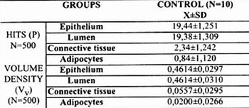
The share of the epithelium, the lumen, the stromal connective tissue and adipocytes in a volume unit of the gland is shown in the Tables 1,2 and 3.

Table 1: STEROLOGICAL ANALYSIS ON THE 7th DAY OF LACTATION
Table 2: STEROLOGICAL ANALYSIS ON THE 14th DAY OF LACTATION
Table 3: STEROLOGICAL ANALYSIS ON THE 21st DAY OF LACTATION

On the 7th day of lactation there was significant difference of the alveolar and ductal epithelium phase (Table 1) which was significantly reduced (p<0.01) in animals treated by lead acetate. In the same animals there were larger lumens of the alveoli and ducti, more abundant connective tissue and grater number of the adipocytes but these differences in comparison to control group, were not significant (p>0.05).

On the 14th day of lactation (Table 2) there was significantly greater presence of the adipocytes phase (P<0.02) and stromal tissue (p<0.05) while the volume of the epithelium of alveoli and ducti was significantly reduced (p<0.01) in study group. There was also an increase of the alveoloductal lumen phase but this was not significant.

On the 2lstday of lactation (Table 3) there were significant differences in epithelial and stromal tissue phases (p<0.01) in two groups, having a significant increase in the epithelium, and significant decrease of stromal tissue in glands of lactating rats treated by lead acetate. There were nonsignificant differences as far as the presence of the adipocytes and alveoloductal lumen volume were concerned (p>0.05) although the values for both phases in study group were above the control values.

DISCUSSION AND CONCLUSIONS

During the stereological analysis of the mammary gland, particularcaution is mandatory because of the non-homogeneous distribution of the tissues in the organ. That’s why the serial sections of thewhole organ have to be used with precise distances between the sections. During the preparatory phase for the stereological analysis, we haven’t found significant differences in volumes using 400 or 600 hits for particular phases as was noted by Lowet al.(1988). We considered the method with 500 hits to be optimal.

Our results have shown that in normal lactating rats the presence of the epithelial phase per volume unit was the greatest onon the 14thday having the tendency of the decrease on the day 21. This fluctuation of the epithelial volume during the lactation was identical in the rats exposed to lead as well. However, in all periods of lactation observed, under the lead influence a relative volume of the mammary epithelium was significantly diminished comparing to controls. Shipman et al. (1987) found that milk production reached its top level on the 14thday being consistent to maximal values of the epithelial presence in our animals.

From the reasons above, the volume changes of the mammary epitheliumin lactating rats should be almost exclusively understood as areflection of the functional activity. This is supported by the results of Joshi et al. (1986) and his generally accepted altitude that the proliferative activity of the mammary epithelium has tobe considered completed in the period of early lactation, i.e. on the 3rd day. Our results have shown that mammary epithelium in the rats exposed to lead consistently follows during the lactation, the control trends. Shipman et al.(1987) emphasized extreme metabolic adaptability of the mammary gland during the lactation period and exposure to toxic substances. Significantly reduced mammary epithelium in lactating rats under lead influence could be probably understood as a consequence of a harmful lead effects on a proliferative activity of mammary epithelium during the pregnancy, what was found out by Mornjakovic et al.(1996), since our animals were exposed to lead not only during the lactation but throughout the pregnancy. This could suggest that the mammary gland was not capable to compensate completely the “defects” in its pathway of proliferation and differentiation that appeared during the pregnancy.

Our results have shown that the mammary glands in control rats had no significant fluctuation as far as luminal presence was concerned and had a trend that followed a fluctuation of the epithelium, i.e. on the day 14 the lumen was maximal and slightly diminished 7 days after. In experimental group this luminal maximum on the 14th day of lactation was not stressed. The basicattitude, that results from the status of the epithelium and lumen of lactating glands in control group, is the existing parallelismin their changes which are synchronized and expressed through the positive relation. Although we haven’t found the significant differences of luminal presence in study group perhaps we should not neglect the fact that it was higher than the control values and it could be the result of nonsynchronization between epithelial volume and luminal volume under lead influence. Munford (1963) found that in normal lactating rats luminal volume reached plateau and remained constant from 8th to 14th day. On contrary, our results showing the maximal luminal volume on the 14th day of lactation, could be contributed to a different strain of rats. We also haven’t limited the number of sucklings to 8 immediatelypost partum, out our rats breast-feeded 10-13 newborns, i.e. their normal litter.

Low et al. (1988) found that the lumen constitued 46% of the total mammary gland volume at the end of lactation in normal lactating rats. This is almost identical to our finding on the day 14. Low et al. analyzed the second pair of thoracic mammary glands. Besides, they haven’t described the treatment prior to sacrifice and it is well-known that the period from the last breast feeding to the sacrifice is very important factor because of the milk accumulation and consecutive distension of the alveoli and ducti.

We know little about direct effects of lead on the alveolar and ductal lumen. Mayne et al. (1968) considered the insuffitient prolactin level to be a factor leading to enlargement of the lumen. Mizuno and Shiba (1969) have found increased alveolar volume incases of refractory myoepithelial cells for oxytocin.

During the lactation our rats were characterized by the fluctuation of the volume of the stromal connective tissue and its blood vessels being inverse to the epithelial volume changes. So, in control group the volume of the stromal tissue was minimal on the 14thday, while its increase was found on the 21st day. We found similar trend in lactating rats treated by lead, but their mammary glands consisted of significantly greater amount of the connective tissue both on the days 14 and 21 in comparsion to controls.

Hay den et al. (1979) have found the presence of very abundant stroma in mammary glands of hypophysectomized gravid rats pointing out the crucial importance of hypophysisovary axis. In lactating rats mammary stroma was enlarged concurrent to milk stasis and damage of lactocytes.

We explain the changes in volume of stromal tissue as a result of disbalance and/or deficit in hormones of the ovary and pituitary gland, since the alteration of the hypopysis-gonadsaxis Juring the lead exposure is well-known (Silbergeld, 1983). An increase of the stromal connective tissue in gravidity, after progesterone d’efici pointed out by El Eterby and Wrobel(1978)could be ai explanation for our lactating rats, intoxicatedby lead if w< accept the fact that lead, according to Wide (1983), dimin ishes progesterone level. Wide (1983) has found th< decrease in progesterone concentration (80% of contro level) during the first trimester of pregnancy after the lea» exposure. There is apossibility of direct lead influence t< mammary connective tissue according to El lander and Han (1987). We have found the collapse of certain number of thi alveoli and invasion of connective tissue in lead treated lac tating rats on the days 14 and 21 (unpublished data).

A participation of the adipocytes per mass unit of th< mammary gland in our normal lactating rats could be con sidered to be constant having low values. The same wa! found in the experimental group. On the 14th day of lac tation, there was significant differencein favour of thi adipocyte phase in study group. Our results have showi that the complete disappearance of the adipocytes didn’ occur even in first part of lactation regardless of leat presence. Anzai et al. (1979) pointed out that bi valen ions depressed the lipolytic activity in the lactating mam mary gland. We don’t know if this mechanism of lea(influence existed in our experiment. There is a possibilit) that lead affects an lipogenic capacity ofthe adipocyte! which is increased inversely to lactocytes and become: manifested during maximal milk secretion, i.e. on the daj 14 of lactation. Our lactating rats exposed to lead had less er number of sucklings (unpublished).

According to Elias et al. (1973) the number of suckling; could affect the structure of the mammary gland. The) have found the lower depletion of the adipocytes in rat! with lesser number of sucklers.

On the basis of given results we conclude: lead change! the quantitative characteristics of the mammary gland, i.e the composition of the volume unit of the organ through all phases of lactation.

LITERATURE