Local Cellular Immune Response in Dysplastic Epithelial Lesions of the Flat Colonic Mucosa

Authors

  • Svjetlana Radović Institute for Pathology, Medical Faculty, Sarajevo
  • Ivan Selak Institute for Pathology, Medical Faculty, Sarajevo
  • I. Bratović Clinic for gastroenterology, Clinical Centre, Sarajevo

DOI:

https://doi.org/10.17305/bjbms.1998.3607

Keywords:

immunocompetent cells, epithelial dysplasia, immunological techniques, intestinal neoplasia

Abstract

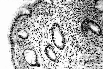
This is an immunohistochemical study of the local cellular immune response characteristic in inflammatory-regenerative and dysplastic flat colonic mucosa. The aim of this study is to determine a possible existence of the mononuclear cellular infiltration specificity which could be important for the prognosis in further development of dysplastic lesion. Biopsy specimens from 170 patients (specimens stained by hematoxylin eosin) were examined. 74 specimens showed inflammatory-regenerative changes and 96 had dysplastic changes (38 with mild dys-plasia, 28 with moderate dysplasia and 30 with severe dysplasia). Three monoclonal antibodies were used for the identification of mononuclear cells in the inflammatory cellular infiltration in the lamina propria of colonic mucosa. The inflammatory cells type and their location in respect of the epithelial cells and lesion itself were analysed and their number was determined by the semi-quantitative method. T lymphocytes were the dominant cells of local immune response in dysplastic lesions while macrophages were less present and B lymphocytes, as rare cells, were present in sporadic cases. It is notified that increase in the extent of dysplasia was followed by increase in the number of macrophages and T lymphocytes in particular. Immediate contact between macrophages and T lymphocytes in epithelial dysplasia was found in a small number of cases and was mostly independent from the intensity of dysplasia. Signs of the direct lytical effect of the mononuclear ceils on dysplastic epithelial cells were not observed in this contact. It seems that epithelial dysplasia does not provoke more significant local immune response which is the most probably a part of the chronic non-specific inflammation that has a negative influence on further development of the lesion. The conclusion is that local immune response in the dysplastic alteration of flat colonic mucosa has no importance in further prognosis of the lesion.

Downloads

Download data is not yet available.
Local Cellular Immune Response in Dysplastic Epithelial Lesions of the Flat Colonic Mucosa

Published

1998-02-20

How to Cite

1.
Radović S, Selak I, Bratović I. Local Cellular Immune Response in Dysplastic Epithelial Lesions of the Flat Colonic Mucosa. Biomol Biomed [Internet]. 1998Feb.20 [cited 2023Jun.6];1(1):12-9. Available from: https://www.bjbms.org/ojs/index.php/bjbms/article/view/3607

Issue

Section

Pathology

INTRODUCTION

Epithelial dysplasia (ED) in flat colonic mucosa is a pre-malignant lesion which presence and increase of the extent, in particular, raise a risk of cancer. In the last ten years, etiology, epidemiology, histology, macroscopic shapes, treatment and prognosis of this lesion were intensively researched. Although epithelial dysplasia is a well defined group of the morphological entities its biological nature is not completely known. True possibility of the malignant alteration of the lesion at its different stages is not known. Almost no attention is dedicated to a morphological manifestation of the local immune response and it is not known whether that can be important for further prognosis and can influence further development of the lesion.

Importance of the local immune response with a reference on further prognosis is up to now mainly investigated in neoplasia (1,2,3,4) and mostly in planocellular head and neck carcinoma (5,6,7,8,9). Contrary to these reports, there is a small number of those who speak about local reaction of stroma in pre-cancerous dysplastic lesions of mucosa. One of the first studies, which dealt with problems of this kind was published in 1971 (10). It presents the classification of hyperplastic lesions of larynx with special emphasis on the imunocompetent cells as an important prognostic factor especially within the group of pre-cancerous lesions.

Due to development of the immunohistochemical techniques and the discovery of highly specific monoclonal antibodies, it is possible, today, to obtain a precise in situ identification of the mononuclear cells in inflammatory infiltration with determination of the incidence, way of distribution and interaction with changed epithelial cells what all together can have a significance in determination of further prognosis of the lesion. Morphological and immunohistochemical analysis of the epithelial dysplasia of fiat colonic mucosa are performed in this study with a special reference on the research of local cellular immune response. We tried to determine the existence of possible relation between the type and the quantity of mononuclear cells infiltration, the way of its distribution at different grades of epithelial dysplasia which presence and quantity were determined on the basis of previously established criteria (11).

MATERIAL AND METHODS

Two or three biopsy specimens of colonic mucosa (always 30 cm far from anus) are taken during the routine endoscopic examination from the patients with established diagnosis of any inflammatory process. The specimens have always been taken from flat mucosa. We took biopsy specimens from 170 patients out of which 108 were males and 62 females. All of them were more than 45 years old. The specimens from 40 patients with ade-nocarcinoma, aged 42 -76, were taken as a control group. The specimens were fixed by a neutral buffered 10% formalin, paraffin-embedded and cut in 5 mm sections and then taken for standard hematoxylin -eosin staining (HE) and immunohistochemica! staining on CD 20 antigen for Β lymphocytes (Monoclonal Mouse Anti-Human Β Cell, CD 20 Clone L26, Lot 083, Code No. Μ 0755 by Dako Corp.), CD 45 RO for Τ lymphocytes (Monoclonal Microscopically, it has been established that 105 patients had chronic ulcerative colitis, 40 patients hadlymphocyt-ic colitis and 25 patients had eosinophilic colitis. Presence of the inflammatory-regenerative and dysplastic changes was investigated in all patients. Histological criteria for a more simple differentiation of the inflammatory-regenerative and dysplastic changes and also for a severity degree of dysplasia are defined. According to these criteria epithelial dysplasia was divided into three grades -mild, moderate and severe dysplasia. Classification of the changes into mentioned categories is based on 19 criteria, graded into degrees of severity from 1 to 4. Within these classification criteria are ihe following: size of the epithelial cells, nuclei shape, nuclei/cytoplasmatic relation, chromasia and stratification of the nuclei, arrangement of chromatin in nuclei, visibility and the number of nucleoli, cytoplasmatic basophilia, presence of the different cellular types in the crypt epithelium, presence and the number of mitosis, irregular growth of the crypt epithelium and its branching, a number of crypts, presence of the “back to back” formations, tendency of the villous formation production on the mucosa surface, inflammatory eellular infiltration in the lamina propria and ciypt abscesses. The total intensity of each alteration was divided with their total number and that value was marked as an index (I). Trend range of the index value for inflammatory-regenerative and dysplastic changes of different severity degree (mild, moderate and severe dysplasia) was mathematically determined (11).

  • 1.3 <| <1.8 for inflammatory-regenerative changes

  • 1.9 <| <2.3 for mild dysplasia

  • 2.4 <| <2.9 for moderate dysplasia

  • 3 <| <3.7 for severe dysplasia

Mouse Anti-Human Τ cell, CD 45 RO Clone UHCL-1, Code No. Μ 742, Lot 064 by Dako Corp.) and CD 68 for macrophages (Monoclonal Mouse Anti-Human Macrophage, CD 68 Clone PG-M1, Code No. Μ 876, Lot 084 by Dako Corp.) (shown in Table 1).

Table 1: Monoclonal antibodies used in the research of immune response in the inflammatory-regenerative and dysplastic changes of flat colonic mucosa in 170 patients

The analysis of immunocompetent cells in the colorectal mucosa includes an estimate of (a) the type of mononu-clear cells (B lymphocytes, T lymphocytes and macrophages) in the lamina propria of the colonic mucosa with inflammatory-regenerative and dysplastic lesion (b) the number of positively stained cells (semiquantitative grading 0= no positively stained cells; 1= stained less than 30% of cells; 2= stained 30 to 80 % of cells; 3= more than 80% cells stained); (c) antigen location in the positive cells (near the lesion, in contact with the crypt epithelium, far from the crypt epithelium; far from lesion).

Biopsy specimens of the colonic mucosa adenocarcinoma are used as a positive control of the immunoreactivity for B and T lymphocytes and macrophages. Presence of the plasma cells (eccentric nucleus with spoke shaped chro-matin, basophilic cytoplasm and bright perinuclear area) and eosinophiles (bilobular nuclei, redorange intracyto-plasmatic granules) are evaluated with standard HE stained specimens.

RESULTS

After the microscopy of the specimens, inflammatory-regenerative changes were found in 74 cases and 96 cases were with epithelial dysplasia.. Mild dysplasia was present in 38 cases while 28 cases showed moderate dysplasia and 30 cases were with a severe dysplasia. (Table 2)

Table 2: Classification of the morphological changes in the colonic mucosa of 170 patients with inflammatory-regenerative and dysplastic lesions of the epithelium

Lymphoreticular cells were found in the lamina propria of colonic mucosa in all these cases with inflammatory -regenerative and dysplastic changes.

CD 45 RO positive cells (Figure 1) -(activating) Τ lymphocytes (including subpopulations CD 4 and CD 8 lymphocytes) were found in the majority of cases with inflammatory -regenerative changed mucosa as a small number of the infiltration cells (Table 3).

Figure 1: Small number of CD 45 RO positive cells in the lamina propria of the inflammatory-regenerative altered colonic mucosa (x 250)
Table 3: Incidence of the findings of immunocompetent cell types in the inflammatoly-regenerative and dysplastic flat colonic mucosa in 170 patients

With appearance of the epithelial dysplasia and increase in its extent, (Figure 2) the number of mononuclear cells increased and among these cells, Τ lymphocytes were numerically superior (Table 3).

Figure 2: Numerous CD 45 ROpositive cells in the laminapropria of the colonic mucosa with developed moderate dysplasia (x 250) The findings with macrophages - CD 68positive cells, which sporadicalIy appear in inflammatory-regenerative mucosa is ahnost identical (Figure 3) while increase in the ED dent (Figure 4) is accompanied by increase in the macrophages number (Table 3).
Figure 3: Smoll number of CD 68 positive cells in the Iominapmprio of the inflammatory-regenerative colonic mucoso (x 125)
Figure 4: Numemur CD 68 poritive cells in the lomrnoproprta of colonic mucoso with developed moderote dysplasia (x 125)

The increase in the number of macrophages is in correlation with increase of the extent of epithelial dysplasia almost on the same way like it is with Τ lymphocytes. Both cell types were found closely to the lesion but the direct contact between them and dysplastic epithelial cells was rarely noticed. In those cases where such contact was noticed a sign of their direct litical effect was not observed (Figure 5).

Figure 5: Dimct contoct between dysplastic epithelial cells and CD 68positive cells (macmphages) u visible at some places but without signs of their direct Iytical effect on epithelial cells (x 250)

Incidence of the Β lymphocytes presence among inflammatory cells, in general, was small (Table 3).

These cells were found in a small number of cases but in the both categories of changes (that was a case with eosyn-ophilic leukocytes and plasmacytes). In the control group which consisted of 40 cases with colorectal adenocarcinoma (19 cases of well differentiated and 21 cases of moderately differentiated adenocarcinoma), no Β lymphocytes were found among the cells of tumor stroma in any case while Τ lymphocytes (Figure 6) and macrophages (Figure 7) were the dominant cells and more numerous in relation to the number of these cells in mucosa dysplasia.

Figure 6: Slroma of adenocarcinoma with the most numerous CD 45 ROpositive cells (x 250)
Figure 7: Stmma of colonic mucasa adenocarcr noma with numerous CD 68 positive cells (x 125)

DISCUSSION

Immunohistochemical analysis of the mononuclear cellular infiltration in the lamina propria of the inflammatory-regenerative changed flat colonic mucosa showed that dominant cells were (activated) T lymphocytes and macrophages while B lymphocytes, which appeared in sporadic cases, were in a small number. It has been noticed that the intensity of cellular infiltration (immune response) was raising together with the increase in extent of dysplasia and was the largest in the cases of well differentiated colonic carcinoma. Intensity and composition of the immune reaction was nearly identical at all patients with the same intensity of epithelial dysplasia. Direct contact between T lymphocytes and dysplastic epithelial cells of the crypts was observed in a small number of cases but without microscopically recognized signs of lytical destruction of dysplastic cells. In the control group, which consisted of the adenocarcinoma specimens, reaction of the stroma was the most intensive with domination of T lymphocytes and macrophages.

According to available data, this kind of research of epithelial dysplasia of flat colonic mucosa have not been done yet and therefore there is no possibility to compare our own results with the results of other workers.

Similar immunohistochemical research is performed at pre-neoplastic lesions of larynx (12). By usage of the series of monoclonal antibodies for different types of leukocytes, this study showed that T lymphocytes (CD 45 RO and CD 43) were dominant cells of the stromal infiltration and also that their quantity was related to increase of the hyperplastic lesion intensity. In this study, as well as in our study, all cell types which could have an active role in the local immune response were found.

Majority of the studies (local immune response) of this kind were performed on the carcinoma tissue (1,2,3,4,7,8,9,13,14) and showed the existence of variations in the composition and number of cells which infiltrated stroma of the tumor. According to the results of some scientists (13,14) macrophages and lymphocytes were represented in stroma in a small number while at the others (7,8) these cells were the dominant ones. In the cases with a small number of the immunocompetent cells in the epithelium and lamina propria, it is concluded that they have no effective role in the prevention and defence from malignant progression.

In the study which dealt with a number and connection between peri -and intratumorus macrophages at larynx carcinoma, Morra did not find a statistically significant connection between the number of these cells and further clinical feature of the tumor.

On the opinion of the same author, the number of macrophages at poorly differentiated carcinomas is in correlation with the possibility of recidive appearance. On the basis of the non-existence of the visible cytotoxic effects of the lymphocytes and macrophages in the direct contact between these cells and cells of the planocetlular carcinoma of the head and neck, Horst concluded that it was about insufficient local immune response. It is stated that the low antigenic ability of these cells, immunosup-pressive cells action, suppressive factors released by the malignant cells and internal defects of the Τ lymphocytes which infiltrated stroma of the lesion may be the reason for this type of response. The functional significance of macrophages in that infiltration is not clear. These cells can cause a strong antitumor response through the action of many biologically active products but also can support the tumor’s growth by the stimulation of angiogenesis (neovasculature) what has been proved experimentally.

On the basis of immunohistochemical research we can speak only about a phenotypic recognition of the inflammatory cells infiltration but not about their active function. On the basis of the intensity and composition of the immunocompetent cells in the stroma of examined pre-cancerous lesion, further conduct of the lesion can not be predicted for sure i.e. the possibility of its development into malignancy. The findings of the T lymphocytes and macrophages which were not in close contact with dys-plastic epithelial cells but dominantly present in the stroma of lesion, would speak in favour of the existence of the local immune response deficit at the pre-neoplastic growth of the tissue. The absence of the signs of the active epithelial cells destruction in the cases where they were in a close contact with the lymphocytes and macrophages would be the evidence for this. It seemed that immunocompetent cells in the epithelium and stroma of the lesion have no effective defensive role in the prevention of the development of the premalignant lesion into malignancy. It seems that epithelial dysplasia in flat colonic mucosa does not provoke substantive immune response. A local inflammatory reaction could be the result of hemotactic action of the cytokines and it represents only a part of the nonspecific chronic inflammatory reaction which, by itself, could provoke the appearance of malignancy.

Conclusion: Our study showed that the intensity of infiltration by the immunocompetent mononuclear cells is correlated with the increase in the extent of dysplasia. The signs of active lytical action of the lymphocytes and macrophages at the occasion of close contact with dysplastic epithelial cells of the mucosa were not found. Previously stated observations indicate that the local immune response can not be taken as a prognostic factor. It seems that epithelial dysplasia does not provoke more significant local immune response but the local immune reaction is the most probably a part of the chronic nonspecific inflammation which has negative influence on further progression of the lesion

References