Intervertebral disc tissue engineering: A brief review

Authors

  • Janja Stergar Institute of Biomedical Sciences, Faculty of Medicine, University of Maribor, Maribor, Slovenia Laboratory of Inorganic Chemistry, Faculty of Chemistry and Chemical Technology, University of Maribor, Maribor, Slovenia
  • Lidija Gradisnik Institute of Biomedical Sciences, Faculty of Medicine, University of Maribor, Maribor, Slovenia Alma Mater Europaea – European Center Maribor (AMEU-ECM), Maribor, Slovenia
  • Tomaz Velnar Institute of Biomedical Sciences, Faculty of Medicine, University of Maribor, Maribor, Slovenia Alma Mater Europaea – European Center Maribor (AMEU-ECM), Maribor, Slovenia Department of Neurosurgery, University Medical Centre Ljubljana, Ljubljana, Slovenia
  • Uros Maver Institute of Biomedical Sciences, Faculty of Medicine, University of Maribor, Maribor, Slovenia Department of Pharmacology, Faculty of Medicine, University of Maribor, Maribor, Slovenia https://orcid.org/0000-0002-2237-3786

DOI:

https://doi.org/10.17305/bjbms.2019.3778

Keywords:

Intervertebral disc, IVD, cell-based therapies, biomaterials, scaffolds, tissue engineering, IVD degeneration, IDD

Abstract

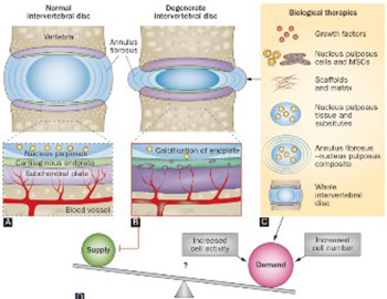
Intervertebral disc (IVD) degeneration (IDD) is associated with low back pain and significantly affects the patient’s quality of life. Degeneration of the IVD alters disk height and the mechanics of the spine, leading to chronic segmental spinal instability. The pathophysiology of IVD disease is still not well understood. Current therapies for IDD include conservative and invasive approaches, but none of those treatments are able to restore the disc structure and function. Recently, tissue engineering techniques emerged as a possible approach to treat IDD, by replacing a damaged IVD with scaffolds and appropriate cells. Advances in manufacturing techniques, material processing and development, surface functionalization, drug delivery systems and cell incorporation furthered the development of tissue engineering therapies. In this review, biomaterial scaffolds and cell-based therapies for IVD regeneration are briefly discussed.

Downloads

Download data is not yet available.

Author Biographies

Tomaz Velnar, Institute of Biomedical Sciences, Faculty of Medicine, University of Maribor, Maribor, Slovenia Alma Mater Europaea – European Center Maribor (AMEU-ECM), Maribor, Slovenia Department of Neurosurgery, University Medical Centre Ljubljana, Ljubljana, Slovenia

Department of Neurosurgery

Uros Maver, Institute of Biomedical Sciences, Faculty of Medicine, University of Maribor, Maribor, Slovenia Department of Pharmacology, Faculty of Medicine, University of Maribor, Maribor, Slovenia

Department of Pharmacology
Intervertebral disc tissue engineering: A brief review

Downloads

Additional Files

Published

2019-05-20

How to Cite

1.
Stergar J, Gradisnik L, Velnar T, Maver U. Intervertebral disc tissue engineering: A brief review. Biomol Biomed [Internet]. 2019May20 [cited 2023Jun.2];19(2):130-7. Available from: https://www.bjbms.org/ojs/index.php/bjbms/article/view/3778

Issue

Section

Reviews

INTRODUCTION

In recent years, our knowledge of human physiology and cell biology has increased exponentially. This significantly impacted the development of tissue engineering in various medical fields, including regenerative techniques for intervertebral disc (IVD) degeneration (IDD). Degenerative discs are among the main causes of chronic segmental spinal instability in both males and females, and significantly affect the quality of life, especially of young patients [1]. IDD may present clinically as axial back pain, spinal stenosis, myelopathy, or radiculopathy [2-4].

Low back pain is associated with IDD [5,6]. The disk height decreases due to degeneration, altering the mechanics of the affected spinal segment. This process accelerates the degeneration of adjacent segments and other spinal structures, such as small joints, ligaments and muscles [5]. In the long term, IDD leads to narrowing of the spinal canal (spinal stenosis) and the subsequent compression of neural tissue. Spinal stenosis is the major cause of pain, especially in the elderly. Considering current demographic trends in Europe [7] such as the increase in the elderly population, the problem of IDD and related conditions becomes more difficult [8]. Current therapies for IDD include conservative and invasive treatments [6], however, none of those approaches can restore the disc structure and function [9,10]. Recently, tissue engineering techniques emerged as a possible approach to treat IDD, by replacing a damaged IVD with scaffolds and appropriate cells [9,11,12].

In this review, biomaterial scaffolds and cell-based therapies for IVD regeneration are briefly discussed.

INTERVERTEBRAL DISC STRUCTURE

IDD and associated back pain are chronic conditions that affect a large number of people worldwide. Thus, prevention and treatment of disk degeneration are the focus of intensive research. Recent findings show the potential of biological methods, such as molecular, cell-based and whole organ tissue engineering therapies, to prevent and manage IDD [13-15]. Because various scaffold materials and cell sources are used in tissue engineering, advances in materials science are particularly important for the development of this field. These include improvements in manufacturing techniques, material processing and development, surface functionalization, drug delivery systems and cell incorporation [16-19].

The IVD is complex in structure and consists of three distinct parts (Figure 1): 1) the fibrocartilaginous annulus fibrosus (AF) with its outer and inner regions, composed of concentrically oriented layers of fibrous tissue; 2) the central nucleus pulposus (NP); and 3) the cartilaginous endplates (EPs) [9,20]. The AF consists of a series of 15 to 25 concentric rings or lamellae, with aligned parallel collagen fibers located within each lamella. Elastic fibers lie between the lamellae and help the IVD to return to its original shape following flexion or extension of the spine. The cells of the AF are elongated and fibroblast-like and are aligned parallel to the collagen fibers. The NP contains a highly hydrated gel-like matrix that is comprised primarily of the proteoglycan aggrecan. A small amount of randomly arranged collagen fibers and radially arranged elastin fibers are embedded in the matrix [20,21,22]. The cells in the NP are spheroidal and chondrocyte-like. The EPs are made up of osseous and hyaline-cartilaginous layers and contain rounded chondrocytes.

FIGURE 1: Schematic depiction of the normal and degenerate IVD structure with possible therapeutic approaches. A) Nutrient pathways in normal disc. B) Nutrient pathways in a degenerate disc (e.g., calcification of cartilaginous endplate, occlusion of marrow spaces, atherosclerosis of vertebral arteries, reduced capillary density etc.). C) Different forms of biological therapies for disc repair. D) Current therapies increase the cell number and/or cellular activity causing nutrient demand to exceed nutrient supply, which is already diminished in degenerate discs. Reprinted by permission from Macmillan Publishers Ltd: Nature Reviews Rheumatology [20], copyright 2014.

The IVD is an avascular structure. Both the cells and extracellular matrix (ECM) are essential for normal IVD function [4]. Regenerative approaches in tissue engineering of IVD aim to restore/preserve the anatomy and function of both AF and NP [23]. Therefore, an ideal scaffold for IVD replacement should have good biocompatibility and moderate porosity, and the shape, structure and mechanical properties similar to the IVD [9,24]. Currently, the research in this area is focused on constructing AF- and NP-scaffolds that have all the properties of the native structures, using various synthetic and natural polymers [12,25,26].

Main challenges in IVD tissue engineering

Mechanical properties of the IVD are important for its proper function. In vivo, the IVD transmits the load imposed on the spine, including spinal tension, torsion, compression and bending [27]. Thus, it is essential for tissue engineered scaffolds to have all the (bio)mechanical properties of the IVD so they can replace a damaged disc in the body [9].

Scaffolds represent one of the key components in tissue engineering, as they provide the structural support for cell attachment/proliferation and ECM accumulation [28]. Tissue engineered scaffolds should be able to withstand the physiological load imposed on the IVD in vivo, in addition to good biocompatibility, moderate porosity and similarity to the IVD in shape, structure and mechanical properties [29]. Moreover, for cell/whole organ tissue engineering therapy to be successful, the local environment in which transplanted cells or tissue engineered IVDs are introduced must be able to support cell growth and proliferation [9,26].

Scaffolds and appropriate source materials

Tissue engineering provides a promising alternative for the restoration of physiological function of a damaged IVD [30]. Because degeneration of the IVD affects both the AF and NP, composite scaffolds that enable simultaneous repair of the two parts should be used [30]. Studies with small animal models showed promising results in IVD regeneration and repair. Various techniques are utilized for the preparation of three-dimensional (3D) biomimetic scaffolds, including solvent casting, leaching method, phase separation, freeze drying, and electrospinning [11,31,32]. However, clinical application of these scaffolds is still limited [33]. Collagen, which is the most abundant protein in mammals, is widely used in biomedical applications and may also be appropriate for IVD regeneration [34,35]. In the extraction of collagen from skin tissue, pepsin is added to remove N- and C-terminal telopeptide components from collagen and to solubilize the collagen derivative [36]. This low-immunogenic derivative of collagen is called atelocollagen. Due to its low antigenicity, atelocollagen is regarded as one of the best basic matrices for implantable materials [37]. An atelocollagen scaffold with honeycomb structure is mechanically stable, easily handled, and supports the growth of a large number of cells (high-density cell cultures). Considering these characteristics, atelocollagen may be useful as a 3D scaffold in tissue engineering [38].

Another important material for scaffold construction is silk fibroin, a protein produced by silkworms and some other insects [39]. Silk has a high resistance to compression and is considered to be one of the strongest natural fibers [31,40]. The stability of the silk fibers is the result of the extensive hydrogen bonding, hydrophobic nature of the protein, and high degree of crystallinity due to the specific organization of β-sheets [41]. There are several benefits of using silk fibroin as a scaffold in IVD bioengineering [42]. Mechanical stability is an important property of a scaffold, and in the case of silk scaffolds, it is the result of compressive and tensile strength of silk protein fibers. In vivo, IVDs are always to some degree under load [24]. Once implanted, the silk scaffold would degrade at a sufficiently slow rate to allow proper tissue development [39,43]. Another benefit of silk is that other peptides can be covalently attached to it, which could potentially enhance cell attachment during AF and NP development [44].

Other biomaterials that may be used as matrix supporting materials in AF and NP tissue engineering include chitosan and alginate, and both are cheap and easily accessible. Moreover, the two polymers yield superior characteristics when combined into a hybrid scaffold [7,45]. Chitosan is derived from the shells of crustaceans [46]. It is a biodegradable and biocompatible polymer with low toxicity and good antimicrobial properties [47,48]. Chitosan-based scaffolds are soft and spongy, have high porosity and pore interconnectivity, and support cell adhesion and growth [46]. Alginate is a linear polysaccharide composed of α-L-guluronic acid (G block) and β-D-mannuronic acid (M block) residues, which are linked together in different sequences. It represents one of the most abundant natural materials and is derived primarily from brown algae and some bacteria [49]. Due to its outstanding properties in terms of biocompatibility, biodegradability, non-antigenicity and chelating ability, alginate is used in a variety of biomedical applications, including tissue engineering (e.g., as a supporting matrix) and drug delivery [45,49-52].

Gellan gum (GG) is a natural polysaccharide produced by the bacterium Sphingomonas elodea [53]. It consists of repeating tetrasaccharide units that are comprised of L-rhamnose, D-glucuronic acid and two D-glucose residues. GG is noncytotoxic and particularly resistant to heat and acid stress [54]. The gelation of this biomaterial leads to the formation of a stable hydrogel structure. At higher temperatures (above ~80 °C), GG exists in a coil conformation. As temperature decreases, a thermoreversible coil to helix transition occurs [55]. Untwined regions of polysaccharide chains link to the oriented bundles of the double helix structures (junction zones) leading to the formation of a 3D gel network, which can be used as a matrix for cell seeding [55,56].

In addition to natural polymers and natural-polymer derived materials, biodegradable synthetic polymers are also utilized as scaffolds in tissue engineering [57,58]. Synthetic polymers have a number of advantages over natural polymers, including highly reproducible synthesis, predictable properties, lack of immunogenicity, and easy processing into desired structures and implants [58]. Synthetic polyesters that are extensively used in AF tissue regeneration include poly(ε-caprolactone) [PCL], polylactide (PLA), polyglycolide (PGA), and copolymers produced from the respective monomers [26,59]. In addition, many other natural and synthetic materials are being investigated as scaffolds for AF, NP and IVD tissue engineering [60].

Tissue engineering of AF and NP

Tissue engineered AF and NP have the potential to repair or replace degenerated tissue in the IVD, thus restoring its functionality. Therefore, considerable research is directed toward developing appropriate scaffolds for AF and NP regeneration [15,42,61]. Many natural and synthetic materials can be used as a supporting matrix in AF and NP scaffolds [25].

Damage to the AF is attributed to numerous factors, such as mechanical stress, biological remodeling, loss of nutrition of cells, and accumulation of cellular waste products [27,42,62]. Silk scaffolds are appropriate for AF engineering due to their mechanical properties and biocompatibility. According to Chang et al. [44], porous silk scaffolds may be used to modulate the phenotype of seeded AF cells, mimicking the native tissue with inner transition and outer zones [44]. AF scaffolds can also be produced from alginate or alginate/chitosan [15,51]. A hybrid alginate/chitosan scaffold has good biocompatibility, promotes AF cell proliferation, supports ECM deposition, and has a slower degradation rate compared to a pure alginate scaffold [51]. The atelocollagen honeycomb-shaped scaffolds, which allow high-density growth and 3D culture of various cells, may be used for IVD regeneration by transplanting the AF cells [37,51]. In addition, Cabraja et al. showed that a 3D polymer of pure polyglycolic acid (PGA) combined with hyaluronan is an excellent scaffold for AF cell redifferentiation [63].

Similar to AF tissue engineering, different types of materials are used for NP scaffolds, such as collagen, alginate, chitosan, fibrin and hyaluronic acid [24]. Hydrogels for NP engineering should possess good mechanical strength, viscoelasticity, swelling capacity, diffusion properties, biocompatibility, and to be able to support cell growth and ECM accumulation [64]. Generally, the main challenge in NP tissue engineering is to find a biomaterial that can withstand large mechanical loads, imposed on the spine in natural conditions [15]. There are two different cell types in the NP: notochordal cells and mature NP cells. The latter are called chondrocyte-like cells due to their rounded shape and secretory activity, and are found primarily in adults [65]. An example of a scaffold appropriate for NP tissue engineering is a chitosan-based hydrogel seeded with IVD cells [15,66]. This scaffold is suitable for cell-based supplementation, to restore NP function in the early stages of IVD degeneration [66]. Halloran et al. showed that an enzymatically cross-linked atelocollagen type II-based scaffold, containing aggrecan and hyaluronan in varying concentrations, has the potential for developing an injectable scaffold seeded with cells for NP regeneration [67]. Moreover, modification of low viscosity alginate with methacrylate groups produces a photo-crosslinkable alginate hydrogel with tunable material properties and the ability to maintain the viability of the encapsulated NP cells [50]. Similarly, porous silk fibroin (SF) scaffolds represent a plausible candidate for tissue engineered NP as they support NP cell attachment, proliferation and infiltration, and the production of ECM [40]. Other scaffolds for NP regeneration include ionic- and photo-crosslinked methacrylated gellan gum hydrogels, which can be used as acellular or cellular tissue engineering scaffolds; polylactide beads; demineralized bone matrix; gelatin microcarriers; gelatin/chondroitin-6-sulfate/hyaluronan tri-copolymers, which are bioactive scaffolds for culturing human NP cells that preserve cell viability and support cell proliferation and ECM production; and biphasic polyurethane scaffolds, which have good swelling capacity in vitro, fast swelling rate after hydration, and dynamic compressive stiffness [15,37,40,50,64,66,67].

Final task: cell integration

For a complete and functional tissue-engineered model of IVD, which includes the cells and ECM, a scaffold needs be able to support the survival and preserve/induce the phenotype of both AF and NP cells. Various cell types are used for regenerative therapy of IDD, and stem cells represent a particularly attractive option [68]. As unspecialized cells capable of long-term self-renewal and lineage-specific differentiation, stem cells can be programmed/induced to differentiate into different types of cells [68-70]. Mesenchymal stem cells (MSCs) are particularly suitable for IVD cell therapy, because they are capable of differentiation into various connective tissue cells and can be obtained relatively easily from a number of sources, including fetal liver, umbilical cord blood, bone marrow, adipose tissue, muscles and dermis [70-81]. MSCs participate in the repair of degenerated disc tissue in several ways, including: 1) directly by differentiation into disc tissue-specific cells to supply lost or damaged cells and promote the formation of the ECM; 2) indirectly by secreting growth factors to enhance tissue regeneration; and 3) by modulating the inflammatory response [82-86]. Two main therapeutic strategies exists for the application of MSCs in tissue repair. In the first approach, undifferentiated MSCs are transplanted, which then undergo differentiation in vivo under the stimulation of local factors. In the second strategy, MSC are induced to differentiate in vitro, prior to transplantation [86]. A number of studies investigated the ability of MSCs to differentiate into NP or AF cells and promote ECM synthesis, using either co-culture systems with growth factors or animal models in which MSCs are injected directly into the IVD [62,87-92]. When injected directly into the IVD MSCs promote ECM synthesis, resulting in restoration of the disc height. Human cell cultures are also used for IVD regeneration. Transplantation of autologous MSCs from bone marrow into a rabbit model of disc degeneration leads to regeneration of IVDs, providing a new hope for the treatment of degenerative disc disease in humans [93,94]. Figure 2 summarizes the most important recent strategies for IVD regeneration.

FIGURE 2: Overview of recent strategies related to intervertebral disc regeneration. Reprinted by permission from Elsevier: Biotechnology Advances [94], copyright 2013.

CONCLUSION

The pathophysiology of IVD disease is still not well understood and IDD remains a significant health problem worldwide. Although still in the experimental phase, regenerative strategies for IDD such as tissue engineering show great promise. Further research will provide new insights into IVD regeneration mechanisms and, hopefully, enable the integration of regenerative therapies for IDD into clinical practice.

Acknowledgements

ACKNOWLEDGMENTS

The authors would like to acknowledge the financial support received from the Slovenian Research Agency (Grant Numbers: P3-0036 and I0-0029).

DECLARATION OF INTERESTS

The authors declare no conflict of interests.

REFERENCES

  1. , , , , (). Intervertebral disc regeneration:A great challenge for tissue engineers. Trends Biotechnol. https://doi.org/10.1016/j.tibtech.2014.05.006
  2. , (). The economic burden of back pain in the UK. Pain. https://doi.org/10.1016/S0304-3959(99)00187-6
  3. , , , , , (). Prevalence and pattern of lumbar magnetic resonance imaging changes in a population study of one thousand forty-three individuals. Spine (Phila Pa 1976). https://doi.org/10.1097/BRS.0b013e3181a01b3f
  4. , , , , (). Cross-sectional magnetic resonance imaging study of lumbar disc degeneration in 200 healthy individuals. J Neurosurg Spine. https://doi.org/10.3171/2009.5.SPINE08675
  5. , , , (). Treatment of the degenerated intervertebral disc;closure, repair and regeneration of the annulus fibrosus. J Tissue Eng Regen Med. https://doi.org/10.1002/term.1866
  6. , , (). Is the spinal cord stimulation an effective treatment for failed back surgery syndrome? Our experience with 21 cases. Acta medico-biotechnica.
  7. , , , , (). Novel chitosan/diclofenac coatings on medical grade stainless steel for hip replacement applications. Sci Rep. https://doi.org/10.1038/srep26653
  8. , , , (). Pathophysiology of the human intervertebral disc. Int J Biochem Cell Biol. https://doi.org/10.1016/j.biocel.2007.12.011
  9. , , , , , (). Intervertebral disc tissue engineering with natural extracellular matrix-derived biphasic composite scaffolds. PloS One. https://doi.org/10.1371/journal.pone.0124774
  10. , , , , (). A review of animal models of intervertebral disc degeneration:Pathophysiology, regeneration, and translation to the clinic. Biomed Res Int. https://doi.org/10.1155/2016/5952165
  11. , , , , , (). Evaluation of various types of scaffold for tissue engineered intervertebral disc. Tissue engineering. https://doi.org/10.1007/978-0-387-34133-0_12
  12. , , , , (). Role of biomechanics in intervertebral disc degeneration and regenerative therapies:What needs repairing in the disc and what are promising biomaterials for its repair?. Spine J. https://doi.org/10.1016/j.spinee.2012.12.002
  13. , , , , , (). Mesenchymal stem cell for prevention and management of intervertebral disc degeneration. Stem Cells Int. https://doi.org/10.1155/2012/921053
  14. , , , , (). Growth factors and anticatabolic substances for prevention and management of intervertebral disc degeneration. Stem Cells Int. https://doi.org/10.1155/2012/897183
  15. , , , , (). Recent advances in biological therapies for disc degeneration:Tissue engineering of the annulus fibrosus, nucleus pulposus and whole intervertebral discs. Curr Opin in Biotechnol. https://doi.org/10.1016/j.copbio.2013.04.012
  16. , , , , (). Advanced therapies of skin injuries. Wien Klin Wochenschr. https://doi.org/10.1007/s00508-015-0859-7
  17. , (). Review of aerogel-based materials in biomedical applications. J Sol-Gel Sci Techn. https://doi.org/10.1007/s10971-016-3968-5
  18. , , , (). The intervertebral disc functional cell models:A new revolution for in vitro toxicology testing. Global Spine J. s-0036-1582628-s-0036-1582628. https://doi.org/10.1055/s-0036-1582628
  19. , , , , , (). Polyester type polyHIPE scaffolds with an interconnected porous structure for cartilage regeneration. Sci Rep. https://doi.org/10.1038/srep28695
  20. , , (). Intervertebral disc regeneration:Do nutrients lead the way?. Nat Rev Rheumatol. https://doi.org/10.1038/nrrheum.2014.91
  21. , , , , , (). Biomimetic method for combining the nucleus pulposus and annulus fibrosus for intervertebral disc tissue engineering. J Tissue Eng Regen Med. https://doi.org/10.1002/term.412
  22. , (). Degeneration of the intervertebral disc. Arthritis Res Ther. https://doi.org/10.1186/ar629
  23. , , , (). Intervertebral disc regeneration:From the degenerative cascade to molecular therapy and tissue engineering. J Tissue Eng Regen Med. https://doi.org/10.1002/term.1719
  24. , , , , , (). Intervertebral disk tissue engineering using biphasic silk composite scaffolds. Tissue Eng Part A. https://doi.org/10.1089/ten.tea.2011.0195
  25. , , (). Mechanical design criteria for intervertebral disc tissue engineering. J Biomech. https://doi.org/10.1016/j.jbiomech.2009.12.001
  26. , , (). Tissue engineering and the intervertebral disc:The challenges. Eur Spine J. https://doi.org/10.1007/s00586-008-0746-2
  27. , , , , , (). Mechanics and biology in intervertebral disc degeneration:A vicious circle. Osteoarthritis Cartilage. https://doi.org/10.1016/j.joca.2015.03.028
  28. , , (). Control of scaffold degradation in tissue engineering:A review. Tissue Eng Part B Rev. https://doi.org/10.1089/ten.teb.2013.0452
  29. , (). A structurally and functionally biomimetic biphasic scaffold for intervertebral disc tissue engineering. PloS One. https://doi.org/10.1371/journal.pone.0131827
  30. , , , , , (). Biomaterials for intervertebral disc regeneration:Past performance and possible future strategies. Eur Cell Mater. https://doi.org/10.22203/eCM.v030a15
  31. , , , , , (). A novel integrated biphasic silk fibroin scaffold for intervertebral disc tissue engineering. Mater Lett. https://doi.org/10.1016/j.matlet.2013.12.029
  32. , , (). Porous 3-D scaffolds from regenerated silk fibroin. Biomacromolecules. https://doi.org/10.1021/bm034327e
  33. , , , , , (). A novel natural ECM-derived biphasic scaffold for intervertebral disc tissue engineering. Mater Lett. https://doi.org/10.1016/j.matlet.2013.04.040
  34. , (). Advancing the cellular and molecular therapy for intervertebral disc disease. Adv Drug Deliv Rev. https://doi.org/10.1016/j.addr.2014.06.009
  35. , , , (). Combining collagen and bioactive glasses for bone tissue engineering:A review. Adv Healthc Mater. https://doi.org/10.1002/adhm.201400302
  36. , , (). Biomimetic composite scaffolds containing bioceramics and collagen/gelatin for bone tissue engineering - A mini review. Int J Biol Macromol. https://doi.org/10.1016/j.ijbiomac.2016.06.043
  37. , , , , , (). Tissue engineering of the intervertebral disc with cultured nucleus pulposus cells using atelocollagen scaffold and growth factors. Spine. https://doi.org/10.1097/BRS.0b013e31823c↛
  38. , , , , (). A honeycomb collagen carrier for cell culture as a tissue engineering scaffold. Artif Organs. https://doi.org/10.1046/j.1525-1594.2001.025003213.x
  39. , , , , , (). Silk structure and degradation. Colloids Surf B Biointerfaces. https://doi.org/10.1016/j.colsurfb.2015.04.040
  40. , , , , , (). Silk fibroin porous scaffolds for nucleus pulposus tissue engineering. Mater Sci Eng C Mater Biol Appl. https://doi.org/10.1016/j.msec.2014.01.012
  41. , , , , , (). Silk-based biomaterials. Biomaterials. https://doi.org/10.1016/S0142-9612(02)00353-8
  42. , , , , , (). Annulus fibrosus tissue engineering using lamellar silk scaffolds. J Tissue Eng Regen Med. https://doi.org/10.1002/term.541
  43. , , , , , (). In vitro degradation of silk fibroin. Biomaterials. https://doi.org/10.1016/j.biomaterials.2004.09.020
  44. , , , , (). Porous silk scaffolds can be used for tissue engineering annulus fibrosus. Eur Spine J. https://doi.org/10.1007/s00586-007-0364-4
  45. , , , , (). Chitosan-alginate hybrid scaffolds for bone tissue engineering. Biomaterials. https://doi.org/10.1016/j.biomaterials.2004.09.062
  46. , , , , , (). Chitosan and its derivatives for tissue engineering applications. Biotechnol Adv. https://doi.org/10.1016/j.biotechadv.2007.07.009
  47. , , , , , (). Gold nanoparticles in the engineering of antibacterial and anticoagulant surfaces. Carbohydr Polym. https://doi.org/10.1016/j.carbpol.2014.08.116
  48. , , , , , (). Interaction and enrichment of protein on cationic polysaccharide surfaces. Colloids Surf B Biointerfaces. https://doi.org/10.1016/j.colsurfb.2014.09.053
  49. (). Alginates in tissue engineering. Biopolymer methods in tissue engineering.
  50. , (). Characterization of photocrosslinked alginate hydrogels for nucleus pulposus cell encapsulation. J Biomed Mater Res A. https://doi.org/10.1002/jbm.a.32191
  51. , (). Developing an alginate/chitosan hybrid fiber scaffold for annulus fibrosus cells. J Biomed Mater Res A. https://doi.org/10.1002/jbm.a.31030
  52. , , , , , (). Functional wound dressing materials with highly tunable drug release properties. RSC Advances. https://doi.org/10.1039/C5RA11972C
  53. , , , (). Tissue engineering with gellan gum. Biomater Sci. https://doi.org/10.1039/C6BM00322B
  54. , , , (). An initial evaluation of gellan gum as a material for tissue engineering applications. J Biomater Appl. https://doi.org/10.1177/0885328207076522
  55. , , , , , (). Gellan gum-based hydrogels for intervertebral disc tissue-engineering applications. J Tissue Eng Regen Med. https://doi.org/10.1002/term.363
  56. , , , , (). Hydrogels and cell based therapies in spinal cord injury regeneration. Stem Cells Int.
  57. , (). Biodegradable synthetic polymers for tissue engineering. Eur Cell Mater. https://doi.org/10.22203/eCM.v005a01
  58. , (). Synthetic biodegradable functional polymers for tissue engineering:A brief review. Sci China Chem. https://doi.org/10.1007/s11426-014-5086-y
  59. , , , , , (). Decellularized bovine intervertebral disc as a natural scaffold for xenogenic cell studies. Acta Biomater. https://doi.org/10.1016/j.actbio.2012.09.005
  60. , , , (). Construction strategy and progress of whole intervertebral disc tissue engineering. Orthop Surg. https://doi.org/10.1111/os.12218
  61. , , (). Identification of novel nucleus pulposus markers:Interspecies variations and implications for cell-based therapies for intervertebral disc degeneration. Bone Joint Res. https://doi.org/10.1302/2046-3758.28.2000184
  62. , , , , , (). Inflammation in intervertebral disc degeneration and regeneration. J R Soc Interface. https://doi.org/10.1098/rsif.2014.1191
  63. , , , , , (). A 3D environment for anulus fibrosus regeneration. J Neurosurg Spine. https://doi.org/10.3171/2012.4.SPINE111095
  64. , , , , (). Composite hydrogel of chitosan-poly(hydroxybutyrate-co-valerate) with chondroitin sulfate nanoparticles for nucleus pulposus tissue engineering. Colloids Surf B, Biointerfaces. https://doi.org/10.1016/j.colsurfb.2015.08.026
  65. , , , , (). Intervertebral disc regeneration:A great challenge for tissue engineers. Trends Biotechnol. https://doi.org/10.1016/j.tibtech.2014.05.006
  66. , , , , , (). The potential of chitosan-based gels containing intervertebral disc cells for nucleus pulposus supplementation. Biomaterials. https://doi.org/10.1016/j.biomaterials.2005.06.037
  67. , , , , , (). An injectable cross-linked scaffold for nucleus pulposus regeneration. Biomaterials. https://doi.org/10.1016/j.biomaterials.2007.10.009
  68. , , , , (). Stem cells sources for intervertebral disc regeneration. World J Stem Cells. https://doi.org/10.4252/wjsc.v8.i5.185
  69. , , , (). Mesenchymal stem cell differentiation and roles in regenerative medicine. Wiley Interdiscip Rev Syst Biol Med. https://doi.org/10.1002/wsbm.26
  70. , , , , , (). Mesenchymal stem cell for prevention and management of intervertebral disc degeneration. Stem Cells Int. https://doi.org/10.1155/2012/921053
  71. , , , , , (). Regeneration of intervertebral discs in a rat disc degeneration model by implanted adipose-tissue-derived stromal cells. Acta Neurochir (Wien). https://doi.org/10.1007/s00701-010-0698-2
  72. , , (). Regeneration of intervertebral disc by mesenchymal stem cells:Potentials, limitations, and future direction. Eur Spine J. https://doi.org/10.1007/s00586-006-0183-z
  73. , , , , (). Mesenchymal stem cells arrest intervertebral disc degeneration through chondrocytic differentiation and stimulation of endogenous cells. Mol Ther. https://doi.org/10.1038/mt.2009.146
  74. , , , , , (). Human mesenchymal stem cell co-culture modulates the immunological properties of human intervertebral disc tissue fragments in vitro. Eur Spine J. https://doi.org/10.1007/s00586-010-1662-9
  75. , , , , , (). Co-culture of human nucleus pulposus cells with multipotent mesenchymal stromal cells from human bone marrow reveals formation of tunnelling nanotubes. Mol Med Rep. https://doi.org/10.3892/mmr.2013.1821
  76. , , , , (). Osteogenic differentiation of bone marrow stromal cells is hindered by the presence of intervertebral disc cells. Arthritis Res Ther. https://doi.org/10.1186/s13075-015-0900-2
  77. , , , , , (). Mesenchymal stem cells in regenerative medicine:Opportunities and challenges for articular cartilage and intervertebral disc tissue engineering. J Cell Physiol. https://doi.org/10.1002/jcp.21915
  78. , , , , (). Mesenchymal stem cell tracking in the intervertebral disc. World J Stem Cells. https://doi.org/10.4252/wjsc.v7.i1.65
  79. , , , , , (). Intervertebral disc cell therapy for regeneration:Mesenchymal stem cell implantation in rat intervertebral discs. Ann Biomed Eng. https://doi.org/10.1023/B:ABME.0000017545.84833.7c
  80. , , , (). Multipotent mesenchymal stem cell treatment for discogenic low back pain and disc degeneration. Stem Cells Int. https://doi.org/10.1155/2016/3908389
  81. , , , , , (). Induction of intervertebral disc-like cells from adult mesenchymal stem cells. Stem Cells. https://doi.org/10.1634/stemcells.2004-0107
  82. , , , , (). Recent advances in mesenchymal stem cell immunomodulation:The role of microvesicles. Cell Transplant. https://doi.org/10.3727/096368913X675728
  83. , , , , (). Growth and differentiation factor-5 contributes to the structural and functional maintenance of the intervertebral disc. Cell Physiol Biochem. https://doi.org/10.1159/000369670
  84. , , , , (). Nonviral gene delivery of growth and differentiation factor 5 to human mesenchymal stem cells injected into a 3D bovine intervertebral disc organ culture system. Stem Cells Int. https://doi.org/10.1155/2013/326828
  85. , , , (). Co-culture induces mesenchymal stem cell differentiation and modulation of the degenerate human nucleus pulposus cell phenotype. Regen Med. https://doi.org/10.2217/rme.10.59
  86. , , , (). Mesenchymal stem cells:Potential application in intervertebral disc regeneration. Transl Pediatr. https://doi.org/10.3978/j.issn.2224-4336.2014.03.05
  87. , (). Stemming the degeneration:IVD stem cells and stem cell regenerative therapy for degenerative disc disease. Adv Stem Cells. https://doi.org/10.5171/2013.724547
  88. , , (). Current trends in biologics delivery to restore intervertebral disc anabolism. Adv Drug Deliv Rev. https://doi.org/10.1016/j.addr.2014.08.008
  89. , , , , , (). Growth differentiation factor 6 and transforming growth factor-beta differentially mediate mesenchymal stem cell differentiation, composition, and micromechanical properties of nucleus pulposus constructs. Arthritis Res Ther. https://doi.org/10.1186/ar4505
  90. , , , , , (). Intervertebral disc regeneration in an ex vivo culture system using mesenchymal stem cells and platelet-rich plasma. Biomaterials. https://doi.org/10.1016/j.biomaterials.2009.07.019
  91. , , , (). An in vitro study investigating the survival and phenotype of mesenchymal stem cells following injection into nucleus pulposus tissue. Arthritis Res Ther. https://doi.org/10.1186/ar2611
  92. , , , , , (). Feasibility of a stem cell therapy for intervertebral disc degeneration. Spine J. https://doi.org/10.1016/j.spinee.2007.09.011
  93. , , , , , (). Regenerative effects of transplanting mesenchymal stem cells embedded in atelocollagen to the degenerated intervertebral disc. Biomaterials. https://doi.org/10.1016/j.biomaterials.2005.06.038
  94. , , , (). Tissue engineering strategies applied in the regeneration of the human intervertebral disk. Biotechnol Adv. https://doi.org/10.1016/j.biotechadv.2013.07.010