An exceptional group of non-small cell lung cancer difficult to diagnose: Evaluation of lipid-poor adrenal lesions
DOI:
https://doi.org/10.17305/bjbms.2019.3837Keywords:
NSCLC, Adrenal lesion, Lipid-poor, 18F-FDG-PET/CT, Diagnostic difficultiesAbstract
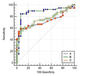
In some non-small cell lung cancer (NSCLC) patients, lipid-poor adrenal adenomas cannot be adequately differentiated from metastases using imaging methods. Invasive diagnostic procedures also have a low negative predictive value (NPV) in such cases. The current study aims to establish a specific and clinically practical metabolic parameter for lipid-poor adrenal lesions (ALs) in NSCLC patients. This diagnostic approach may prevent unnecessary abdominal enhanced computed tomography (CT), magnetic resonance imaging, or invasive diagnostic procedures. Sixty-four NSCLC patients with 69 lipid-poor ALs and 28 control patients with 30 benign lipid-poor ALs, who underwent FDG-PET/CT, were retrospectively reviewed. Two morphological and four metabolic parameters were analyzed in FDG-PET/CT images of NSCLC and control patients. Baseline and post-chemotherapy images of 64 NSCLC patients were re-evaluated according to the PERCIST 1.0. In cases where ALs could not be differentiated, follow-up FDG-PET/CT images were re-examined. The receiver operating characteristic (ROC) curve method was used for the evaluation of diagnostic parameters. Out of 69 ALs, 39 were determined as metastatic lesions (adrenal metastasis), while 30 lesions were considered non-metastatic (adrenal adenomas). The mean attenuation value, SUVmax AL/SUVmax primary tumor, SUVmax, SUVmax AL/liver, and SUVmax AL/SUVmean liver were significantly different between metastatic and benign ALs from NSCLC patients. The SUVmax AL/SUVmean liver ≥1.81 had the best positive (PPV, 94.3%) and negative (NPV, 82.4%) predictive values, and the highest specificity (93.3%), sensitivity (84.6%) and accuracy (86.9%). Lipid-poor ALs with SUVmax AL/SUVmean liver ≥1.81 can be accepted as malignant in NSCLC. However, if SUVmax AL/SUVmean liver is <1.81, a pathologic examination is required. Utilizing this cut-off value to decide on adrenal core biopsy may prevent its unnecessary use. Moreover, this diagnostic approach can save time and reduce the healthcare costs.
Citations
Downloads

Downloads
Additional Files
Published
Issue
Section
Categories
How to Cite
Accepted 2019-01-02
Published 2019-05-20


